QUOTE FOR TUESDAY:

“Some types of glaucoma are caused by other medical conditions — but for other people, the doctor doesn’t find another condition that causes it. When the doctor doesn’t find another cause, it’s called primary glaucoma.

Sometimes glaucoma is caused by another medical condition — this is called secondary glaucoma.”

National Eye Institute – NIH (Types of Glaucoma | National Eye Institute)

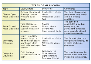
Part I National Glaucoma Awareness: Definition of Glaucoma, the Primary Types of Glaucoma and their symptoms.

   glaucoma2                      

 Glaucoma-Table

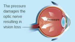
A common eye condition in which the fluid pressure inside the eye rises to a level higher than healthy for that eye. If untreated, it may damage the optic nerve, causing the loss of vision or even blindness. The elderly, African-Americans, and people with family histories of the disease are at greatest risk.

Glaucoma is a multi-factorial, complex eye disease with specific characteristics such as optic nerve damage and visual field loss. While increased pressure inside the eye (called intraocular pressure or IOP) is usually present, even patients with normal range IOP can develop glaucoma.

There is no specific level of elevated eye pressure that definitely leads to glaucoma; conversely, there is no lower level of IOP that will absolutely eliminate a person’s risk of developing glaucoma. That is why early diagnosis and treatment of glaucoma is the key to preventing vision loss.

Eye pressure is measured in millimeters of mercury (mm Hg). Normal eye pressure ranges from 12-22 mm Hg, and eye pressure of greater than 22 mm Hg is considered higher than normal. When the IOP is higher than normal but the person does not show signs of glaucoma, this is referred to as ocular hypertension.

High eye pressure alone does not cause glaucoma. However, it is a significant risk factor. Individuals diagnosed with high eye pressure should have regular comprehensive eye examinations by an eyecare professional to check for signs of the onset of glaucoma.

A person with elevated IOP is referred to as a glaucoma suspect, because of the concern that the elevated eye pressure might lead to glaucoma. The term glaucoma suspect is also used to describe those who have other findings that could potentially, now or in the future, indicate glaucoma. For example, a suspicious optic nerve, or even a strong family history of glaucoma, could put someone in the category of a glaucoma suspect.

Vision loss from glaucoma occurs when the eye pressure is too high for the specific individual and damages the optic nerve. Any resultant damage cannot be reversed. The peripheral (side) vision is usually affected first. The changes in vision may be so gradual that they are not noticed until a lot of vision loss has already occurred.

In time, if the glaucoma is not treated, central vision will also be decreased and then lost; this is how visual impairment from glaucoma is most often noticed. The good news is that glaucoma can be managed if detected early, and with medical and/or surgical treatment, most people with glaucoma will not lose their sight.

Most common signs and symptoms of Glaucoma:

There are several forms of glaucoma; the two most common forms are primary open-angle glaucoma (POAG) and angle-closure glaucoma (ACG). Open-angle glaucoma is often called “the sneak thief of sight” because it has no symptoms until significant vision loss has occurred.

Types of Glaucoma:

1-Open Angle Glaucoma

2-Angle Closure Glaucoma

3-Normal Tension Glaucoma

1-Open-Angle Glaucoma

Open-angle glaucoma, the most common form of glaucoma, accounting for at least 90% of all glaucoma cases:

  • Is caused by the slow clogging of the drainage canals, resulting in increased eye pressure
  • Has a wide and open angle between the iris and cornea
  • Develops slowly and is a lifelong condition
  • Has symptoms and damage that are not noticed.

“Open-angle” means that the angle where the iris meets the cornea is as wide and open as it should be. Open-angle glaucoma is also called primary or chronic glaucoma. It is the most common type of glaucoma, affecting about three million Americans.

Symptoms of Open-Angle Glaucoma

There are typically no early warning signs or symptoms of open-angle glaucoma. It develops slowly and sometimes without noticeable sight loss for many years.

Most people who have open-angle glaucoma feel fine and do not notice a change in their vision at first because the initial loss of vision is of side or peripheral vision, and the visual acuity or sharpness of vision is maintained until late in the disease.

By the time a patient is aware of vision loss, the disease is usually quite advanced. Vision loss from glaucoma is not reversible with treatment, even with surgery.

Because open-angle glaucoma has few warning signs or symptoms before damage has occurred, it is important to see a doctor for regular eye examinations. If glaucoma is detected during an eye exam, your eye doctor can prescribe a preventative treatment to help protect your vision.

In open-angle glaucoma, the angle in your eye where the iris meets the cornea is as wide and open as it should be, but the eye’s drainage canals become clogged over time, causing an increase in internal eye pressure and subsequent damage to the optic nerve. It is the most common type of glaucoma, affecting about four million Americans, many of whom do not know they have the disease.

You are at increased risk of glaucoma if your parents or siblings have the disease, if you are African-American or Latino, and possibly if you are diabetic or have cardiovascular disease. The risk of glaucoma also increases with age.

2-Angle-Closure Glaucoma

Angle-closure glaucoma, a less common form of glaucoma:

  • Is caused by blocked drainage canals, resulting in a sudden rise in intraocular pressure
  • Has a closed or narrow angle between the iris and cornea
  • Develops very quickly
  • Has symptoms and damage that are usually very noticeable
  • Demands immediate medical attention.

It is also called acute glaucoma or narrow-angle glaucoma. Unlike open-angle glaucoma, angle-closure glaucoma is a result of the angle between the iris and cornea closing.

Symptoms of Angle-Closure Glaucoma

  • Hazy or blurred vision
  • The appearance of rainbow-colored circles around bright lights
  • Severe eye and head pain
  • Nausea or vomiting (accompanying severe eye pain)
  • Sudden sight loss

Angle-closure glaucoma is caused by blocked drainage canals in the eye, resulting in a sudden rise in intraocular pressure. This is a much more rare form of glaucoma, which develops very quickly and demands immediate medical attention

In contrast with open-angle glaucoma, symptoms of acute angle-closure glaucoma are very noticeable and damage occurs quickly. If you experience any of these symptoms, seek immediate care from an ophthalmologist.

If you are diagnosed with glaucoma, it is important to set a regular schedule of examinations with your eye doctor to monitor your condition and make sure that your prescribed treatment is effectively maintaining a safe eye pressure.

3-Normal-tension glaucoma

Normal-tension glaucoma is a type of open-angle glaucoma that happens in people with normal eye pressure.

You may be at higher risk for normal-tension glaucoma if you:

  • Are of Japanese ancestry
  • Have a family history of normal-tension glaucoma
  • Have had certain heart problems, like an irregular heartbeat
  • Have low blood pressure

Signs and symptoms of Normal-Tension Glaucoma:

  • Reduced vision
  • Loss of peripheral vision
  • Bulging or swollen cornea
  • Pupil dilation that doesn’t change with light adjustments
  • Redness in the white (sclera) of the eye

Experts don’t know what causes normal-tension glaucoma, but research shows that treatments that lower eye pressure can help slow the disease and stop vision loss.

For Treatments to ALL 3 primary types of Glaucoma listed above are:

Medicines, laser treatment, surgery.

Tomorrow stay tune for part II on glaucoma awareness giving other types of glaucoma, how its diagnosed & the risks.

QUOTE FOR MONDAY:

“In most children, the cause of tetralogy of Fallot isn’t known. It’s a common type of heart defect. It may be seen more commonly in children with Down syndrome or DiGeorge syndrome. Some children can have other heart defects along with tetralogy of Fallot.  Infants and young children with unrepaired tetralogy of Fallot are often blue (cyanotic-not enough oxygen getting to their tissues).”

AHA American Heart Association (www.heart.org/en/health-topics/congenital-heart-defects/about-congenital-heart-defects)

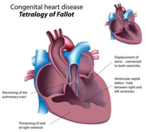
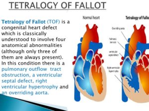
Part III Congenital Birth Defects Cardiac-Tetralogy of Fallot

What is it?

A heart defect that features four problems.

They are:

  • a hole between the lower chambers of the heart
  • an obstruction from the heart to the lungs
  • The aorta (blood vessel) lies over the hole in the lower chambers
  • The muscle surrounding the lower right chamber becomes overly thickened

More information for parents of children with Tetralogy of Fallot

What causes it?

In most children, the cause of tetralogy of Fallot isn’t known. It’s a common type of heart defect. It may be seen more commonly in children with Down syndrome or DiGeorge syndrome. Some children can have other heart defects along with tetralogy of Fallot.

How does it affect the heart?

Normally the left side of the heart only pumps blood to the body, and the heart’s right side only pumps blood to the lungs. In a child with tetralogy of Fallot, blood can travel across the hole (VSD) from the right pumping chamber (right ventricle) to the left pumping chamber (left ventricle) and out into the body artery (aorta). Obstruction in the pulmonary valve leading from the right ventricle to the lung artery prevents the normal amount of blood from being pumped to the lungs. Sometimes the pulmonary valve is completely obstructed (pulmonary atresia).

How does tetralogy of Fallot affect my child?

Infants and young children with unrepaired tetralogy of Fallot are often blue (cyanotic). The reason is that some oxygen-poor blood is pumped to the body through the hole in the wall between the right and left ventricle instead of being pumped to the lungs.

What can be done about tetralogy of Fallot?

Tetralogy of Fallot is treated surgically. A temporary operation may be done at first if the baby is small or if there are other problems. Complete repair comes later. Sometimes the first operation is a complete repair.

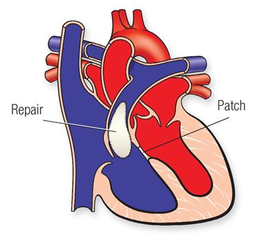
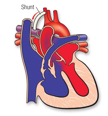
Tetralogy of Fallot patch diagramTetralogy of Fallot shunt diagram

Temporary Operation

In some infants, a shunt operation may be done first to provide adequate blood flow to the lungs. This is not open-heart surgery and doesn’t fix the inside of the heart. The shunt is usually a small tube of synthetic material sewn between a body artery (or the aorta) and the pulmonary artery. The shunt is closed when a complete repair is done later.

Complete Repair

Complete repair tends to be done early in life. The surgeon closes the ventricular septal defect with a patch and opens the right ventricular outflow tract by removing some thickened muscle below the pulmonary valve, repairing or removing the obstructed pulmonary valve and, if needed, enlarging the branch pulmonary arteries that go to each lung.

Sometimes a tube is placed between the right ventricle and the pulmonary artery. This is sometimes called a Rastelli repair. It’s similar to the type of repair used for some other heart defects.

Will my child’s activities be limited?

Your child may need to limit physical activity, particularly for competitive sports, if there is leftover obstruction or leak in the pulmonary valve, which is common after repair. Children with decreased heart function or rhythm disturbances may need to limit their activity more.

If the tetralogy has been repaired with surgery, and there’s no obstruction or leak in the pulmonary valve, your child may be able to participate in normal activities without much increased risk.

Your child’s pediatric cardiologist will help decide if your child needs limits on physical activity.

What will my child need in the future?

If your child has had tetralogy of Fallot repaired, he or she will need regular follow-up with a pediatric cardiologist. As an adult, your child will need lifelong regular follow-up with a cardiologist who’s had special training in congenital heart defects.

Some long-term problems can include leftover or worsening obstruction between the right pumping chamber and the lung arteries. Children with repaired tetralogy of Fallot have a higher risk of heart rhythm disturbances called arrhythmias. Sometimes these may cause dizziness or fainting.

Generally, the long-term outlook is good, but some children may need medicines, heart catheterization or even more surgery.

What about preventing endocarditis?

Children with tetralogy of Fallot are at increased risk for endocarditis. Some children, including those have had a valve replacement, still have a shunt or have leaks around surgical patches, and need to take antibiotics before certain dental procedures to help prevent endocarditis.

QUOTE FOR THE WEEKEND:

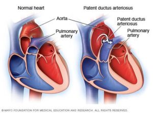
“The ductus arteriosus is a normal fetal artery connecting the aorta and the main lung artery (pulmonary artery). The ductus allows blood to detour away from the lungs before birth.

Every baby is born with a ductus arteriosus. After birth, the opening is no longer needed and it usually narrows and closes within the first few days.

Sometimes, the ductus doesn’t close after birth. Failure of the ductus to close is common in premature infants but rare in full-term babies. In most children, the cause of Patent Ductus Arteriosus-PDA isn’t known. Some children can have other heart defects along with the PDA.

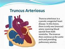
Truncus arteriosus occurs when the two large arteries carrying blood away from the heart don’t form properly and one large artery is present instead. This artery (the truncus) sits over a large opening or hole in the wall between the two pumping chambers (ventricular septal defect). With only one artery, there is no specific path to the lungs for oxygen before returning to the heart to deliver oxygen to the body.”

American Heart Association-AHA   (www.heart.org/en/health-topics/congenital-heart-defects/about-congenital-heart-defects)

Part II Congenital Cardiac Defects Awareness Month-Patent ductus arteriosus & Truncus arteriosus

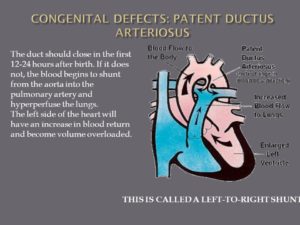
The duct should close in the first hours after birth. If it does not, the blood begins to shunt from the aorta into the pulmonary artery and hyperperfuse the lungs. The left side of the heart will have an increase in blood return and become volume overloaded. Too much blood is going to the lungs. RA – RV – Lungs _ LA – LV – Aorta now blood shunts backwards because pressure in L side higher than R so pressure in aorta in higher it backflows (it is already oxygenated) and prevents the blood that needs to be oxyenate doesn’t get there. Dispalces blood that needs to be oxygenated. Mixed blood in oxygenation. L sided heart failure. L to R shunt. THIS IS CALLED A LEFT-TO-RIGHT SHUNT.

-1 Patent ductus arteriosus (PDA).

Simply put, this is a hole in your baby’s aorta that doesn’t close.

Aorta is the largest artery in the body, the aorta arises from the left ventricle of the heart, goes up (ascends) a little ways, bends over (arches), then goes down (descends) through the chest and through the abdomen to where ends by dividing into two arteries called the common iliac arteries that go to the legs which is called the femoral artery than.

Anatomically, the aorta is traditionally divided into the ascending aorta, the aortic arch, and the descending aorta. The descending aorta is, in turn, subdivided into the thoracic aorta (that descends within the chest) and the abdominal aorta (that descends within the belly).

The aorta gives off branches that go to the head and neck, the arms, the major organs in the chest and abdomen, and the legs. It serves to supply them all with oxygenated blood. The aorta is the central conduit from the heart to the body. A hole in the aorta causes many problems.

During pregnancy, the hole allows your baby’s blood to bypass his lungs and get oxygen from your umbilical cord. After he’s born, he starts to get oxygen from his own lungs, and the hole has to close.

If it doesn’t, it’s called patent ductus arteriosus, or PDA. Small PDAs may get better on their own. A larger one could need surgery.

 2-Truncus arteriosus.

This is when your baby is born with one major artery instead of two that carry blood to the rest of his body. He will need surgery as an infant to repair the defect, and may need more procedures later in life.

I-transposition of the great arteries. This means that the right and left chambers of your baby’s heart are reversed. His blood still flows normally, but over time, his right ventricle doesn’t work as well because it must pump harder.

D-transposition of the great arteries. In this condition, the two main arteries of your baby’s heart are reversed. His blood doesn’t move through the lungs to get oxygen, and oxygen-rich blood doesn’t flow throughout his body. He will have to have surgery to repair this condition, usually within the first month of his life.

Single ventricle defects. Babies are sometimes born with a small lower chamber of the heart, or with one valve missing. Different types of single ventricle defects include:

  • Hypoplastic left heart syndrome: Your baby has an undeveloped aorta and lower left chamber, or ventricle.
  • Pulmonary atresia/intact ventricular septum: Your baby has no pulmonary valve, which controls blood flow from the heart to the lungs.
  • Tricuspid atresia: Your baby has no tricuspid valve, which should be between the upper and lower chambers of the right side of his heart.

Which Type?

In some cases, your doctor can spot congenital heart problems when your baby is still in the womb. But he can’t always diagnose the defect until after birth and until your baby shows signs of a problem.

Many mild congenital heart defects are diagnosed in childhood or even later because they don’t cause any obvious symptoms. Some people don’t find out they have them until they’re adults.

Whatever the type of congenital heart defect, rest assured that with advances in diagnostic tools and treatments, there’s a much greater chance of a long, normal life than ever before.

Heart transplants are recommended for children who have serious heart problems. These children are not able to live without having their heart replaced. Illnesses that affect the heart in this way include complex congenital heart disease, present at birth at times. They also include heart muscle disease (cardiomyopathy).


QUOTE FOR FRIDAY:

“Every 4.5 minutes a baby is born with a condition that affects the structure or function of their body. Collectively, these conditions are referred to as birth defects. They can vary widely in how and where they affect the body and include things from cleft lip to heart problems. While medical advancements have greatly improved health and survival, many of these conditions are lifelong and require lifelong care.

Achieving the best possible health will look different for different people. No two people living with these conditions are exactly alike. Everyone’s journey is unique, shaped by their specific condition, individual strengths, and the support system in place. However, a community of support can help people with these conditions no matter what health experiences they are navigating.”

Centers for Disease Control and Prevention – CDC

(Birth Defects Awareness Month | Birth Defects | CDC)

Part I Congenital Cardiac Defects Awareness Month-Hole defects, valve defects, atresia defects, and Epstein’s anomaly of the heart.

Your heart has 4 chambers=

2 uppers=atriums & 2 lowers=ventricles.

Congenital heart defects:

1.  Types of Hole Defects in the heart:

This a problem with your heart that you’re born with.

-They’re the most common kind of birth defect.

There are at least 18 different types of congenital heart defects. Most affect the walls, valves, or blood vessels of your heart. Some are serious and may need several surgeries and treatments.

1-Heart Septal Defects=Hole(s) in the Heart

This means you’re born with a hole in the wall, or septum of the heart that separates the left and right sides of your heart. The hole lets blood from the two sides mix.  This allows more oxygenated (L side of the heart) and more carbon dioxide blood (Rt side of the heart) mix together causing many problems.

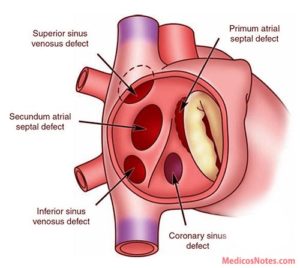
a Atrial Septal Defect (ASD)

An ASD is a hole in the wall between the upper chambers or the right and left atrium, of your heart.

Some ASDs close on their own. Your doctor may need to repair a medium or large ASD with open-heart surgery or another procedure.

The cardiology surgeon might seal the hole with a minimally invasive catheter procedure. The MD inserts a small tube, or catheter, in your blood vessel all the way to your heart. Then he can cover the hole with a variety of devices.

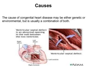
b Ventricular Septal Defect (VSD)

A ventricular septal defect (pronounced ven·tric·u·lar sep·tal de·fect) (VSD) is a birth defect of the heart in which there is a hole in the wall (septum) that separates the two lower chambers (ventricles) of the heart. This wall also is called the ventricular septum.

A VSD is a hole in the part of your septum that separates your heart’s lower chambers that we call ventricles. If you have a VSD, blood gets pumped back to your lungs instead of to your body.

A small VSD may also close on its own. But if yours is larger, you may need surgery to repair it.  Similar to ASD surgery, just a different spot on the chambers.

c Complete Atrioventricular Canal Defect (CAVC)

This is the most serious septal defect. It’s when you have a hole in your heart that affects all four chambers.

Complete atrioventricular canal (CAVC) defect is a severe congenital heart disease in which there is a large hole in the tissue (the septum) that separates the left and right sides of the heart. The hole is in the center of the heart, where the upper chambers and lower chambers meet.

As the heart formed abnormally, the valves that separate the upper and lower chambers also developed abnormally. In a normal heart, two valves separate the upper and lower chambers of the heart: the tricuspid valve separates the right chambers and the mitral valve the left chambers. In a child with a complete atrioventricular canal defect, there is one large valve, and it may not close correctly.

As a result of the abnormal passageway between the two sides of the heart, blood from both sides mix, and too much blood circulates back to the lungs before it travels through the body. This means the heart works harder than it should have to, often becoming enlarged and damaged if the problems aren’t repaired.

A CAVC prevents oxygen-rich blood from going to the right places in your body. Your doctor can repair it with patches. But some people need more than one surgery to treat it.

2-Types of Heart Valve Defects

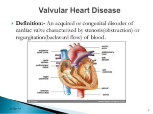
Valves control the flow of blood through your heart’s ventricles and arteries. And some minor heart defects can involve the valves.  Your heart has four valves  aortic, pulmonic, tricuspid, and mitral valve.  All four of them can develop different kinds of heart valve disease, this would include stenosis, regurgitation, and atresia.

a Stenosis.

When your cardiac valves become involved with stenosis where they narrow or stiffen, and won’t open or allow properly from a little amount to completely closed.  Ranging the blood to pass easily to not at all depending on the extent of the stenosis to the valve.  This makes treatment range from none and just medication if needed at all to surgery later in life or immediately born, again depending on the extent of cardiac valvular stenosis.

Aortic Valve Stenosis:

Aortic valve stenosis is most common in the world and in elderly, becoming more and more common after age 65. Several diseases can also cause it to develop in people when they reach middle age. The bicuspid aortic valve is the most common cause of aortic stenosis in patients less than the age of 70 years in developed countries. Rheumatic valve disease is the most common cause in developing countries.  Although some people have aortic stenosis because of a congenital heart defect called a bicuspid aortic valve, this condition more commonly develops during aging as calcium or scarring damages the valve, that is a common cause for this condition in elderly pts since it takes time to develop calcium build up in causing the stenosis.  This in turn restricts the amount of blood flowing getting through the aorta to deliver that oxygenated blood to the body.  The calcium build up takes time to put an affect on the aorta with symptoms as well; so this is why this is commonly seen in elders.

Aortic stenosis is one of the most common and serious valve disease problems. Know the aorta is the main artery bringing blood from the heart to the body’s tissues/organs. Aortic stenosis is a narrowing of the aortic valve opening, and can sometimes be referred to as a failing heart valve. Aortic stenosis restricts the blood flow from the left ventricle to the aorta and may also affect the pressure in the left atrium due to the blood flow regurgitating backwards from the stenosis to the L Ventricle up to the L atrium.

The condition may range from mild to severe; the longer you have it the more severe the symptoms and intensity it would have on the heart, especially left untreated.

Over time, aortic valve stenosis causes your heart’s left ventricle to pump harder to push blood through the narrowed aortic valve. The extra effort may cause the left ventricle to thicken, enlarge and weaken. If not addressed, this form of heart valve disease may lead to heart failure known as CHF.  This heart failure would start on the Left Side of the Heart first=L CHF and if left untreated it would in time effect the Right Side of the Heart=R CHF as well.  The symptoms of L CHF versus R CHF would be different at first and in another topic later this year (over extends this topic for today).

Pulmonary Valve Stenosis:

Pulmonary valve stenosis is a heart valve disorder that involves the pulmonary valve. This is the valve separating the right lower chamber or the right ventricle (one of the chambers in the heart) and the pulmonary artery.  Pulmonary artery is one of the few arteries with high carbon dioxide and low oxygen level in the blood stream. This blood flow normally is being sent to the lungs for more oxygen where it will send the blood flow to the left side of the heart to go through the aorta and sent throughout the body to give high oxygen to all organs.  This is how we survive; with out oxygen we would be going through cellular starvation and die.   The pulmonary artery carries oxygen-poor blood to the lungs  as its ending function. Stenosis, or narrowing in the pulmonary valve occurs when the valve cannot open wide enough to its normal capacity or in some cases completely stenosis, not able to open or close at all.

Treatment surgery at some time in the person’s lifetime.  It would all depend on the severity of the stenosis condition.

Mitral Valve Stenosis:

Mitral valve stenosis — sometimes called mitral stenosis — is a narrowing of the valve between the two left heart chambers. The narrowed valve reduces or blocks blood flow into the heart’s main pumping chamber. The heart’s main pumping chamber is the lower left heart chamber, also called the left ventricle.

There are two types of mitral valve regurgitation:

  • Degenerative mitral regurgitation: This occurs when the mitral valve itself is dysfunctional. The flaps may droop or bulge and do not close tightly.
  • Functional mitral regurgitation: Functional mitral regurgitation happens when an issue outside of the valve (such as diseases of the left ventricle) causes the leakage. You may have normal valve flaps and still be diagnosed with functional mitral regurgitation.

Mitral valve stenosis can make you tired and short of breath. Other symptoms may include irregular heartbeats, dizziness, chest pain or coughing up blood. Some people don’t notice symptoms.

Mitral valve stenosis can be caused by a complication by a sore throat with strep throat called rheumatic fever. Rheumatic fever is now rare in the United States.

Treatment for mitral valve stenosis may include medication or mitral valve repair or replacement surgery.  Some people only need regular health checkups. Treatment depends on the severity of the condition and whether it’s getting worse. Untreated, mitral valve stenosis can lead to serious heart complications.

Tricuspid Valve Stenosis:

Tricuspid stenosis (TS) is narrowing of the tricuspid orifice that obstructs blood flow from the right atrium to the right ventricle. Almost all cases result from rheumatic fever. Symptoms include a fluttering discomfort in the neck, fatigue, cold skin, and right upper quadrant abdominal discomfort. Jugular pulsations are prominent, and a presystolic murmur is often heard at the left sternal edge in the fourth intercostal space and is increased during inspiration. Diagnosis is by echocardiography. TS is usually benign, requiring no specific treatment, but symptomatic patients may benefit from surgery.

b Regurgitation.

A damaged or diseased valve can severely compromise the ability of the heart in inefficiently which in turn does not let  oxygen or carbon dioxide to be used or removed from the body in the way its suppose to be and in time effects the heart and can lead to heart failure if left untreated. Instead of the valve having stenosis it is now not closing completely when it should allowing regurgitation to occur.

Your valves don’t close tightly, the valve affected will allow regurgitation due to the valve not working efficiently with opening and closing the way it should during your heart beat (lub dub-the sound you hear of the heart when auscultating through the stethoscope, the RN or doctor uses.) Instead this lets your blood flow or leak backward r/t the valve partially open or to completely opened when the valve should be closely closed at that time and results into regurgitation.  Remember the cardiac valves open and close as their function to allow blood flowing with a heart pumping to deliver oxygen to the blood from red blood cells that carry O2 and remove carbon dioxide blood through red blood cells carrying it out of the blood stream back to the lungs to get more O2 for the RBCs to carrying it to organs / tissues that need it to survive.  Depending on the extent of valvular regurgitation will decide on treatment which ranges from surgery later in life or immediately when born.

It does the opposite of valve stenosis in that stenosis is a narrowing of the vessel whereas regurgitation is due to a valve remaining open partially or completely when the valve is suppose to be closing to allow blood fill up in the chamber of the heart and common one is mitral valve regurgitation but there are others like tricuspid, pulmonary and ventricular valve regurgitation.

Mitrial Valve Regurgitation:

Mitral valve regurgitation is the most common type of heart valve disease. In this condition, the valve between the left heart chambers doesn’t close fully. Blood leaks backward across the valve. If the leakage is severe, not enough blood moves through the heart or to the rest of the body. Mitral valve regurgitation can make you feel very tired or short of breath.

Other names for mitral valve regurgitation are:

  • Mitral regurgitation (MR).
  • Mitral insufficiency.
  • Mitral incompetence.

Treatment of mitral valve regurgitation may include regular health checkups, medicines or surgery. You may not need treatment if the condition is mild.

Severe mitral valve regurgitation often requires a catheter procedure or heart surgery to repair or replace the mitral valve. Without proper treatment, severe mitral valve regurgitation can cause heart rhythm problems or heart failure.

Tricuspid Valve Regurgitation:

Tricuspid valve disease is a type of heart valve disease (valvular heart disease). The valve between the two right heart chambers (right atrium and right ventricle) doesn’t work properly. As a result, the heart must work harder to send blood to the lungs and the rest of the body.

Tricuspid valve disease often occurs with other heart valve problems.

Symptoms and treatments of tricuspid valve disease vary, depending on the specific valve condition. Treatment may include monitoring, medication, or valve repair or valve replacement.

The most common cause of tricuspid regurgitation is enlargement of the right ventricle. Pressure from heart conditions, such as heart failure, pulmonary hypertension and cardiomyopathy, cause the ventricle to expand. The result is a misshapen tricuspid valve that is leaky and cannot close properly.

Aortic Valve Regurgitation:

Aortic valve regurgitation — also called aortic regurgitation — is a type of heart valve disease. The valve between the lower left heart chamber and the body’s main artery doesn’t close tightly. As a result, some of the blood pumped out of the heart’s main pumping chamber, called the left ventricle, leaks backward.

The leakage may prevent the heart from doing a good enough job of pumping blood to the rest of the body. You may feel tired and short of breath.

Aortic valve regurgitation can develop suddenly or over many years. Once the condition becomes severe, surgery often is needed to repair or replace the valve.

Pulmonary Valve Regurgitation:

Pulmonary valve disease affects the valve between the heart’s lower right chamber and the artery that delivers blood to the lungs. That artery is called the pulmonary artery. The valve is called the pulmonary valve.

A diseased pulmonary valve doesn’t work properly. Pulmonary valve disease changes how blood flows from the heart to the lungs.

The pulmonary valve usually acts like a one-way door from the lower right heart chamber to the lungs. Blood flows from the chamber through the pulmonary valve. It then goes to the pulmonary artery and into the lungs. Blood picks up oxygen in the lungs to take to the body.

Many types of pulmonary valve disease are due to heart conditions present at birth. Treatment depends on the type and severity of pulmonary valve disease.

c Atresia.

Tricuspid Atresia:

This happens when your valve isn’t formed right or has no opening to let your blood pass through. It causes more complicated heart problems. Tricuspid atresia (pronounced try-CUSP-id uh-TREE-zhuh) is a birth defect of the heart where the valve that controls blood flow from the right upper chamber of the heart to the right lower chamber of the heart doesn’t form at all.  In babies with this defect, blood can’t flow correctly through the heart and to the rest of the body.

Aortic Atresia:

Aortic valvular atresia is a congenital condition in which the aortic valvular cusps are fused at birth. It frequently forms as a spectrum of malformations of the left ventricular outflow tract (LVOT). The atresia can be characterized as sub-valvular, valvular, or supra-valvular, depending on the site of the anomaly. Most commonly, the defect presents as aortic stenosis, though in rare cases, it can manifest as complete atresia.

Pulmonary Atresia

Pulmonary atresia is a birth defect of the pulmonary valve, which is the valve that controls blood flow from the right ventricle (lower right chamber of the heart) to the main pulmonary artery (the blood vessel that carries blood from the heart to the lungs). Pulmonary atresia is when this valve didn’t form at all, and no blood can go from the right ventricle of the heart out to the lungs. Because a baby with pulmonary atresia may need surgery or other procedures soon after birth, this birth defect is considered a critical congenital heart defect (critical CHD). Congenital means present at birth.

In a baby without a congenital heart defect, the right side of the heart pumps oxygen-poor blood from the heart to the lungs through the pulmonary artery. The blood that comes back from the lungs is oxygen-rich and can then be pumped to the rest of the body. In babies with pulmonary atresia, the pulmonary valve that usually controls the blood flowing through the pulmonary artery is not formed, so blood is unable to get directly from the right ventricle to the lungs.

In pulmonary atresia, since blood cannot directly flow from the right ventricle of the heart out to the pulmonary artery, blood must use other routes to bypass the unformed pulmonary valve. The foramen ovale, a natural opening between the right and left upper chambers of the heart during pregnancy that usually closes after the baby is born, often remains open to allow blood flow to the lungs. Additionally, doctors may give medicine to the baby to keep the baby’s patent ductus arteriosus open after the baby’s birth. The patent ductus arteriosus is the blood vessel that allows blood to move around the baby’s lungs before the baby is born and it also usually closes after birth.

d Ebstein’s anomaly.

This is a defect in another heart valve, the tricuspid valve-between the top and bottom of the right side of the heart, which may keep it from closing tightly. Ebstein anomaly is a rare heart problem present at birth. This means it’s a congenital heart defect. The tricuspid valve is incorrectly formed and located lower than usual in the heart. The condition may occur with a hole between the two upper chambers of the heart, called an atrial septal defect.Babies who have Ebstein’s also often have an atrial septal defect (ASD).

In people with Ebstein anomaly, the heart can grow larger. The condition can lead to heart failure.

Treatment of Ebstein anomaly depends on the symptoms. Some people without symptoms only need regular health checkups. Others may need medicines and surgery.

Keep in mind a baby can be born with more than one cardiac defect depending on the pt’s heart formation during pregnancy or at birth or in some cases symptoms arise later in life and that could be the time the patient is diagnosed with the heart condition they have.  Symptoms help tell the doctor there is a problem so diagnosing gets involved until the etiology is found.  If it is your heart your MD will find the defect no problem.

Part II tomorrow on other defects!


QUOTE FOR THURSDAY:

“Congenital heart disease is a heart problem that a person is born with. It means that the heart’s structure — for example, its walls or valves — did not develop properly before birth. More than a million American adults live with congenital heart disease. Many were diagnosed as infants and had surgery or other treatment as a child. Others don’t know they have congenital heart disease until they develop symptoms as adults, like shortness of breath or an abnormal heartbeat.

No matter the cause or the age of diagnosis, people with congenital heart disease need continued care and treatment throughout their lives. It can be difficult for adults to find heart specialists familiar with their particular anatomy and problems–NewYork-Presbyterian has one of the few programs in the country with the depth and breadth of expertise to address the complexity of congenital heart disease in adults.

Our highly trained heart specialists understand congenital heart disease’s unique physiological, anatomical, and clinical features at all ages. Your care team includes physicians, surgeons, and genetic experts specializing in adult congenital heart disease. They’re experts in echocardiography, interventional cardiac catheterization, interventional electrophysiology, complex congenital heart surgery, and heart transplantation.

With this team approach, we address the complex needs of people with adult congenital heart disease at three centers where you can receive treatment in a hospital setting designed for adults without having to stay in a children’s hospital:

  • Schneeweiss Adult Congenital Heart Center at New York-Presbyterian/Columbia University Irving Medical Center is one of the nation’s oldest, largest, and most experienced adult congenital heart disease programs.
  • Cornell Center for Adult Congenital Heart Disease at New York-Presbyterian Weill Cornell Medical Center
  • Ronald O. Perelman Heart Institute, NYP/Weill Cornell”

New York Presbyterian Hospital

(Congenital Heart Disease | NewYork-Presbyterian)

What to know in Prevention for Congenital Defects.

Though not all birth defects can be avoided, prenatal care and awareness of past or current conditions can help with prevention.

  • Prenatal care. Taking a daily prenatal vitamin that includes at least 400 micrograms of folic acid can help prevent a variety of birth defects. You should be taking prenatal vitamins if you’re of reproductive age, when you’re actively trying to conceive and/or as soon as you find out you are pregnant. In addition to taking your prenatal vitamin, avoiding alcohol, tobacco and illegal drugs can significantly help prevent birth defects and pregnancy complications.
  • Awareness of past or current conditions. If you previously had a pregnancy with a birth defect, it’s important to find out the most likely causes because it can help your physician plan preventive measures for your next pregnancy. For example, spina bifida is caused by a deficiency in folate, so if your previous pregnancy had spina bifida, you can take a high dose of folate to help prevent future spina bifida diagnoses.

Are all birth defects discovered before a baby is born?

It’s not always possible to detect all birth defects in utero. However, high-resolution ultrasounds done by certified prenatal ultrasound groups make it possible to diagnose defects that will cause a significant impact before birth.

Baschat says: “At the Center for Fetal Therapy, we recommend that pregnant women have the first-trimester nuchal translucency scan between 11 and 14 weeks and the anatomy scan between 18 and 20 weeks. These two ultrasounds provide us with the best opportunity for detecting birth defects.”

In recent years, some pregnant women have turned down the first-trimester ultrasound because a maternal screening blood test now exists for Down syndrome. The ultrasound is still strongly recommended, though, because there are many other serious birth defects that can be detected this early in pregnancy.

Is it possible to treat birth defects while a baby is in utero?

Absolutely. While there are many different types of birth defects, it’s extremely important to try to correct those that damage vital organs before the baby is born. The Center for Fetal Therapy specializes in treating several of these defects in utero, including:

  • Congenital diaphragmatic hernia. This condition, in which a hole in the diaphragm allows abdominal content to enter the chest and restrict lung development, can be significantly helped in utero through fetoscopic endotracheal occlusion, a surgery that improves lung function and significantly increases survival rates.
  • Lower urinary tract obstruction. This occurs when the flow of urine is blocked from exiting the fetus’ body, leading to permanent kidney damage. Relieving this obstruction before birth protects the kidneys.

Fetal treatments also exist for conditions that make the baby unhealthy, even if they are not considered to be birth defects. For example, if a fetus has an irregular heartbeat, you can give the mother medication that will cross the placenta and treat the fetus.

How successful are in utero treatments for fetal conditions?

By treating fetal conditions in utero rather than waiting until after birth, fetuses are given significantly better chances of survival and a reduced need for major surgery after birth. For example, with a condition like twin-to-twin transfusion syndrome, in which identical twins develop a blood volume imbalance, both babies could die without any intervention. By performing laser surgery in utero, there is approximately a 95 percent chance that at least one baby will survive.

Explains Baschat, “Success rates vary depending on the condition, available treatments and the individual patient, but overall, where fetal interventions are available, we see a much higher rate of survival for affected fetuses.”

If a baby has fetal surgery, will he/she need different care after birth?

This will depend on the individual condition and type of surgery performed. For all fetal surgeries, your baby needs to be delivered at a hospital where pediatric subspecialties are in-house so the baby’s care can be managed after birth. Baschat says: “Many of the treatments we perform require patients to deliver at the highest-level multispecialty hospital, like The Johns Hopkins Hospital. This way, all prenatal and postnatal care is available to you in one location.”

If you have one child affected by a birth defect, will all of your future children have that same condition?

All future children will not definitively be affected by the same birth defect, but it will depend on what the cause was. If the birth defect was caused by a genetic mutation, there may be a higher likelihood of recurrence, but if you seek care from a specialized center, maternal-fetal medicine specialists and genetic counselors can work with you to assess future risk.

The risk of some conditions can be determined before you become pregnant through genetic testing. If that doesn’t exist for the condition in question, maternal-fetal medicine specialists and genetic counselors can genetically test your fetus during pregnancy to see if she or he exhibits the mutation that affected your previous child.

Are there communities in which you can speak with other families who have had children affected by birth defects?

There are many forums online, whether on the web or social media platforms, in which parents come together. “At our center, we reach out to previous patients to see if they are interested in communicating with new patients affected by the same condition,” Baschat explains. “They can empathize about the specific birth defect, but they can also provide guidance and advice about the experience of working with our center throughout the pregnancy and after the baby is born.”

In the future, what advances will take place in treating disease before birth?

Through prenatal diagnosis and understanding fetal disease better, physicians are learning more about what harms the fetus and what may be of benefit before birth. Additionally, new uses for minimally invasive surgical equipment are regularly being discovered.

There are also stem cell therapies, genomic medicine and a whole host of related treatments that are currently used for children and adults. One day, these may also apply to the fetus. “While it’s hard to predict the future, we’re currently doing things we couldn’t have foreseen 10 years ago, so we’re very hopeful for continued advancements in fetal care,” says Baschat.