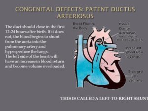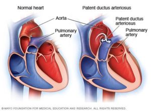
The duct should close in the first hours after birth. If it does not, the blood begins to shunt from the aorta into the pulmonary artery and hyperperfuse the lungs. The left side of the heart will have an increase in blood return and become volume overloaded. Too much blood is going to the lungs. RA – RV – Lungs _ LA – LV – Aorta now blood shunts backwards because pressure in L side higher than R so pressure in aorta in higher it backflows (it is already oxygenated) and prevents the blood that needs to be oxyenate doesn’t get there. Dispalces blood that needs to be oxygenated. Mixed blood in oxygenation. L sided heart failure. L to R shunt. THIS IS CALLED A LEFT-TO-RIGHT SHUNT.
-1 Patent ductus arteriosus (PDA).
Simply put, this is a hole in your baby’s aorta that doesn’t close.
Aorta is the largest artery in the body, the aorta arises from the left ventricle of the heart, goes up (ascends) a little ways, bends over (arches), then goes down (descends) through the chest and through the abdomen to where ends by dividing into two arteries called the common iliac arteries that go to the legs which is called the femoral artery than.
Anatomically, the aorta is traditionally divided into the ascending aorta, the aortic arch, and the descending aorta. The descending aorta is, in turn, subdivided into the thoracic aorta (that descends within the chest) and the abdominal aorta (that descends within the belly).
The aorta gives off branches that go to the head and neck, the arms, the major organs in the chest and abdomen, and the legs. It serves to supply them all with oxygenated blood. The aorta is the central conduit from the heart to the body. A hole in the aorta causes many problems.
During pregnancy, the hole allows your baby’s blood to bypass his lungs and get oxygen from your umbilical cord. After he’s born, he starts to get oxygen from his own lungs, and the hole has to close.
If it doesn’t, it’s called patent ductus arteriosus, or PDA. Small PDAs may get better on their own. A larger one could need surgery.
-2Truncus arteriosus.
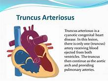
This is when your baby is born with one major artery instead of two that carry blood to the rest of his body. He will need surgery as an infant to repair the defect, and may need more procedures later in life.
I-transposition of the great arteries. This means that the right and left chambers of your baby’s heart are reversed. His blood still flows normally, but over time, his right ventricle doesn’t work as well because it must pump harder.
D-transposition of the great arteries. In this condition, the two main arteries of your baby’s heart are reversed. His blood doesn’t move through the lungs to get oxygen, and oxygen-rich blood doesn’t flow throughout his body. He will have to have surgery to repair this condition, usually within the first month of his life.
Single ventricle defects. Babies are sometimes born with a small lower chamber of the heart, or with one valve missing. Different types of single ventricle defects include:
- Hypoplastic left heart syndrome: Your baby has an undeveloped aorta and lower left chamber, or ventricle.
- Pulmonary atresia/intact ventricular septum: Your baby has no pulmonary valve, which controls blood flow from the heart to the lungs.
- Tricuspid atresia: Your baby has no tricuspid valve, which should be between the upper and lower chambers of the right side of his heart.
Which Type?
In some cases, your doctor can spot congenital heart problems when your baby is still in the womb. But he can’t always diagnose the defect until after birth and until your baby shows signs of a problem.
Many mild congenital heart defects are diagnosed in childhood or even later because they don’t cause any obvious symptoms. Some people don’t find out they have them until they’re adults.
Whatever the type of congenital heart defect, rest assured that with advances in diagnostic tools and treatments, there’s a much greater chance of a long, normal life than ever before.
Heart transplants are recommended for children who have serious heart problems. These children are not able to live without having their heart replaced. Illnesses that affect the heart in this way include complex congenital heart disease, present at birth at times. They also include heart muscle disease (cardiomyopathy).

