Archives

Part III Types of unmodified risk factors (risk factors we can’t change) for breast cancer!

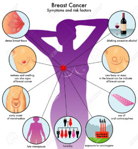
Unmodified Risk factors for Breast Cancer and those are factors we can’t change putting us at risk for breast cancer, which are:

DES Exposure

In the 1950s and 1960s, many pregnant women took a synthetic form of estrogen called diethylstilbestrol (DES) to prevent miscarriage. Many of these women’s daughters eventually developed vaginal and cervical cancer at a rate that seemed higher than normal, and studies found that DES exposure was indeed associated with an increased risk of these types of cancer.

Because of the exposure to additional estrogen, women who were exposed to DES in utero also may be at higher risk for breast cancer. A study published in October 2002 found that in women who were 40 years and older, breast cancer risk was in fact increased if a woman had been exposed to DES.

Because DES is a banned substance, people are no longer at risk for new exposures; however, if you know or suspect that your mother or grandmother took DES while pregnant, you should notify your doctor.

Age at Menstruation

A woman’s amount of exposure to estrogen and progesterone during her lifetime is believed to be a risk factor. The longer a woman is exposed, the more likely she is to develop breast cancer. Therefore, if a woman begins menstruation before age 12, she is believed to be at slightly higher risk.

Age at First Birth

It has been observed that women who have their first child after age 29, or who do not have any children, are at slightly higher risk for breast cancer than women who have their first child before age 29. It has been proposed that breast changes during pregnancy may have protective effects against cancer development because risk of breast cancer appears to decrease with each additional childbirth.

It is important to note that evidence suggests the opposite is true for women who have a family history of breast cancer. In other words, women who have a family history of breast cancer are at lower risk if they have no children or have their children at a later age.

Age at Menopause

Women who go through menopause after the age of 54 have a slightly higher risk of breast cancer than women who go through menopause at age 54 or younger. Their higher risk may be related to their higher lifetime exposure to estrogen and progesterone.

Atypical Hyperplasia or Atypia

Either atypical hyperplasia or atypia indicates the growth of abnormal cells in the breast. The diagnosis of atypical hyperplasia can be made from a core biopsy or excisional biopsy, and has been correlated with an increased risk of breast cancer.

The diagnosis of atypia can be made from nipple aspiration, ductal lavage, or fine needle aspiration (FNA), and also indicates an increased breast cancer risk. Although these cells are not yet cancerous, they do raise a woman’s risk of eventually developing breast cancer. While biopsies and FNAs are usually reserved for when there is a current indication that a woman might have breast cancer, nipple aspiration and ductal lavage are methods that may help assess a woman’s future risk of breast cancer.

Breast Density

Studies have consistently shown that higher breast density is linked with increased risk of breast cancer. Research is examining whether breast density may be modifiable by changing women’s hormones or diet. One medication that has been demonstrated to reduce breast density is tamoxifen.

Serum Estradiol Level

Estradiol is the predominant form of estrogen circulating in the body. ‘Serum estradiol’ refers to the amount of estradiol in the blood, so a woman’s level of serum estradiol may be measured with a simple blood test.

In postmenopausal women, higher hormone levels in the blood have been associated with an increased risk of breast cancer.
QUOTE FOR THE WEEKEND:

“A risk factor is anything that increases your chances of getting a disease, such as cancer. But having a risk factor, or even many, does not mean that you are sure to get the disease. While you can’t change some breast cancer risk factors—family history, sex, race and aging, for example—there are some risk factors that you can control like exercise, diet, and weight. Studies have shown that risk for breast cancer is due to a combination of factors. The main factors that influence risk include being a woman and getting older. Most breast cancers are found in women who are 50 years old or older. ”

Centers for Disease Control and Prevention CDC (https://www.cdc.gov/breast-cancer/risk-factors/index.html)

Part II Breast Cancer Awareness Month-Know the symptoms, if its metastatic or not, the common treatments for it.


Know the symptoms of breast cancer are:

CDC says this:

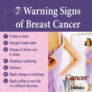
“Different people have different symptoms of breast cancer. Some people do not have any signs or symptoms at all.

Some warning signs of breast cancer are—

  • New lump in the breast or underarm (armpit).
  • Thickening or swelling of part of the breast.
  • Irritation or dimpling of breast skin.
  • Redness or flaky skin in the nipple area or the breast.
  • Pulling in of the nipple or pain in the nipple area.
  • Nipple discharge other than breast milk, including blood.
  • Any change in the size or the shape of the breast.
  • Pain in any area of the breast.”

Is there a link between birth control pills and breast cancer?

A number of older studies suggested that birth control pills slightly increased the risk of breast cancer, especially among younger women. In these studies, however, 10 years after discontinuing birth control pills women’s risk of breast cancer returned to the same level as that of women who never used oral contraceptives. Current evidence does not support an increase in breast cancer with birth control pills.

Be vigilant about breast cancer detection. If you notice any changes in your breasts, such as a new lump or skin changes, consult your doctor. Also, ask your doctor when to begin mammograms and other screenings.

Once you’ve been diagnosed with breast cancer, your doctor works to find out the specifics of your tumor. Using a tissue sample from your breast biopsy or using your tumor if you’ve already undergone surgery, your medical team determines your breast cancer type. This information helps your doctor decide which treatment options are most appropriate for you.  The biopsy helps what’s used to determine your breast cancer type.

Is your cancer invasive or noninvasive?

Whether your cancer is invasive or noninvasive helps your doctor determine whether your cancer may have spread beyond your breast, which treatments are more appropriate for you, and your risk of developing cancer in the same breast or your other breast.

  • Noninvasive (in situ) breast cancer. In situ breast cancer refers to cancer in which the cells have remained within their place of origin — they haven’t spread to breast tissue around the duct or lobule. One type of noninvasive cancer called ductal carcinoma in situ (DCIS) is considered a precancerous lesion. This means that if it were left in the body, DCIS could eventually develop into an invasive cancer. Another type of noninvasive cancer called lobular carcinoma in situ (LCIS) isn’t considered precancerous because it won’t eventually evolve into invasive cancer. LCIS does, however, increase the risk of cancer in both breasts.
  • Invasive breast cancer. Invasive (infiltrating) breast cancers spread outside the membrane that lines a duct or lobule, invading the surrounding tissues. The cancer cells can then travel to other parts of your body, such as the lymph nodes. If your breast cancer is stage I, II, III or IV, you have invasive breast cancer.

In what part of the breast did your cancer begin?

The type of tissue where your breast cancer arises determines how the cancer behaves and what treatments are most effective. Parts of the breast where cancer begins include:

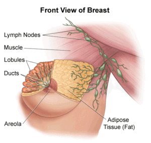
  • Milk ducts. Ductal carcinoma is the most common type of breast cancer. This type of cancer forms in the lining of a milk duct within your breast. The ducts carry breast milk from the lobules, where it’s made, to the nipple.
  • Milk-producing lobules. Lobular carcinoma starts in the lobules of the breast, where breast milk is produced. The lobules are connected to the ducts, which carry breast milk to the nipple.
  • Connective tissues. Rarely breast cancer can begin in the connective tissue that’s made up of muscles, fat and blood vessels. Cancer that begins in the connective tissue is called sarcoma. Examples of sarcomas that can occur in the breast include phyllodes tumor and angiosarcoma.

FYI a complication that can occur with advanced cancer that many of you may be unaware of; bone metastasis.

Bone metastasis occurs when cancer cells spread from their original site to a location in the bone. The most common types of cancer more likely to spread to bone include breast, prostate and lung cancers.

Bone metastasis can occur in any bone, but more commonly occurs in the pelvis and spine. Bone metastasis may be the first sign that you have cancer, or it may occur years after your cancer treatment is completed, ex. Hodgkins Disease.

Signs and symptoms of bone metastasis may include the following:

  • Bone pain (back and pelvic pain are most common)
  • Unexplained broken bones
  • Loss of urine and/or bowel function
  • Weakness in the legs
  • High levels of calcium in the blood (hypercalcemia), which can cause nausea, vomiting and confusion

The most common problem with metastatic bone cancer is pain and fractures. Metastatic bone cancer usually can’t be cured, but instead the goal is to provide pain relief and control further spread. Treatment can make a big difference and may include the following:

  • Medications to repair and build new bone — These medications are similar to those used by people with osteoporosis and can help in building and strengthening your bone.
  • Chemotherapy — Given as a pill or through a vein, used to control and treat cancer that has spread to the bone.
  • Traditional radiation therapy — Radiation is given as external beam therapy to treat the cancer in the bone.
  • Hormone therapy — Medications are used to block hormones (for breast and prostate cancers) that help control the spread of cancer to the bone.
  • Surgery — Used to fix a fracture and stabilize a break from the cancer in the bone.
  • Cryoablation — A special technique that freezes the cancer cells.
  • Radiofrequency ablation — A special technique that heats the cancer cells.
  • Chemoradiation — A form of internal radiation that is given through the vein and travels to the site of bone metastasis and targets the cancer cells.
  • Pain medications — Medications provided with the goal of relieving and controlling pain from bone metastasis.
  • Physical therapy — Exercises may be prescribed to assist in strengthening muscles and providing any assistive devices that may help you (cane, walker, crutches, etc.).

If you’re living with metastatic bone cancer, you may find help and resources from a website called Bone Health in Focus. It was established with partners including BreastCancer.org, the National Lung Cancer Partnership and Us TOO International Prostate Cancer Education & Support Network to offer resources that help patients and caregivers understand more about cancer that has spread to the bone (find the site at www.bonehealthinfocus.com).

Mayo Clinic information on cancer that has spread to the bone can be found at http://www.mayoclinic.org/diseases-conditions/bone-metastasis/basics/definition/con-20035450.

Are you living with cancer that has spread to the bone? Feel free to share your experiences with each other on the this blog striveforgoodhealth.com or on TheMayoclinic.org.

Make the changes in your lifestyles including diet if you want to prevent cancer, live long and have a productive life.

REFERENCE: Mayoclinic.org

Article reviewed 10/18/2022


QUOTE FOR FRIDAY:

“Breast cancer caused 670 000 deaths globally in 2022.

Roughly half of all breast cancers occur in women with no specific risk factors other than sex and age.

Breast cancer was the most common cancer in women in 157 countries out of 185 in 2022.

Breast cancer occurs in every country in the world.”

World Health Organization (https://www.who.int/news-room/fact-sheets/detail/breast-cancer)

Part I Breast Cancer Awareness Month: Know the different types of this cancer!


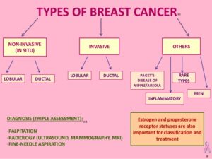
There are many types of breast cancer. The most common types are ductal carcinoma in situ, or invasive ductal carcinoma, and invasive lobular carcinoma.

When a biopsy is done to find out the specific type of breast cancer, the pathologist will also check if the cancer has spread into the surrounding tissues. The following terms are used to describe the extent of the cancer:

  • In situ breast cancers have not spread.
  • Invasive or infiltrating cancers have  spread (invaded) into the surrounding breast tissue.

The type of breast cancer is determined by the specific cells in the breast that are affected. Most breast cancers are carcinomas. Carcinomas are tumors that start in the epithelial cells that line organs and tissues throughout the body. Sometimes, an even more specific term is used. For example, most breast cancers are a type of carcinoma called adenocarcinoma, which starts in cells that make up glands (glandular tissue). Breast adenocarcinomas start in the ducts (the milk ducts) or the lobules (milk-producing glands).

There are other, less common, types of breast cancers, too, such as sarcomas, phyllodes, Paget disease, and angiosarcomas which start in the cells of the muscle, fat, or connective tissue.

Sometimes a single breast tumor can be a combination of different types. And in some very rare types of breast cancer, the cancer cells may not form a lump or tumor at all.

Common kinds of breast cancer

The most common kinds of breast cancer are carcinomas, and are named based on where they form and how far they have spread.

These general kinds of breast cancer below can be further described with the terms outlined above.

In situ cancers

Ductal carcinoma in situ (DCIS; also known as intraductal carcinoma) is a non-invasive or pre-invasive breast cancer.

Lobular carcinoma in situ (LCIS) may also be called lobular neoplasia. This breast change is not a cancer, though the name can be confusing. In LCIS, cells that look like cancer cells are growing in the lobules of the milk-producing glands of the breast, but they don’t grow through the wall of the lobules.

Invasive (infiltrating) breast cancer

Breast cancers that have spread into surrounding breast tissue are known as invasive breast cancer. There are many different kinds of invasive breast cancer, but the most common are called invasive ductal carcinoma and invasive lobular carcinoma.

Less common types of breast cancer

Inflammatory breast cancer

This is an uncommon type of invasive breast cancer. It accounts for about 1% to 5% of all breast cancers.

Paget disease of the nipple

This starts in the breast ducts and spreads to the skin of the nipple and then to the areola(the dark circle around the nipple). It is rare, accounting for only about 1-3% of all cases of breast cancer.

Phyllodes tumor

Phyllodes tumors are rare breast tumors. They develop in the connective tissue (stroma) of the breast, in contrast to carcinomas, which develop in the ducts or lobules. Most are benign, but there are others that are malignant (cancer).

Angiosarcoma

Sarcomas of the breast are rare making up less than 1% of all breast cancers. Angiosarcoma starts in cells that line blood vessels or lymph vessels. It can involve the breast tissue or the skin of the breast. Some may be related to prior radiation therapy in that area.


QUOTE FOR THURSDAY:

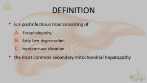
“Reye’s syndrome is a serious condition that causes swelling in the liver and brain. It can occur at any age but usually affects children and teenagers after a viral infection, most commonly the flu or chickenpox. Reye’s syndrome is rare. The condition also is known as Reye syndrome.

Symptoms such as confusion, seizures and loss of consciousness need emergency treatment. Early diagnosis and treatment of Reye’s syndrome can save a child’s life.”

MAYO CLINIC (https://www.mayoclinic.org/diseases-conditions/reyes-syndrome/symptoms-causes/syc-20377255)

Reye’s Syndrome

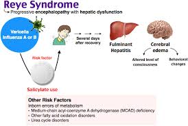
Reye’s syndrome is a rare but serious disease that causes swelling in the liver and brain. It can affect people of any age, but it is most often seen in children and teenagers recovering from a virus such as the flu or chickenpox.Studies have found that the main risk factor for Reye’s syndrome is taking aspirin or other related drugs, called salicylates.Because of this, doctors recommend that children and teenagers recovering from viral infections should avoid taking aspirin.

What Causes It?

Doctors don’t fully understand what causes Reye’s syndrome. They do know that some people are prone to get it when they take aspirin for a virus.

Others have a greater chance of getting it if they:

  • Have a disorder that affects how their bodies break down fatty acids
  • Have been exposed to certain toxins, including paint thinners and products to kill insects and weeds

When Reye’s syndrome strikes, cells throughout your body become swollen and build up fats. In turn, your blood sugar levels drop. Ammonia and acid levels in the blood rise. These changes can hit many organs, such as the brain and liver, where severe swelling can occur.

Symptoms

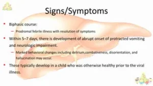
The signs of Reye’s typically appear 3 to 5 days after the start of a viral infection.

In children younger than age 2, early symptoms may include diarrhea and rapid breathing. In older children and teenagers, early symptoms may include ongoing vomiting and unusual sleepinessAs the syndrome goes on, symptoms can become more severe, and may include:

  • Personality changes (more irritable or aggressive)
  • Confusion or hallucinations
  • Weakness or inability to move arms or legs
  • Seizure or convulsions
  • Extreme tiredness
  • Loss of consciousness

Reye’s can be life-threatening. You should call 911 if you see these severe symptoms. Early diagnosis and treatment is crucial.

The syndrome can be mistaken for other conditions, including meningitis (a swelling of membranes covering the brain and spinal cord), a diabetes reaction, or poisoning.

Diagnosis

Doctors don’t have a specific test for Reye’s. They usually do urine and blood tests. They also screen for disorders involving fatty acids.

Other tests may include:

  • Spinal taps (a needle is inserted into a space below the end of the spinal cord to collect fluid)
  • Liver biopsies (a needle is pushed through the abdomen into the liver to get a sample of tissue)
  • Skin biopsies (a doctor scrapes a small skin sample to test)
  • CT or MRI scans (which can also rule out other problems)

Is There a Treatment?

There’s no single treatment that will stop Reye’s syndrome, but doctors can do some things to make sure it is managed. They can also try to prevent more severe symptoms and see that brain swelling is held down. These steps include:

  • Intravenous (IV) fluids that includes glucose and an electrolyte solution may be given through an intravenous (IV) line.
  • Diuretics to help your body get rid of salt and water (and stop swelling)
  • Medications to prevent bleeding
  • Vitamin K, plasma, and platelets (tiny blood cells that help form clots) in instances of liver bleeding
  • Cooling blankets. This intervention helps maintain internal body temperature at a safe level.


QUOTE FOR WEDNESDAY:

“Heart attack and sudden cardiac arrest are both serious medical emergencies, but they are not the same thing. A heart attack happens when blood flow to the heart is blocked. A cardiac arrest occurs when the heart suddenly stops beating. The only way to save a person’s life in cardiac arrest is to restart the heart using CPR and an AED.”

American Heart Association (https://www.heart.org/en/health-topics/house-calls/differences-between-heart-attack-and-cardiac-arrest)

WHAT IS THE DIFFERENCE BETWEEN CARDIAC ARREST VS HEART ATTACK.


Cardiac arrest is the abrupt loss of heart function in a person who may or may not have diagnosed heart disease. The time and mode of death are unexpected. It occurs instantly or shortly after symptoms appear.

Each year, more than 420,000 emergency medical services-assessed out-of-hospital cardiac arrests occur in the United States.

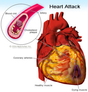
No. The term “heart attack” is often mistakenly used to describe cardiac arrest. While a heart attack may cause cardiac arrest and sudden death, the terms don’t mean the same thing. Heart attacks are caused by a blockage that stops blood flow to the heart. A heart attack (or myocardial infarction) refers to death of heart muscle tissue due to the loss of blood supply, not necessarily resulting in the death of the heart attack victim.

Cardiac arrest is caused when the heart’s electrical system malfunctions. In cardiac arrest death results when the heart suddenly stops working properly. This may be caused by abnormal, or irregular, heart rhythms (called arrhythmias). A common arrhythmia in cardiac arrest is ventricular fibrillation. This is when the heart’s lower chambers suddenly start beating chaotically and don’t pump blood. Death occurs within minutes after the heart stops. Cardiac arrest may be reversed if CPR (cardiopulmonary resuscitation) is performed and a defibrillator is used to shock the heart and restore a normal heart rhythm within a few minutes.

Heart Attack and Cardiac Arrest

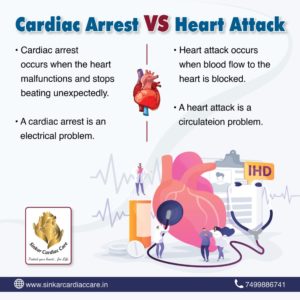
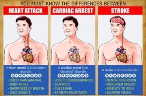
People often use these terms interchangeably, but they are not synonyms. A heart attack is when blood flow to the heart is blocked, and sudden cardiac arrest is when the heart malfunctions and suddenly stops beating unexpectedly. A heart attack is a “circulation” problem and sudden cardiac arrest is an “electrical” problem.

What is a heart attack? A heart attack occurs when a blocked artery prevents oxygen-rich blood from reaching a section of the heart. If the blocked artery is not reopened quickly, the part of the heart normally nourished by that artery begins to die. The longer a person goes without treatment, the greater the damage. Symptoms of a heart attack may be immediate and intense. More often, though, symptoms start slowly and persist for hours, days or weeks before a heart attack. Unlike with sudden cardiac arrest, the heart usually does not stop beating during a heart attack. The heart attack symptoms in women can be different than men.  A heart attack actually caused scarring to the heart since it causes damaging to the heart muscle tissue.

What is cardiac arrest? Sudden cardiac arrest occurs suddenly and often without warning. It is triggered by an electrical malfunction in the heart that causes an irregular heartbeat (arrhythmia). With its pumping action disrupted, the heart cannot pump blood to the brain, lungs and other organs. Seconds later, a person loses consciousness and has no pulse. Death occurs within minutes if the victim does not receive treatment.

Fast action can save lives. Find out what to do if someone experiences a heart attack or cardiac arrestFast action can save lives. Find out what to do if someone experiences a heart attack or cardiac arrest.Fast action can save lives. Find out what to do if someone experiences a heart attack or cardiac arrest.

What to do: Heart Attack Even if you’re not sure it’s a heart attack, don’t wait more than five minutes to call 9-1-1 or your emergency response number. Every minute matters! It’s best to call EMS to get to the emergency room right away. Emergency medical services staff can begin treatment when they arrive — up to an hour sooner than if someone gets to the hospital by car. EMS staff are also trained to revive someone whose heart has stopped. Patients with chest pain who arrive by ambulance usually receive faster treatment at the hospital, too.

What to do: Sudden Cardiac Arrest Cardiac arrest is reversible in most victims if it’s treated within a few minutes. First, call 9-1-1 for emergency medical services. Then get an automated external defibrillator if one is available and use it as soon as it arrives. Begin CPR immediately and continue until professional emergency medical services arrive. If two people are available to help, one should begin CPR immediately while the other calls 9-1-1 and finds an AED.  Learn CPR  you may just save someone one day being at the right place at the right time.

Sudden cardiac arrest is a leading cause of death unfortunately – nearly 400,000 out-of-hospital cardiac arrests occur annually in the United States. By performing Hands-Only CPR to the beat of the classic disco song “Stayin’ Alive,” you can double or even triple a victim’s chance of survival.

Learn the two easy steps to save a life CPR, go to heart.org/handsonlycpr.


QUOTE FOR TUESDAY:

“Sudden Cardiac Arrest is a leading cause of death in the United States, taking the lives of more than 356,0001 people each year, including more than 23,0002 youth under age 18.  Sudden Cardiac Arrest is a life threatening emergency and it can lead to death in minutes if there is no help immediately to the person having the cardiac arrest. The sudden cardiac arrest survival rate is less than 10%. A bystander administering hands-only CPR can triple a person’s chance of survival, and using an AED in the first minute of collapse can increase survival to 90%.”

Stop Cardiac Arrest (https://stopcardiacarrest.org/)