Archives

Part I Sarcoma Awareness Month-What sarcoma is and the causes!

What is Sarcoma?

Sarcoma is a rare but deadly form of cancer affecting connective tissues such as fat, muscle, blood vessels, nerve, bone, deep skin and cartilage. There are roughly 15,000 new cases in the United States each year, with a very low median age of 36 (for bone sarcomas). Survival rates are low, even in comparison to other rare cancers. Rhabdomyosarcoma, a relatively common sarcoma, has a five-year survival rate of 65%, even if caught before metastasis. If caught after metastasis, the five-year survival rate lowers to approximately 30%. This is all to say that while sarcomas are rare, they are extremely devastating.

Sarcomas are a diverse and sporadic group of tumors that have minimal hereditary influence. They are generally classified into two major groups; the group of tumor-specific reoccurring genetic mutations via specific and aberrant chromosome translocation, and the group of non-reoccurring mutations which are based on severe genetic and chromosomal instability. Both groups have altered cell growth-factor signaling pathways. As a result, the introduction of drugs which can normalize growth-factor receptors and proteins are of great interest and the primary treatment given. With improvements in these drugs, it is believed that sarcoma can move from a deadly disease to a chronic but non-life-threatening disease.

Unfortunately, the specificity of the first group of sarcomas, where specific chromosomes are improperly translocated, can often permit the sarcoma to prevail after standard treatment. This is obviously problematic but, if overcome, could spark the development of a whole new field of cancer treatment. In other words, if scientists can discover how to isolate and guide specific chromosomal arrangement, they will able to address many of the hard to treat cancers of today.

With genetic engineering it is theoretically possible to prevent cancer before it begins. However, even without moving this far into the future, the improper chromosomal translocation can provide a sort of name-tagging of the cancer cell. With the cancer cells identified, it can guide modalities aimed at destroying diseased tissue while sparing healthy tissue.

Sarcoma is an under-researched cancer whose research can help to elevate cancer treatment, as a whole. Not only would breakthroughs save the lives of countless children, it could also advance medicine, as a whole. By bringing awareness to sarcoma and the potential development its research can spark, this potential can become a reality.

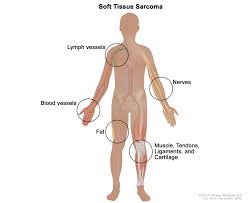
Soft tissue sarcomas may develop in any tissue that connects, supports or surrounds other structures and organs in the body. Some examples of where soft tissues sarcomas can develop are muscles, fascia (the tough membrane that surrounds muscles), tendons, fat, blood vessels, nerves and synovial tissues (connective tissue that makes up the membranes surrounding joints).

Soft tissue sarcomas are rare in adults, accounting for less than 1 percent of all new cases of cancer. The American Cancer Society reports that about 12,750 new cases of soft tissue sarcoma will be diagnosed in 2019 (7,240 cases in males and 5,510 cases in females). Sarcomas can be found almost anywhere in the body.

According to the National Cancer Institute, about 50 percent of soft tissue sarcoma cases occur in the extremities (arms and legs), 40 percent occur in the trunk (back and chest), and 10 percent occur in the head and neck.

Causes of Sarcoma:

The American Cancer Society states the following on causes of Sarcoma:

“Scientists don’t know exactly what causes most soft tissue sarcomas , but they have found some risk factors that can make a person more likely to develop these cancers. And research has shown that some of these risk factors affect the genes in cells in the soft tissues.

Researchers have made great progress in understanding how certain changes in DNA (pieces of genes) can cause normal cells to become cancer. DNA carries the instructions for nearly everything our cells do. We usually look like our parents because they are the source of our DNA. But DNA affects more than just the way we look.

The DNA is made of genes. Genes carry the recipes for making proteins, the molecules that control all cell functions. Some genes contain instructions for proteins that control when our cells grow and divide.

  • Certain genes that promote cell division are called oncogenes.
  • Others that slow down cell division or cause cells to die at the right time are called tumor suppressor genes.

Cancers can be caused by DNA mutations (defects) that turn on oncogenes or turn off tumor suppressor genes.

Many family cancer syndromes have been found in which inherited DNA mutations cause a very high risk of developing breast, colon, kidney, eye, or other cancers. Some of these syndromes are also linked to an increased risk of developing soft tissue sarcomas.

DNA mutations in soft tissue sarcoma are common. But they’re usually acquired during life rather than having been inherited before birth. Acquired mutations may result from exposure to radiation or cancer-causing chemicals. In most sarcomas, they occur for no apparent reason.

Researchers still don’t know why most soft tissue sarcomas develop in people who have no apparent risk factors.”
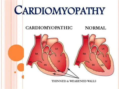
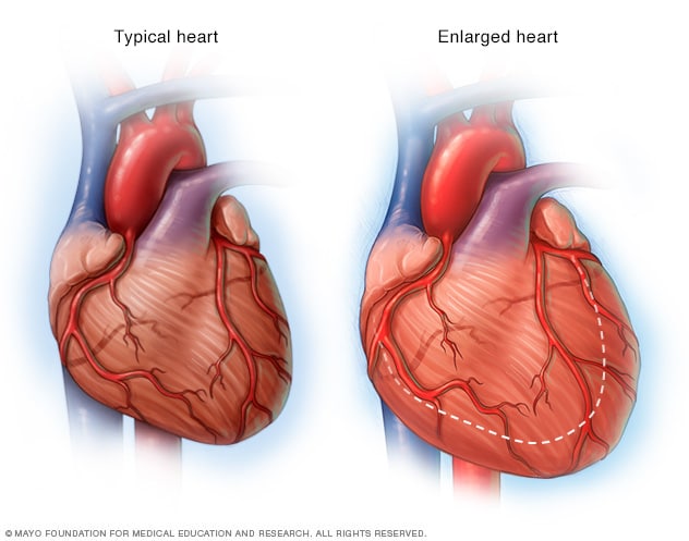
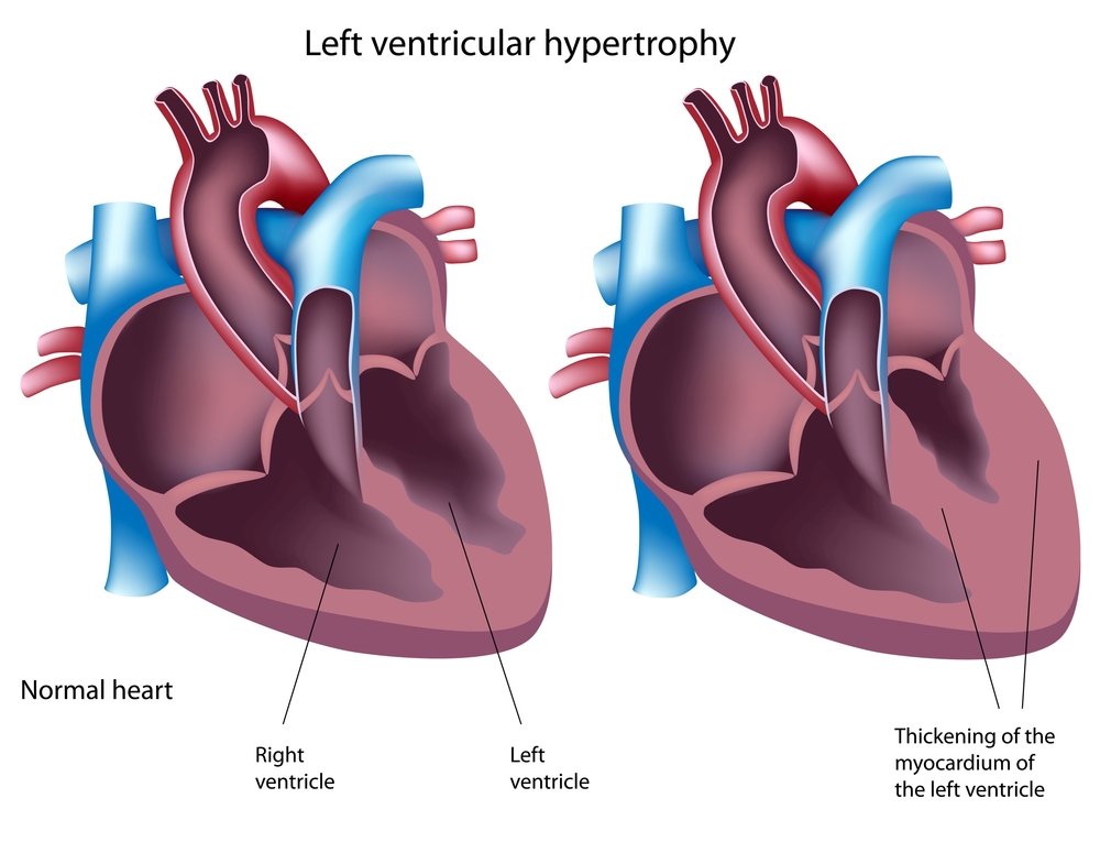
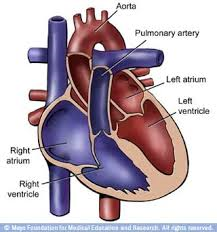
QUOTE FOR WEDNESDAY:

“Cardiomyopathy can be caused by various factors, including genetic predisposition, viral infections, alcohol abuse, high blood pressure, autoimmune diseases, and certain medications. Complications of cardiomyopathy can include arrhythmias, heart failure, stroke, heart valve disease, cardiac arrest, and cardiogenic shock. Early detection and management are crucial to prevent these complications.”

National Heart, Lung and Blood Institute (Cardiomyopathy – Causes and Risk Factors | NHLBI, NIH)

Part III Cardiomyopathy – Types of it, risk factors in getting it, complications from the diagnosis, and how to prevent it!

The main types of cardiomyopathy include dilated, hypertrophic and restrictive cardiomyopathy.

Types of cardiomyopathy include:

  • Dilated cardiomyopathy. In this type of cardiomyopathy, the pumping ability of your heart’s main pumping chamber — the left ventricle — becomes enlarged (dilated) and can’t effectively pump blood out of the heart.Although this type can affect people of all ages, it occurs most often in middle-aged people and is more likely to affect men. The most common cause is coronary artery disease or heart attack.
  • Hypertrophic cardiomyopathy. This type involves abnormal thickening of your heart muscle, particularly affecting the muscle of your heart’s main pumping chamber (left ventricle). The thickened heart muscle can make it harder for the heart to work properly.Hypertrophic cardiomyopathy can develop at any age, but the condition tends to be more severe if it becomes apparent during childhood. Most affected people have a family history of the disease, and some genetic mutations have been linked to hypertrophic cardiomyopathy.
  • Restrictive cardiomyopathy. In this type, the heart muscle becomes rigid and less elastic, so it can’t expand and fill with blood between heartbeats. This least common type of cardiomyopathy can occur at any age, but it most often affects older people.Restrictive cardiomyopathy can occur for no known reason (idiopathic), or it can by caused by a disease elsewhere in the body that affects the heart, such as when iron builds up in the heart muscle (hemochromatosis).
  • Arrhythmogenic right ventricular dysplasia. In this rare type of cardiomyopathy, the muscle in the lower right heart chamber (right ventricle) is replaced by scar tissue, which can lead to heart rhythm problems. It’s often caused by genetic mutations.
  • Unclassified cardiomyopathy. Other types of cardiomyopathy fall into this category.

Risk factors

There are a number of factors that can increase your risk of cardiomyopathy, including:

  • Family history of cardiomyopathy, heart failure and sudden cardiac arrest
  • Long-term high blood pressure
  • Conditions that affect the heart, including a past heart attack, coronary artery disease or an infection in the heart (ischemic cardiomyopathy)
  • Obesity, which makes the heart work harder
  • Long-term alcohol abuse
  • Illicit drug use, such as cocaine, amphetamines and anabolic steroids
  • Certain chemotherapy drugs and radiation therapy for cancer
  • Certain diseases, such as diabetes, an under- or overactive thyroid gland, or a disorder that causes the body to store excess iron (hemochromatosis)
  • Other conditions that affect the heart, such as a disorder that causes the buildup of abnormal proteins (amyloidosis), a disease that causes inflammation and can cause lumps of cells to grow in the heart and other organs (sarcoidosis), or connective tissue disorders

Complications

Cardiomyopathy can lead to other heart conditions, including:

  • Heart failure. Your heart can’t pump enough blood to meet your body’s needs. Untreated, heart failure can be life-threatening.
  • Blood clots. Because your heart can’t pump effectively, blood clots might form in your heart. If clots enter your bloodstream, they can block the blood flow to other organs, including your heart and brain.
  • Valve problems. Because cardiomyopathy causes the heart to enlarge, the heart valves might not close properly. This can lead to a backward flow of blood.
  • Cardiac arrest and sudden death. Cardiomyopathy can lead to abnormal heart rhythms. These abnormal heart rhythms can result in fainting or, in some cases, sudden death if your heart stops beating effectively.

Prevention

In many cases, you can’t prevent cardiomyopathy. Let your doctor know if you have a family history of the condition.

You can help reduce your chance of cardiomyopathy and other types of heart disease by living a heart-healthy lifestyle and making lifestyle choices such as:

  • Avoiding the use of alcohol or cocaine
  • Controlling high blood pressure, high cholesterol and diabetes
  • Eating a healthy diet
  • Getting regular exercise
  • Getting enough sleep
  • Reducing your stress

QUOTE FOR TUESDAY:

“Cardiomyopathy (kahr-dee-o-my-OP-uh-thee) is a disease of the heart muscle. It causes the heart to have a harder time pumping blood to the rest of the body, which can lead to symptoms of heart failure. Cardiomyopathy also can lead to some other serious heart conditions.

There are various types of cardiomyopathy in all age groups.”

MAYO CLINIC (Cardiomyopathy – Symptoms and causes – Mayo Clinic)


Part II Types of Cardiomyopathy, including how it’s diagnosed and the various treatments in all ages!


C

Heart Enlarged


Cardiomyopathy is cardiomyopathy, meaning rather in adult or child the same problems in each age group occur but degree of the disease varies for all, it is case by case.  What is cardiomyopathy?

Cardiomyopathy is a disease of the heart muscle characterized by an abnormally large, thick or stiff heart muscle. It may affect only the heart’s lower chambers (ventricles) or both the lower and upper chambers (atria).  For an adult there just more wear and tear on the organ due to age and severity of the disease.

Cardiomyopathy causes damage to tissue around the heart, as well as heart muscle cells. In severe cases, the heart becomes so weak that it can’t pump blood properly. This can lead to heart failure or irregular heartbeats (arrhythmias). In some cases, cardiomyopathy also involves a buildup of scar tissue or fat within the heart muscle. In rare cases, the heart muscle can’t relax and blood can’t fill the heart properly.

Remember the natural pacemaker in humans is located in the upper right chamber (Rt. atrium) in the upper right region called the sinus node.  The sinus node initiates our impulses from that right atrium across to the Lt. atrium and works the impulse sensation down thru the A-V node-atrioventricular node (site between upper and lower chambers in the heart located at the inferior end of the interatrial septum ending at the top of the ventricles in the heart)).   From the A-V node the impulse sensation continues on from A-V node to the end of the lower right and left lower chambers (called the ventricles) of the heart that continues up the ventricles and passing through the bundle of His to the bundle branches/Purkinje fibers (The bundle of His is an important part of the electrical conduction system of the heart, as it transmits impulses from the A-V node, located at the inferior end of the interatrial septum, to the ventricles of the heart).   Than the impulse sensation goes located in the inner ventricular walls of the heart radiating over the outside of the ventricles = Purkinje Fibers. This whole process allows the heart beat to occur sounding “LubDub”.

This entire impulse conduction process allows the heart to fill up the chambers with blood first in upper chambers to lower ones to releasing the blood in the ventricles (the Rt Ventricle pumps blood to the heart to get more oxygen from lungs that sends this fresh oxygenated blood to the left side of the heart.  The left ventricle pumps the filling of its blood into our blood stream called Cardiac Output in creating the heart to beat.  Stroke Volume x Heart Rate=our Cardiac Output (SVxHR=CO).  Stroke volume is the amount of blood circulated by the heart with each beat  x HR = beat in 60 secs.

Cardiomyopathy is very often a “time-will-tell” disease. Symptoms can vary and the progression of the disease can be unpredictable.  There are many forms of cardiomyopathy; just like in the adult.

What are the types of cardiomyopathy?

The main types include:

Dilated cardiomyopathy (DCM)

DCM is the most common type and occurs when the main pumping chamber of the heart muscle is too stretched out (dilated). Dilated cardiomyopathy makes the heart unable to pump blood effectively.

Hypertrophic cardiomyopathy (HCM)

HCM makes the heart muscle too thick. Usually, the thickening occurs in the muscle of the left ventricle in the heart, often involving the wall between the heart’s two ventricles.

Restrictive cardiomyopathy

Restrictive cardiomyopathy is a rare type of cardiomyopathy that causes the heart muscle to become very rigid or stiff. This makes it difficult for the ventricles of the heart to properly fill with blood.

Arrhythmogenic right ventricular cardiomyopathy (ARVC)

ARVC is a rare form of cardiomyopathy that affects only one in 5,000 people. It occurs when the muscle of the heart’s right ventricle is replaced by thick or fatty scar tissue. The scarring “scrambles” electrical signals within the heart and can make it difficult for the heart to pump blood.

Remember the sinus node (natural pacemaker) of the heart.

How Cardiomyopathy is diagnosed:

Because the symptoms of cardiomyopathy can be diverse, it is often misdiagnosed as asthma, an infection or a gastrointestinal problem but the tests similar to adults tests being ruled out for this disease.  Remember its the same problem at a different age.

In children who have no symptoms, cardiomyopathy is sometimes diagnosed when the child has a chest x-ray that shows an enlarged heart or an echocardiogram for another reason.

If your child’s doctor suspects cardiomyopathy, he or she may order one or more of the following tests, from least invasive to most invasive tests:

  • blood tests
  • chest x-rays
  • electrocardiogram  (EKG or ECG)
  • echocardiogram (cardiac ultrasound)
  • cardiac magnetic resonance imaging (MRI)
  • stress echocardiography, which uses ultrasound and heart-rate monitoring to assess your child’s heart function just before and just after exercise
  • stress test, or exercise test
  • cardiac catheterization
  • coronary angiography

What are the treatment (Rx) options for cardiomyopathy, again similar to adults Rx.?

Your child’s treatment options will be determined by the type of cardiomyopathy he or she has, as well as the specific symptoms. A child with no symptoms might not need medication or other treatment right away. Instead, the cardiologist will monitor your child to gauge the progression of the disease.

A child with more serious symptoms may need additional tests to give the treatment team more detailed information about how the cardiomyopathy is affecting the heart and the rest of the body. .

Many children with cardiomyopathy do well with medication alone. Others need surgery to improve the function of valves, and in the most severe cases, some children need a heart transplant.

Medication for cardiomyopathy

There are several different types of medications for cardiomyopathy, depending on which type your child has and the symptoms.

  • Angiotestin converting enzyme (ACE) inhibitors are drugs that dilate blood vessels in the body, fighting the constricting effect caused by heart failure.
  • Antiarrhythmic medications combat the abnormal heart rhythms caused by irregular electrical activity within the heart.
  • Beta blockers block certain chemicals from binding to nerve receptors in the heart, slowing the heart rate and lowering blood pressure.
  • Blood thinners or anticoagulants help prevent the formation of blood clots, especially in children with the dilated form of cardiomyopathy.
  • Diuretics prevent the buildup of fluid in the body and can help breathing by reducing fluid in the lungs. These drugs may also be helpful in treating scar tissue on the heart.

Surgical treatments for cardiomyopathy

There are several options for treating cardiomyopathy using surgery or minimally-invasive procedures.

Defibrillators

For some children with cardiomyopathy — particularly HCM — an implantable cardioverter-defibrillator (ICD) can be a life-saving option. This tiny instrument, about the size of a deck of cards, is placed in the chest to monitor the child’s heartbeat. If the child has an arrhythmia, the defibrillator will administer a precise electrical pulse to restore normal heart rhythm.

Pacemakers

Pacemaker implantation is minimally invasive option for some children with cardiomyopathy. The pacemaker — a small electronic device — is inserted directly under the skin, where it sends electric signals to the child’s heart, controlling and monitoring the heart rate. The procedure can be performed under local anesthesia in a matter of hours.

Radiofrequency ablation

Radiofrequency ablation is another type of minimally invasive treatment that can be very effective for some children with cardiomyopathy due to arrhythmia. A small, needle-like probe is inserted into the scarred tissue of the heart muscle, sending out radiofrequency waves that burns away the scar tissue and the arrhythmia.

Surgical removal of some heart muscle

In serious cases of hypertrophic cardiomyopathy, the treatment team may perform a surgery called a septal myectomy or septal myomectomy. This procedure involves the removal of a portion of the thickened muscle in the heart, widening the channel in the heart’s ventricle that leads to the aortic valve. The procedure has a very good success rate, and most children have improved blood flow throughout the heart and body.

Ventricular assist device

In certain types of cardiomyopathy, a ventricular-assist device (VAD) can be used to help recover the heart and normalize the heart’s function. After the heart’s function has returned to normal, the VAD will be removed and the child will be monitored for any further heart problems. This approach using the VAD is called a “bridge to recovery.”

Heart transplant

Children with the most severe cases of cardiomyopathy may need a heart transplant if other methods don’t manage symptoms. While your child is waiting for an available heart, a VAD may be used to support the heart. In many cases, children can return to school and other activities while waiting for a transplant.

So similar to how adults are treated also with cardiomyopathy.  The age is the pretty much the difference.


QUOTE FOR MONDAY:

“In the United States, approximately 1.1 million people, have some form of cardiomyopathy.  In 2021, there were 14,770 hospitalizations for cardiomyopathy as the principal diagnosis, and it accounted for 23% of all heart failures.  Some people with cardiomyopathy don’t ever get symptoms. For others, symptoms appear as the condition becomes worse. ”

American Heart Association (2024-Statistics-At-A-Glance-final_2024.pdf)

Part I Cardiomyopathy – what it is, the symptoms, when to see a doctor and causes!

Cardiomyopathy (kahr-dee-o-my-OP-uh-thee) is a disease of the heart muscle that makes it harder for your heart to pump blood to the rest of your body. Cardiomyopathy can lead to heart failure.

Symptoms

There might be no signs or symptoms in the early stages of cardiomyopathy. But as the condition advances, signs and symptoms usually appear, including:

  • Breathlessness with exertion or even at rest
  • Swelling of the legs, ankles and feet
  • Bloating of the abdomen due to fluid buildup
  • Cough while lying down
  • Fatigue
  • Heartbeats that feel rapid, pounding or fluttering
  • Chest discomfort or pressure
  • Dizziness, lightheadedness and fainting

Signs and symptoms tend to get worse unless treated. In some people, the condition worsens quickly; in others, it might not worsen for a long time.

When to see a doctor

See your doctor if you have one or more signs or symptoms associated with cardiomyopathy. Call 911 or your local emergency number if you have severe difficulty breathing, fainting or chest pain that lasts for more than a few minutes.

Because some types of cardiomyopathy can be hereditary, if you have it your doctor might advise that your family members be checked.

Causes

Often the cause of the cardiomyopathy is unknown. In some people, however, it’s the result of another condition (acquired) or passed on from a parent (inherited).

Contributing factors for acquired cardiomyopathy include:

  • Long-term high blood pressure
  • Heart tissue damage from a heart attack
  • Chronic rapid heart rate
  • Heart valve problems
  • Metabolic disorders, such as obesity, thyroid disease or diabetes
  • Nutritional deficiencies of essential vitamins or minerals, such as thiamin (vitamin B-1)
  • Pregnancy complications
  • Drinking too much alcohol over many years
  • Use of cocaine, amphetamines or anabolic steroids
  • Use of some chemotherapy drugs and radiation to treat cancer
  • Certain infections, especially those that inflame the heart
  • Iron buildup in your heart muscle (hemochromatosis)
  • A condition that causes inflammation and can cause lumps of cells to grow in the heart and other organs (sarcoidosis)
  • A disorder that causes the buildup of abnormal proteins (amyloidosis)
  • Connective tissue disorders

QUOTE FOR THE WEEKEND:

“Health care providers can treat hemochromatosis safely and effectively by removing blood from the body on a regular basis. This is similar to donating blood. The process is known as phlebotomy.

The goal of phlebotomy is to lower your iron levels. The amount of blood removed and how often it’s removed depend on your age, your overall health and the severity of iron overload.”

MAYO CLINIC (https://www.mayoclinic.org/diseases-conditions/hemochromatosis/diagnosis-treatment/drc-20351448)

Part II Hemochromatosis – How its Diagnosed & Rx.

Diagnosis

It can be tricky for your doctor to diagnose hemochromatosis, because other conditions have the same symptoms. He might want you to get tested if:

  • You’re having symptoms.
  • You have one of the problems listed above.
  • A family member has the disorder.

There are some other ways your doctor can figure out if you have it:

Checking your history. He’ll ask about your family and if anyone has hemochromatosis or signs of it. He might also ask about things like arthritis and liver disease, which might mean you or someone in your family has hemochromatosis but doesn’t know it.

Physical exam. Your doctor will examine your body. This involves using a stethoscope to listen to what’s going on inside. He might also tap on different parts of your body.

Blood tests. Two tests can give your doctor a clue about hemochromatosis:

  • Transferrin saturation. This shows how much iron is stuck to transferrin, a protein that carries iron in your blood.
  • Serum ferritin. This test measures the amount of ferritin, a protein that stores iron, in your blood. – If either of these show you have more iron than you should, your doctor might order a third test to see if you have a gene that causes hemochromatosis.
  • Liver biopsy. Your doctor will take a small piece of your liver. He’ll look at it under a microscope to see if there’s any liver damage.
  • MRI. This is a scan that uses magnets and radio waves to take a picture of your organs.

    Treatment

    If you have primary hemochromatosis, doctors treat it by removing blood from your body on a regular basis. It’s alot like donating blood. Your doctor will insert a needle into a vein in your arm or leg. The blood flows through the needle and into a tube that’s attached to a bag.

    • Initial treatment schedule. In the beginning, you may have a pint (about 470 milliliters) of blood taken once or twice a week — usually in a hospital or your provider’s office. While you lean back in a chair, a needle is inserted into a vein in your arm. The blood flows from the needle into a tube that’s attached to a blood bag. The process of removing blood is referred to as therapeutic blood removal.  In the Initial treatment. You’ll visit your doctor’s office or a hospital once or twice a week to have your blood drawn. You may have up to a pint taken at a time.
    • Maintenance treatment schedule. Once your iron levels go down, blood can be removed less often, typically every 2 to 3 months. Some people may maintain typical iron levels without having any blood taken. Some may need to have blood removed monthly. The schedule depends on how quickly iron builds up in your body.  So Maintenance treatment starts when once your blood iron levels have gone back to normal, you’ll still have to have to have blood taken, but not as often. It’ll be based on how fast iron builds back up in your body.

    The goal is to remove some of your blood so that your iron levels return to normal. This could take up to a year or more. Blood removal is divided into two parts: initial treatment and maintenance treatment.


QUOTE FOR FRIDAY:

“Hemochromatosis is a metabolic disorder in which your organs accumulate excess iron, leading to organ damage. Hereditary hemochromatosis affects one in 300 people in the United States.”

John Hopkins Medicine (https://www.hopkinsmedicine.org/health/conditions-and-diseases/hemochromatosis)