Archives

QUOTE FOR WEDNESDAY:

“The human brain has 86 billion neurons, give or take — on the same order as the number of stars in the Milky Way. If you look at the synapses, the connections between neurons, the numbers start to get beyond comprehension pretty quickly. The number of synapses in the human brain is estimated to be nearly a quadrillion, or 1,000,000,000,000,000. And each individual synapse contains different molecular switches. If you want to think about the brain in terms of an electrical system, a single synapse is not equivalent to a transistor — it would be more like a thousand transistors.

To make things more complicated, not all neurons are created equal. Scientists still don’t know how many different kinds of neurons we have, but it’s likely in the hundreds. Synapses themselves aren’t all the same either. And that’s not even taking into account all the other cells in our brain. Besides neurons, our brains contain lots of blood vessels and a third class of brain cells known collectively as glia — many of which are even more poorly understood than neurons.

Scientists are making progress breaking those numbers down into something more comprehensible. At the level of individual brain cells, research teams at the Allen Institute and elsewhere are making headway into sorting the cells into different categories, defined as “cell types,” as well as being able to record electrical activity from living human neurons using creative new techniques.

Many neuroscientists study the brain of the lab mouse, in part with the hopes of understanding basic principles of the mammalian brain that could apply to our brains too.

The mammal the octopus (cephalopods related to squid and cuttlefish-The octopus is classified as one branch of mollusks. — and its brain — are fascinating. For an animal with a brain, they’re about as different from humans as it gets. Octopuses have about half a billion neurons, more than five times as many as the lab mouse. But unlike in our nervous systems, more than half of those neurons are in the octopus’ arms. The animals have incredible autonomous control over their limbs — similar in some ways to our own spinal cords that send messages to out body to move.  Understanding the octopus brain isn’t just interesting in its own right, it could also help us understand broad general principles of large brains and animals who can learn and remember complicated behaviors — like us.”

The human brain is probably one of the most, if not the most, complicated brains in any living species.”

Allen Institute (https://alleninstitute.org/news/why-is-the-human-brain-so-difficult-to-understand-we-asked-4-neuroscientists/)

QUOTE 2 – “The human brain can be subdivided according to various criteria. It can be explained in terms of evolution, as, like all vertebrates, it consists of an end brain, interbrain, midbrain, hindbrain and medullary brain. Anatomically, the areas known as the cerebrum, interbrain and cerebellum, as well as the brain stem, are particularly noticeable. Particularly striking is the cerebral cortex, which forms part of the end brain. During evolution, it has grown so strongly that it surrounds almost the entire brain. With its ridges and coils, the cortex gives the brain its walnut-like look.”

MAX-PLANCK-GESELLSCHAFT (https://www.mpg.de/brain)

Knowing how the brain functions to understand this month’s awareness on Aphasia!

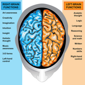
IIlustration body part,human brain left and right functions
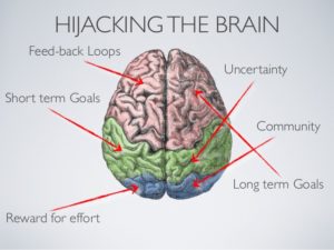
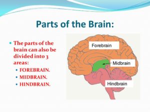


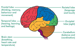
The brain is like a committee of experts. All the parts of the brain work together, but each part has its own special properties. The brain can be divided into three basic units: 1 the forebrain, 2 the midbrain, and 3 the hindbrain.

1-THE CEREBRUM (The Forebrain) AND ITS FUNCTIONS:  Knowing what part of the cerebrum, if the brain injury is their, can explain the reasons for the symptoms the individual is having.

1-The forebrain is the largest and most highly developed part of the human brain: it consists primarily of the cerebrum and the structures hidden beneath it, which is the inner brain.

THE REGIONS (The 4 LOBES) THAT MAKE UP THE CEREBRUM:
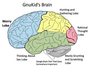


The cerebrum, the large, outer part of the brain, controls reading, thinking, learning, speech, emotions and planned muscle movements like walking. It also controls vision, hearing and other senses. The cerebrum is divided two cerebral hemispheres (halves): left and right. The right half controls the left side of the body. The left half controls the right side of the body.

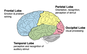
Each hemisphere has four sections, called lobes: frontal, parietal, temporal and occipital.  A lobe simply means a part of an organ (earlobe for example).  Each lobe controls specific functions. For example, the frontal lobe controls personality, decision-making and reasoning, while the temporal lobe controls, memory, speech, and sense of smell.

The frontal lobe is the largest lobe of the brain.  The frontal lobe are the last parts of the brain develop as a person ages and the part of the human brain that is most different from other mammals and primates.  The last part to mature is the prefrontal lobe. This happens during adolescence. Many things affect brain development including genetics, individual and environmental factors.  We learn to become adults in our frontal lobes.   You choose between good and bad actions; override and suppress socially unacceptable responses; and determine similarities and differences between objects or situations. The frontal lobe is considered to be the moral center of the brain because it is responsible for advanced decision making processes. It also plays an important role in retaining emotional memories derived from the limbic system, and modifying those emotions to fit socially accepted norms.  The frontal lobes are considered our emotional control center and home to our personality. There is no other part of the brain where lesions can cause such a wide variety of symptoms (Kolb & Wishaw, 1990). The frontal lobes are involved in motor function, problem solving, spontaneity, memory, language, initiation, judgment, impulse control, and social and sexual behavior. Frontal lobe damage effects one or more of these areas depending on the severity of the damage.  The frontal lobes are extremely vulnerable to injury due to their location at the front of the cranium, proximity to the sphenoid wing and their large size. MRI studies have shown that the frontal area is the most common region of injury following mild to moderate traumatic brain injury.

The parietal lobes can be divided into two functional regions. One involves sensation and perception and the other is concerned with integrating sensory input, primarily with the visual system. The first function integrates sensory information to form a single perception (cognition).  The parietal lobes have an important role in integrating our senses. In most people the left side parietal lobe is thought of as dominant because of the way it structures information to allow us to read & write, make calculations, perceive objects normally and produce language. Damage to the dominant parietal lobe can lead to Gerstmann’s syndrome (e.g. can’t tell left from right, can’t point to named fingers), apraxia and sensory impairment (e.g. touch, pain). Damage to the non-dominant lobe, usually the right side of the brain, will result in different problems. This non-dominant lobe receives information from the occipital lobe and helps provide us with a ‘picture’ of the world around us. Damage may result in an inability to recognize faces, surroundings or objects (visual agnosia). So, someone may recognize your voice, but not your appearance (you sound like my daughter, but you’re not her). Damage to the parietal lobe depends on severity and location of the area. Because this lobe also has a role in helping us locate objects in our personal space, any damage can lead to problems in skilled movements (constructional apraxia) leading to difficulties in drawing or picking objects up.

The temporal lobes they are in the section of the brain located on the sides of the head behind the temples and cheekbones.   It’s responsible for processing auditory information from the ears (hearing).   The temporal lobes play an important role in organizing sensory input, auditory perception, language and speech production, as well as short term memory association and formation. The Temporal Lobe mainly revolves around hearing and selective listening. It receives sensory information such as sounds and speech from the ears. It is also the key to being able to comprehend, or understand meaningful speech. In fact, we would not be able to understand someone talking to us, if it wasn’t for the temporal lobe. This lobe is special because it makes sense of the all the different sounds and pitches (different types of sound) being transmitted from the sensory receptors of the ears. Temporal Lobes Kolb & Wishaw (1990) have identified eight principle symptoms of temporal lobe damage: 1) disturbance of auditory sensation and perception, 2) disturbance of selective attention of auditory and visual input, 3) disorders of visual perception, 4) impaired organization and categorization of verbal material, 5) disturbance of language comprehension, 6) impaired long-term memory, 7) altered personality and affective behavior, 8) altered sexual behavior. These can be due to tumors on the right or left side of the temporal lobe, due to seizures in the temporal lobe and if seizures regularly happen to this individual in the temporal region, which causes lack of oxygen to that area of that area of the brain it will effect one or more of the functions of that lobe which we discussed earlier, listed above.

-The last region or lobe that makes up the cerebrum is the occipital lobe. The occipital lobe is important to being able to correctly understand what our eyes are seeing. These lobes have to be very fast to process the rapid information that our eyes are sending. This is similar to how the temporal lobe makes sense of auditory information, the occipital lobe makes sense of visual information so that we are able to understand it. If our occipital lobe was impaired or injured we would not be able to correctly process visual signals, thus visual confusion would result.

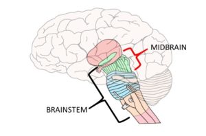
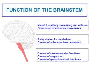
2-Midbrain – The uppermost part of the brainstem is the midbrain, which controls some reflex actions and is part of the circuit involved in the control of eye movements and other voluntary movements.
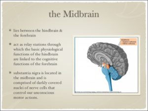
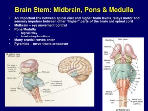


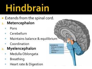
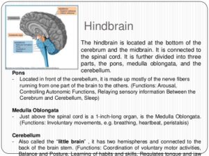
3-The hindbrain includes the upper part of the spinal cord, the brain stem, and a wrinkled ball of tissue called the cerebellum. The hindbrain controls the body’s vital functions such as respiration and heart rate. The cerebellum coordinates movement and is involved in learned rote movements. Rote means “mechanical or habitual repetition of something to be learned.”. Rote learning is flashcards, times tables, any kind of memorization-based learning. Rote movement applies to activities we do in a mechanical, repetitive way. Running, for example.  When you play the piano or hit a tennis ball you are activating the cerebellum= balance/coordination.


Knowing how the brain functions to understand this month’s awareness Aphasia (which we will discuss tomorrow).

QUOTE FOR TUESDAY:

“While people are accustomed to dealing with runny noses and scratchy throats in the fall and winter, many are experiencing the same symptoms this summer.

This could be due to a number of reasons, according to Dr. Judy Tung, section chief of Adult Internal Medicine at NewYork-Presbyterian/Weill Cornell Medical Center. Cold and flu viruses are continuing to circulate — in fact, in late April and early May, New York state saw an unusual spike in influenza — coinciding with summer allergies, not to mention an uptick in COVID-19 cases due to the rise of Omicron subvariants.

Summer cold symptoms are common and confusing this year not only because of COVID but also because of the late flu peak.  Remember Influenza activity is usually is from October to May but can be all year.

So remember colds are not unusual to have in the summer.”

Dr.  Tung from New York Presbyterian/Weill Cornell Medical Center  (https://healthmatters.nyp.org/what-to-know-about-the-surge-in-summer-colds/)


Colds in the summer!

While people are accustomed to dealing with runny noses and scratchy throats in the fall and winter, many are experiencing the same symptoms this summer.

This could be due to a number of reasons, according to Dr. Judy Tung, section chief of Adult Internal Medicine at NewYork-Presbyterian/Weill Cornell Medical Center. Cold and flu viruses are continuing to circulate — in fact, in late April and early May, New York state saw an unusual spike in influenza — coinciding with summer allergies, not to mention an uptick in COVID-19 cases due to the rise of Omicron subvariants.

“Summer cold symptoms are common and confusing this year not only because of COVID but also because of the late flu peak,” says Dr. Tung.

To understand what viruses are circulating now and how to tell the difference between a summer cold, allergies, and COVID-19, Health Matters spoke with Dr. Tung, who is also associate dean for faculty development at Weill Cornell Medicine.

What have you seen in the past recent years with the flu and colds?

We started to see a resurgence of flu at the end of 2021, and then a big drop in cases at the beginning of 2022, during the initial Omicron surge. But with the relaxation of masking and distancing during covid and some still today through out the months when the mask lifted to have to use, influenza experienced a late peak.

As for colds, this summer a lot more GI symptoms that accompany colds — vomiting and diarrhea in addition to fever, congestion and cough. This probably speaks to a dominance of enterovirus, a common summer cold virus that can produce more GI symptoms or pink eye symptoms than rhinovirus, which is more dominant in the winter. This can be confusing, because COVID also causes GI symptoms.

Why are we usually able to avoid bad colds in the summer? Why are colds lasting longer?One theory for why colds are lasting longer is that the immune system got a little forgetful, not having been exposed to the most current viral strains, and therefore is less prepared to fight them off. The immune system builds antibodies and other memory white blood cells to fight off pathogens after being exposed to them. When our immune systems are exposed to cold viruses all year long, they are “on the ready.” That didn’t happen last few years because of all the precautions people took to protect themselves against COVID with masks and distancing.

What are the biggest differences in symptoms between common colds, allergies, and COVID-19?
COVID is associated with loss of smell and taste, or unusual tastes that are not common in uncomplicated colds. Sinus infections can do this, but regular colds typically don’t affect smell or taste to the degree we see in COVID-19.

Allergies can really feel like a cold, down to the body aches when allergies are severe. Allergies do not produce fever and normally take many days of postnasal dripping to cause a cough, whereas colds and COVID can move to coughing swiftly.

What’s the best way to care for summer colds?
There is little difference in the way we care for summer and winter colds — drink fluids and get plenty of rest. One advantage of summer is that you can open windows to ensure that shared space is well ventilated, especially if there is a member in the household who is sick.

With the rise of the Omicron subvariants, what is important to keep in mind when you come down with what seems to be an ordinary cold or allergy symptoms?
It is important to get tested for COVID if you have cold symptoms — not because you are going to get gravely ill, but because you may inadvertently pass it along to someone who could get gravely ill.

Vaccination and boosting definitely protect people from severe COVID infection, preventing hospitalization and death. However, Omicron is highly infectious, and there is increasing evidence that while the vaccines are still proving to protect us against severe COVID, they are not as effective against stopping us from getting infected or reinfected.

Furthermore, there is some recent evidence that while Omicron is definitely milder than Delta, it is more contagious and may linger for longer, so people stay masked for 10 to 14 days and to use home antigen tests and look for a negative test to guide on when you can relax with masking again.


QUOTE FOR MONDAY:

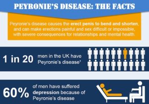
“Medical experts estimate that about 6% to 10% of people between the ages of 40 and 70 with penises have Peyronie’s disease. It can affect anyone with a penis, but it’s less common at other ages.

Peyronie’s disease may be even more widespread because many people may feel too embarrassed to talk about it with a healthcare provider. It’s a good idea to talk to a healthcare provider any time you have concerns about your sexual health.

Peyronie’s disease can be painful. Pain most commonly occurs during the acute stage. But it may continue in the chronic stage. The severity varies from person to person.

The cause of Peyronie’s disease isn’t always clear.”

Cleveland Clinic (https://my.clevelandclinic.org/health/diseases/10044-peyronies-disease)

QUOTE FOR THE WEEKEND:

“Peyronie’s disease is a connective tissue disorder of the penis that can be likened to Dupuytren’s contracture of the hand. It is characterized by the triad of bent erections, pain in the penis with erections and palpable penile plaque. Peyronie’s disease is quite common, affecting as many as one in 11 men, despite the lack of public awareness. The penis is composed of the same connective tissue as every other joint in the body.”

John Hopkin’s Medicine (https://www.hopkinsmedicine.org/health/conditions-and-diseases/peyronie-disease)

Part I Peyronie’s Disease – what it is, what causes it, how common is it, and who is more likely to develop this condition.

Some men have a penis that curves to the side, upward or downward when erect. This is common, and a bent penis in most men isn’t a problem. Generally, a bent penis is only a cause for concern if your erections are painful or if the curvature of your penis interferes with sex.

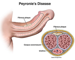
Peyronie’s disease is a disorder in which scar tissue, called a plaque, forms in the penis—the male organ used for urination and sex. The plaque builds up inside the tissues of a thick, elastic membrane called the tunica albuginea. The most common area for the plaque is on the top or bottom of the penis. As the plaque builds up, the penis will curve or bend, which can cause painful erections. Curves in the penis can make sexual intercourse painful, difficult, or impossible. Peyronie’s disease begins with inflammation, or swelling, which can become a hard scar.

The plaque that develops in Peyronie’s disease is not the same plaque that can develop in a person’s arteries. The plaque seen in Peyronie’s disease is benign, or noncancerous, and is not a tumor. Peyronie’s disease is not contagious or caused by any known transmittable disease.

Early researchers thought Peyronie’s disease was a form of impotence, now called erectile dysfunction (ED). ED happens when a man is unable to achieve or keep an erection firm enough for sexual intercourse. Some men with Peyronie’s disease may have ED. Usually men with Peyronie’s disease are referred to a urologist—a doctor who specializes in sexual and urinary problems.

How does an erection occur?

An erection occurs when blood flow increases into the penis, making it expand and become firm. Two long chambers inside the penis, called the corpora cavernosa, contain a spongy tissue that draws blood into the chambers. The spongy tissue contains smooth muscles, fibrous tissues, spaces, veins, and arteries. The tunica albuginea encases the corpora cavernosa. The urethra, which is the tube that carries urine and semen outside of the body, runs along the underside of the corpora cavernosa in the middle of a third chamber called the corpus spongiosum.

An erection requires a precise sequence of events:

  • An erection begins with sensory or mental stimulation, or both. The stimulus may be physical contact or a sexual image or thought.
  • When the brain senses a sexual urge, it sends impulses to local nerves in the penis that cause the muscles of the corpora cavernosa to relax. As a result, blood flows in through the arteries and fills the spaces in the corpora cavernosa like water filling a sponge.
  • The blood creates pressure in the corpora cavernosa, making the penis expand.
  • The tunica albuginea helps trap the blood in the corpora cavernosa, thereby sustaining the erection.
  • The erection ends after climax or after the sexual urge has passed. The muscles in the penis contract to stop the inflow of blood. The veins open and the extra blood flows out of the penis and back into the body.

What causes Peyronie’s disease?

Medical experts do not know the exact cause of Peyronie’s disease. Many believe that Peyronie’s disease may be the result of

  • acute injury to the penis
  • chronic, or repeated, injury to the penis
  • autoimmune disease—a disorder in which the body’s immune system attacks the body’s own cells and organs

Injury to the Penis

Medical experts believe that hitting or bending the penis may injure the tissues inside. A man may injure the penis during sex, athletic activity, or an accident. Injury ruptures blood vessels, which leads to bleeding and swelling inside the layers of the tunica albuginea. Swelling inside the penis will block blood flow through the layers of tissue inside the penis. When the blood can’t flow normally, clots can form and trap immune system cells. As the injury heals, the immune system cells may release substances that lead to the formation of too much scar tissue. The scar tissue builds up and forms a plaque inside the penis. The plaque reduces the elasticity of tissues and flexibility of the penis during erection, leading to curvature. The plaque may further harden because of calcification––the process in which calcium builds up in body tissue.

Autoimmune Disease

Some medical experts believe that Peyronie’s disease may be part of an autoimmune disease. Normally, the immune system is the body’s way of protecting itself from infection by identifying and destroying bacteria, viruses, and other potentially harmful foreign substances. Men who have autoimmune diseases may develop Peyronie’s disease when the immune system attacks cells in the penis. This can lead to inflammation in the penis and can cause scarring. Medical experts do not know what causes autoimmune diseases. Some of the autoimmune diseases associated with Peyronie’s disease affect connective tissues. Connective tissue is specialized tissue that supports, joins, or separates different types of tissues and organs of the body.

How common is Peyronie’s disease?

Researchers estimate that Peyronie’s disease may affect 1 to 23 percent of men between 40 and 70 years of age.1 However, the actual occurrence of Peyronie’s disease may be higher due to men’s embarrassment and health care providers’ limited reporting.1 The disease is rare in young men, although it has been reported in men in their 30s.1 The chance of developing Peyronie’s disease increases with age.

Who is more likely to develop Peyronie’s disease?

The following factors may increase a man’s chance of developing Peyronie’s disease:

  • vigorous sexual or nonsexual activities that cause microscopic injury to the penis
  • certain connective tissue and autoimmune disorders
  • a family history of Peyronie’s disease
  • aging

Vigorous Sexual and Nonsexual Activities

Men whose sexual or nonsexual activities cause microscopic injury to the penis are more likely to develop Peyronie’s disease.

Connective Tissue and Autoimmune Disorders

Men who have certain connective tissue and autoimmune disorders may have a higher chance of developing Peyronie’s disease. A common example is a condition known as Dupuytren’s disease, an abnormal cordlike thickening across the palm of the hand. Dupuytren’s disease is also known as Dupuytren’s contracture. Although Dupuytren’s disease is fairly common in older men, only about 15 percent of men with Peyronie’s disease will also have Dupuytren’s disease.2 Other connective tissue disorders associated with Peyronie’s disease include

  • plantar fasciitis––inflammation of the plantar fascia, thick tissue on the bottom of the foot that connects the heel bone to the toes and creates the arch of the foot
  • scleroderma––abnormal growth of connective tissue, causing it to get thick and hard; scleroderma can cause swelling or pain in muscles and joints

Autoimmune disorders associated with Peyronie’s disease include

  • systemic lupus erythematosus––inflammation and damage to various body tissues, including the joints, skin, kidneys, heart, lungs, blood vessels, and brain
  • Sjögren’s syndrome––inflammation and damage to the glands that make tears and saliva
  • Behcet’s syndrome––inflammation of the blood vessels

Family History of Peyronie’s Disease

Medical experts believe that Peyronie’s disease may run in some families. For example, a man whose father or brother has Peyronie’s disease may have an increased chance of getting the disease.

Aging

The chance of getting Peyronie’s disease increases with age. Age-related changes in the elasticity of tissues in the penis may cause it to be more easily injured and less likely to heal well.


QUOTE FOR FRIDAY:

“In 2021, 4.3% of adults aged ≥18 years reported being bothered a lot by headache or migraine in the past 3 months with the percentage among women (6.2%) higher than that among men (2.2%). Percentages were higher among women than men in all age groups: 7.4% versus 2.5% in adults aged 18–44 years, 6.7% versus 2.4% in those aged 45–64 years, and 3.1% versus 1.5% in those aged ≥65 years. Among men and women, the percentage of those bothered a lot by headache or migraine in the past 3 months was lowest among those aged ≥65 years.”

Source: National Center for Health Statistics, National Health Interview Survey, 2021. https://www.cdc.gov/nchs/nhis.htm Center for Disease Control and Prevention

National Headache & Migraine Month – Tension Headaches vs. Migraines

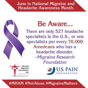
June is National Migraine & Headache Awareness Month (MHAM), an opportunity to raise awareness about migraine and other headache diseases. Migraine impacts forty million people in the United Statesone billion people across the globe, and is recognized as the #2 cause of disability worldwide. Currently, about 16 million people with migraines in the U.S. are undiagnosed. Approximately 400,000 Americans experience cluster headaches, recognized as one of the most painful diseases a person can have.

The word “headache” is a broad term used to describe pain in the scalp, head and neck. There are many different types of headaches. They may be primary conditions such as tension headaches, migraines and cluster headaches, or they may occur due to underlying health conditions.

Headaches are very common, most people experience them to some degree during their life.

“Headache disorders are amongst the world’s most debilitating conditions globally,” says Susan Broner, M.D., medical director of the Weill Cornell Medicine Headache Program. “In fact, migraine itself is the second most disabling condition in the world in terms of years lost to disability. And if you look at populations of people under 50, it’s the first most disabling condition yet many people go undiagnosed and untreated.”

Severe or recurring headaches of any type can significantly impact daily life. Learning about them can help you communicate your concerns more clearly to your primary care provider.

Tension Headaches:

Tension headaches are the most common type of headache. People often experience occasional tension headaches and don’t seek medical care. However, if you have tension headaches 15 days per month or more, you should consult with your primary care provider.

Causes of Tension Headaches

These headaches are caused by tense muscles around the head and neck, often due to stress, anxiety or depression. Tension headaches may also be triggered by:

  • Alcohol
  • Caffeine or caffeine withdrawal
  • Dental problems such as frequently grinding your teeth or clenching your jaw
  • Eyestrain
  • Keeping your head in one position for a long time
  • Not getting enough sleep

Treating and Preventing Tension Headaches

Occasional tension headaches can often be prevented by:

  • Exercising regularly
  • Getting enough sleep
  • Maintaining good posture while seated and taking breaks from sitting
  • Managing daily stress

Chronic tension-type headaches are typically treated with stress reduction techniques such as meditation or cognitive behavioral therapy and biofeedback.

Over-the-counter pain medications (e.g., ibuprofen or acetaminophen) may be used to decrease pain. Muscle relaxers or prescription antidepressants may also be recommended in some cases.

Migraine Headaches:

Migraines are a severe, recurring type of headache that is often debilitating. About 12% of people in the United States have migraines and they are more common in women.

Causes of Migraines

The exact cause of migraines is unknown. But researchers believe that genetics plays a part. Having certain medical conditions may increase your risk of developing migraines, including:

  • Anxiety
  • Bipolar disorder
  • Depression
  • Epilepsy
  • Sleep disorders

Migraines can be triggered by several factors. Caffeine is one of the more common triggers people encounter.  Caffeine is always a mystery to people because many people find that it helps a headache,

There is currently no cure for migraine, but we are in a renaissance of new treatments, therapies, and approaches to managing the disease.  If you have been struggling in silence, now is the time to speak up, educate yourself, and seek care. You might just change your life for the better.

During the Covid pandemic, there was a significant rise in telemedicine. According to a survey by the Headache and Migraine Policy Forum and MigraineAgain, 78% of migraine and headache patients used telemedicine after the start of the pandemic, in comparison to just 22% before the pandemic. This trend was ushering in a new era of care, where patients could interact with health professionals from the convenience and comfort of their homes. The survey also revealed that there had been a nearly 70% rise in the number of migraine attacks during the pandemic and 84% of people had more stress managing their disease.

On the treatment front, there are new options for patients to explore. “Migraine and headache patients now have more options due to a wave of innovation in acute and preventive care,” noted Dr. William Young, Professor of Neurology, Thomas Jefferson University and Medical Advisor for the Coalition for Headache and Migraine Patients. “These include CGRP monoclonal antibodies, gepants, lasmiditan, and several neuromodulation devices.”

It is so much easier to for many humans to feel since they don’t see anything a migraine or general headache is not so bad but when you experience you may feel different when seeing those people with that symptom.  Headache specialists play a vital role in migraine advocacy and awareness efforts. The symptoms of migraine often cause patients living with the disease to withdraw from their daily lives.. Others may refrain from identifying themselves as a migraine patient, due to stigma surrounding the disease and a lack of compassion surrounding its symptoms. For these patients and so many more, we as healthcare professionals must continue to advocate on their behalf.

A disease awareness month plays a vital role to raise public knowledge, address stigma and build a stronger community of patient advocates.

QUOTE FOR THURSDAY:

“On an x-ray, the spine of a person with scoliosis looks like an “S” or “C” instead of a straight line. Providers use a special measurement tool to measure the angle of the curve, called a Cobb angle. A slight curve may be normal. Scoliosis is diagnosed when the Cobb angle is 10 degrees or greater.

Scoliosis usually isn’t life-threatening or painful, and those who have it can live normal and active lives. While there are no known ways to prevent the development of scoliosis, early diagnosis and treatment can help prevent the condition from getting worse.”.

Gillette Children’s (https://www.gillettechildrens.org/conditions-care/scoliosis-idiopathic-neuromuscular-and-congenital/what-is-scoliosis)