Archive | February 2017

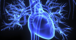
How the heart works anatomically.


The heart works like a pump and beats 100,000 times a day.  For the heart to do its function therapeutically it needs another organ to be involved called the lungs or the body could not live.  If one of these organs gets damaged in time the other organ gets affected with no treatment the person will die sooner in life (Ex. Heart Failure in time affects the lungs to some degree, even respiratory failure).  A good metaphor for this is the car; if the engine gets damaged with no repair, in time the transmission will get affected; with no repair at all the car will die.

One of the main functions of the red blood cells (RBCs) is to carry oxygen throughout the body to give all tissues our food to stay alive, that would be oxygen.  For this to take place this is done through the heart beating nonstop which allows the flow of blood to be running continuous in our blood stream (circulatory system) and at the same time this allows our cells in the blood stream to get to the lungs for oxygen and carbon dioxide exchange.   These 3 functions could not take place if the heart wasn’t pumping blood to and from the heart/lungs 24 hrs a day.  Your pulse or heart rate does the pumping.

This is what takes place when the heart pumps our blood:  

The red blood cells carry oxygen and remove the ending result of oxygen used by our tissues, called carbon dioxide=CO2.   To get rid of the CO2 (a toxin) in our body the gas is carried from our body by carrying CO2 to the lungs through the RBCs.  The high CO2 with low oxygen (O2)  concentration RBCs go to one side of the heart (being the right side) to get CO2 removed in exchange for more O2 in a red blood cell (RBC) this is done at the base of the lungs.  This is done through breathing.   Then these RBCs get highly oxygenated which then proceed to the left side of the heart.  These RBCs get pumped throughout the body where the tissues utilize the oxygen in the RBCs by transferring O2 from the RBCs to the tissue as their food to stay alive.  Without oxygen our tissues would starve and we would die.  For the oxygen to be transported to the tissues of the body (as their food) it works like this:  After the right side of the heart pumps blood to the lungs with high CO2 concentration RBCs, while we inhale to allow more oxygen in the cells at the bottom of the lungs an exchange of gas concentration in the RBCs takes place.  The cells release CO2 which is released out of our body through exhaling but also when we inhale more 02 is transported into the RBCs to take another trip around the body releasing the high 02 levels to the tissues that need to utilize it BUT to do this the blood now is sent to the left side of the heart.  For all this to take place the heart pumps to transport the oxygen throughout the body giving nutrition to the tissues, without it=cellular starvation but at the same time sends high CO2 RBCs to the right side for a 02/CO2 exchange at the lungs by going through the right side of the heart.   

The heart has two sides, separated by an inner wall called the septum.  The septum divides the right from the left side of the heart since the right and left chambers do different functions, as described above.  Remember the right side of the heart is sending RBCs with highier CO2 concentration and a low oxygen concentration.  You have these RBCs coming from above the heart finally entering the Rt upper chamber (Right Atrium=RA) called the Superior Vena Cava with below the heart the Inferior Vena Cava the meet into each other dumping the high CO2 blood into the RA.  Then the blood goes through a valve called the tricuspid valve dumping the blood into the Rt.lower chamber called Rt. Ventricle and through the pulmonary valve going via the pulmonary artery (one of the few arteries with high CO2 concentration, usually arteries high in 02 concentration) dumping the blood at the base of the lungs for 02 and CO2 exchange when we inhale and exhale.  After the gas exchange takes place the red blood cells become higher oxygenated in levels and much lower in carbon dioxide levels.  Inside a cell is never 100%  02 or CO2.  Now this high oxygenated blood goes to the left side of the heart leaving the lungs via the pulmonary vein (one of the few veins high in O2).  The blood dumps now in the L upper chamber=Lt. atrium down through the mitral valve to the left ventricle proceeding to the aorta where the high concentrated RBCs go throughout the body dispensing oxygen to our tissues where it is needed.

If you look the right side sends blood from the heart to lungs back to heart=short distance so the muscle on the right side is thin compared to the left side.  The reason for this is the left side of the heart receives the oxygen-rich blood from the lungs and pumps it to throughout the body.  The left side of the heart has to dispense the high (O2) blood throughout the body so that side of the heart works out more in pumping; so the muscle on the left side is larger than the right side.  The muscle of the heart is called myocardium (myo=muscle and cardium=the heart).

Let’s review the heart anatomy.  It has 4 chambers in it.  Two chambers are on the right side and another two on the left.  The right side of the heart has only higher concentrated CO2-carbon dioxide in the blood going through that side to get to the lungs for more oxygen. To do this the blood first is entered to the lungs from the right side of the heart.  Here it removes the toxin CO2 from our body through us breathing to get more 02-oxygen into our RBCs (red blood cells).  Then this blood enters to the left side of the heart  where it has more high oxygenated blood entering that side to have our food, being O2, get dispensed throughout our tissues to stay alive.  Without 02 we would go through cellular/tissue starvation.  The heart moves the oxygen throughout the body via its pumping action whereas the lungs provide us the oxygen via inhaling with removing the toxin CO2 from the RBCs via exhaling.

Now to do all these functions from pumping to getting 02 and C02 properly dispensed in and out of our bodies how does the heart do this?????????

Well he don’t fill the heart up with gas but instead has a conduction pathway that operates through electrical activity taking place by certain electrolytes.  We have sodium, potassium and calcium channels that are the major ones in doing this.

We also have a natural pacemaker of the heart and this is called the SA NODE.  It sits in the the upper Rt. chamber (atrium) of the heart.  This natural pacemaker conducts impulses from the top to the bottom of the heart with these electrolytes (mentioned above) involved to let this conduction take place in allowing the heart to beat; allowing the heart to pump the blood throughout our system to give oxygen to all tissues and remove carbon dioxide from the body bringing it to the lungs where we expel it via breathing.  Inspiration brings the 02 via the lungs to the bottom of them to get dispensed into our red blood cells at the same time during expiration removes the C02 from the RBCs to be expelled out via the lungs to the exit port our mouth.  Knowing the anatomy of the heart is so important especially to understand heart disease. 

We know there is a right and left side of the heart and each side having 2 chambers with completely different functions, which we will discuss another day.

QUOTE FOR THURSDAY:

Heart disease continues to be the number one killer; cancer, the number 2 killer, not far behind. The tragic aspect of these deadly diseases is that they could all be cured, I do believe, if we had sufficient funding.”

Arlen Specter ((February 12, 1930 – October 14, 2012) was a United States Senator from Pennsylvania


QUOTE FOR WEDNESDAY:

“Catheter ablation for SVT utilizing radiofrequency ablation (electrocautery injury) was developed in the 1980’s and has since revolutionized the treatment of SVT.”

UNC Healthcare (Center for vascular and heart care)


Part II Ablation and other treatments for dysrhythmias.

TREATMENT APPROACHES


In some cases, arrhythmias may not require treatment. Other arrhythmias can be controlled by treating the underlying cause. Arrhythmias that cause symptoms may require one or more of the following treatments to reduce the number or duration of arrhythmic events.

Medications. Common medications for suppressing arrhythmias include:

  • Beta-blockers;
  • Calcium channel blockers;
  • Digitalis; and
  • Antiarrhythmic agents.

Digitalis should not be used for certain arrhythmias, such as WPW syndrome. People with atrial fibrillation are typically prescribed an anticoagulant to minimize their risk of clotting and stroke.

Cardioversion. This procedure restores a normal heartbeat by transmitting a brief electric shock through the chest to the heart. Usually an outpatient procedure that is performed in a hospital while the patient is under heavy sedation or anesthesia, it is commonly used to treat:

  • Atrial fibrillation;
  • Atrial flutter; and
  • Ventricular arrhythmias.

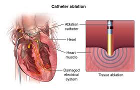
Radiofrequency Catheter ablation.(diagram above is ablation) A catheter with an electrode tip is positioned on the affected area. The catheter delivers energy to destroy tissue that is interfering with the normal transmission of electrical impulses through the heart. It is most commonly used for:

  • SVT – Supra Ventricular Tachycardia (Pulse over 100);
  • Atrial fibrillation-particularly newly diagnosed;
  • Atrial flutter; and
  • Certain types of ventricular arrhythmias.

Catheter ablation for SVT utilizing radiofrequency ablation (electrocautery injury) was developed in the 1980’s and has since revolutionized the treatment of SVT. With catheter ablation, a procedure is performed entirely through intravenous catheters inserted into the veins in the leg and sometime the shoulder. It is a minimally invasive procedure. That is, no open heart surgery is needed. Generally, procedures can be performed on an outpatient basis. Overall cure rates with catheter ablation is >90% and can be as high as 96-98% depending on the specific type of SVT.

During a catheter ablation procedure, catheters (long wire electrodes) are advanced through the veins in the leg up to the heart.

Various measurements of the electrical system are performed. If a person is in normal rhythm at the time of the procedure, an attempt is made then to reproduce the SVT by pacing the heart through the catheters. Occasionally an intravenous medicine called isoproterenol is required to “rev up” the heart in order to reproduce the SVT. Once the SVT is reproduced, the specific type of SVT can be diagnosed using the catheters in the heart.

Once the SVT is diagnosed, to cure the SVT, an ablation catheter is advanced to the heart. An ablation catheter is capable of delivering small radiofrequency lesions (electrocautery burns) on the order of 4-5 mm in diameter. These radiofrequency lesions have no long-term adverse consequences. Depending on the type of SVT, these radiofrequency lesions are delivered in various locations of the heart.

Occasionally, more complex diagnostic and ablation techniques are required for catheter ablation of SVT. This may be the case particularly in patients with other heart problems or a history of heart surgery. In such situations, sophisticated 3-dimensional mapping techniques using a balloon catheter may be used to identify the location necessary to successful ablate the SVT

Pacemaker. A small electronic device that is surgically implanted under the skin near the collarbone. A pacemaker regulates a slow or erratic heartbeat by sending rhythmic electrical charges to the right atrium and right ventricle. Pacemakers are frequently used to treat Sick Sinus Syndrome.

Maze procedure. A physician makes multiple incisions through the atrium. The resulting scar tissue conducts impulses through the heart’s electrical system in a way that allows normal conduction but does not sustain atrial fibrillation. Since it is a form of cardiac surgery, it is reserved for those patients who have undergone a failed catheter ablation or as an add-on for those having a surgical procedure for another condition.


QUOTE FOR TUESDAY:

“Cardiac arrhythmia, also known as cardiac dysrhythmia or irregular heartbeat, is a group of conditions in which the heartbeat is irregular, too fast, or too slow.”

Susheel K. Kodali, MD is the co-director of the Heart Valve Center at NewYork-Presbyterian/ Columbia University Medical Center.

QUOTE FOR THE WEEKEND:

Atrial Fibrillation with RVR (rapid ventricular rate) is a major cause of stroke, especially in the elderly. Although the causes are diverse, hypertension is common.

AHA (American Heart Association)

HOW TO KEEP YOUR MEMORY SHARP AS A TACK WITH AGE.

 How to keep your brain sharp as a tack.

brain4

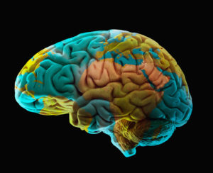
Ways to keep your brain sharp as a tack despite the brain shrinking as we grow older

–  Get plenty of aerobic exercise, at least 20 minutes every other day,” said Professor Klemm who is the author the book Memory Power 101.

People who stay physically fit tend to stay mentally sharp and hold their cognitive abilities well into their seventies and eighties. A 2012 study of 691 seniors in the journal Neurology found that seniors who reported high levels of physical activity at age 70 had less brain shrinkage at age 73 than seniors who reported less physical activity. Exercise may decrease memory loss by improving blood flow to the brain.

– Brain Exercise

Train your attentiveness and focus. The most common mental problem with aging is distractibility, which inevitably interferes with memory. An example is when you open the refrigerator door and suddenly realize you forgot what you went to the fridge for,” said Klemm. He recommends challenging your brain with games like chess or Sudoku. Dr. Gandy recommends


– Learn a New Skill

Some research shows that learning a new language or learning to play a musical instrument may help prevent memory loss and improve cognitive abilities. A 2011 study published in the journal Neuropsychology found that people who had instrumental musical training retained their memory and had

 The study included 70 seniors between age 60 and 83. The study found that the more years of musical training a person had, the better their cognitive performance was with age.

– Be More Sociable

Klemm and Gandy agree that social engagement is important in preventing memory loss. “Social engagement, along with physical and mental stimulation, all release substances in the brain that strengthen nerve connections called synapses,” said Gandy. A 2012 study published in the journal Neuropsychology followed 952 seniors for 12 years to see if

Social engagement protected seniors from memory loss and decline in communication skills. They concluded that being socially active reduced these declines and that seniors who showed declines tended to become less socially engaged.

– Get Your Antioxidants

Composition of human brain model and world map

Composition of human brain model and world map

Antioxidant vitamins may benefit memory byblocking free radicals that contribute to cell aging. Over the years, some large studies have found that antioxidant vitamins C and E may protect against cognitive decline. Gandy said that vitamins could help but cautions that they only help in cases of vitamin deficiency. You can also get plenty of antioxidants naturally in your diet. “They’re in any dark-colored fruit, berry, or vegetable. Also, take vitamin D3 and resveratrol pills,” advised Klemm.


–  Learn to Meditate

Stress and anxiety may decrease memory and cognitive ability, so take steps to reduce these negatives. “Take up meditation, yoga, or another type of mind-body exercise that reduces stress,” said Klemm. A 2010 study in the journal Consciousness and Cognition found that just four days of meditation training could significantly

To reduce anxiety and improve memory and cognition. In the study, 24 volunteers took meditation training and 25 listened to a recorded book. Both groups had improved mood, but the meditation group also had better memory, less stress, and clearer thinking.

The gradual mental decline that typically begins in middle age — what some refer to as “getting rusty” — is largely due to altered connections between brain cells. Putting your brain to work on new and different challenges can help build up those neural connections. It’s frequently referred to as the “use it or lose it” principle.

“There is data that suggests keeping your mind active is important,” said Dr. Burns. Scientific evidence linking low levels of education to a higher risk of Alzheimer’s later in life further supports the idea that mental stimulation keeps the mind sharper longer. “We need to develop tools or exercises that will keep the brain from deteriorating,” said Dr. Burns.

Research has suggested that certain activities such as reading, puzzles and crafts may reduce the risk of mild cognitive impairment, which could signal the onset of Alzheimer’s.