Archive | April 2024

QUOTE FOR THURSDAY:

“Head and neck cancers usually begin in the squamous cells that line the mucosal surfaces inside the mouth, nose, throat, and salivary glands. They are often referred to as squamous cell carcinomas of the head and neck.

Head and neck cancers account for nearly 4 percent of all cancers in the United States, according to the National Cancer Institute (NCI).

Counting cancers of the oral cavity, pharynx, and larynx, the NCI estimates that about 66,920 cases occurred in the U.S. in 2023. About 15,400 people died from these diseases, according to the NCI’s Surveillance, Epidemiology and End Results (SEER) program.

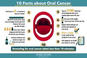
Head and neck cancer is more common among men than women. These cancers are also diagnosed more often among people over age 50 than among younger people.

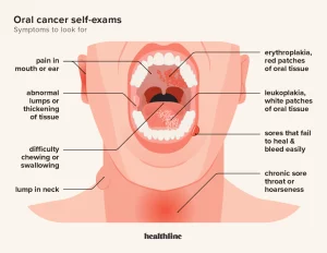
Symptoms of head and neck cancers may include a lump or a sore that doesn’t heal. In addition, symptoms can include sore throat that doesn’t go away, difficulty in swallowing, and a hoarse voice.

Alcohol and tobacco use are major risk factors for head and neck cancers. Also, infection with cancer-causing types of human papillomavirus (HPV) increases a person’s risk of certain head and neck cancers.”

American Association for Cancer Research – AACR (https://www.aacr.org/patients-caregivers/awareness-months/head-and-neck-cancer-awareness-month/)

Oral, Head, and Neck Cancer Month Awareness!


  ORAL CANCER RECONCONSTRUCTION SURVIVORS
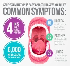
KNOW THE SYMPTOMS

Most oral, head, and neck cancers begin in the cells that make up the moist tissues (mucosal surfaces) of the head and neck, including in the mouth, nose, and throat. Like other cancers, oral, head, and neck cancers occur when abnormal cells begin to grow and divide uncontrollably and form a mass, called a malignant tumor.

The National Cancer Institute estimates that 85 percent of oral, head, and neck cancers are linked to tobacco use. Men (age 50 or older) who use both tobacco and alcohol are at the highest risk.

Cancers of the brain, eye, thyroid, as well as the skin, bones, muscles and nerves of the head and neck are not included under the heading of “oral, head and neck” cancer.

Doctors identify oral, head, and neck cancers by the area in which the cancer begins:

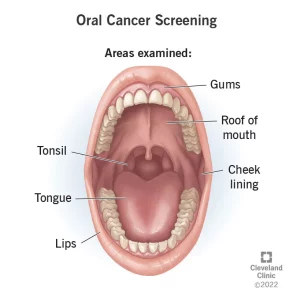
Lip and oral cavity cancer occurs on the lips, the front two-thirds of the tongue, the gums, the lining of the cheeks and lips, the floor of the mouth under the tongue, the palate, and the area behind the wisdom teeth.

Where does oral cancer affect:

Oral cancer (mouth cancer) is the broad term for cancer that affects the inside of your mouth. Oral cancer can look like a common problem with your lips or in your mouth, like white patches or sores that bleed. Oral cancer (mouth cancer) is the most common form of head and neck cancer. It typically affects people age 60 and older. Oral cancer affects your lips and the first parts of your tongue, mouth roof and floor. It also affects your oropharynx — the last part of your tongue and roof of your mouth, your tonsils, the sides and back of your throat that can include your sinuses, and pharynx (throat). The difference between a common problem and potential cancer is these changes don’t go away. Left untreated, oral cancer can spread throughout your mouth and throat to other areas of your head and neck. Approximately 63% of people with oral cavity cancer are alive five years after diagnosis.

 It can be life-threatening if not diagnosed and treated early.

When it is caught early, like other cancers is much easier for doctors to treat when caught early. Yet most people get a diagnosis when their condition is too advanced to treat effectively. If you see your dentist or doctor regularly and learn how to spot suspicious changes, you’ll have a much better shot at an early diagnosis.

Overall, about 11 people in 100,000 will develop oral cancer during their lifetime. Men are more likely than women to develop oral cancer. People who are white are more likely to develop oral cancer than people who are Black.

Risk factors in developing oral cancer include:

  • Smoking. Cigarette, cigar, or pipe smokers are six times more likely than nonsmokers to develop oral cancers.
  • Smokeless tobacco use. Users of dip, snuff, or chewing tobacco products are 50 times more likely to develop cancers of the cheek, gums, and lining of the lips.
  • Excessive consumption of alcohol. Oral cancers are about six times more common in drinkers than in nondrinkers.  Using alcohol and tobacco together increases your chances even more.
  • Family history of cancer.
  • Excessive sun exposure, especially at a young age. Ultraviolet radiation from the sun can cause lip cancers.
  • Human papillomavirus (HPV). Certain HPV strains are etiologic risk factors for Oropharyngeal Squamous Cell Carcinoma (OSCC). Almost everyone who’s sexually active will get HPV at some point in life. A specific type of this virus is causing a growing number of otherwise healthy men under 50 to get cancers in the back of their mouths and throats from oral sex. The more people you and your partners have sex with, the bigger your risk.
  • Age. Oral cancers can take years to grow. Most people find they have it after age 55. But more younger men are getting cancers linked to HPV.
  • Gender. Men are at least twice as likely as women to get oral cancer. It could be because men drink and smoke more than women do.
  • Poor diet. Studies have found a link between oral cancer and not eating enough vegetables and fruits.

It is important to note that over 25% of all oral cancers occur in people who do not smoke and who only drink alcohol occasionally.

The overall 5-year survival rate for patients with an early diagnosis of oral cavity and pharynx cancers is 84%. If the cancer has spread to nearby tissues, organs, or lymph nodes, the 5-year survival rate drops to 65%.

How it is diagnosed?

As part of your routine dental exam, your dentist will conduct an oral cancer screening exam.  Your dentist knows what a healthy mouth should look like and probably has the best chance of spotting any cancer. Experts recommend getting checked every year starting at age 18, and sooner if you start smoking or having sex.

More specifically, your dentist will feel for any lumps or irregular tissue changes in your neck, head, face, and oral cavity. When examining your mouth, your dentist will look for any sores or discolored tissue as well as check for any signs and symptoms mentioned above.

A biopsy may be needed to determine the makeup of a suspicious-looking area. There are different types of biopsies and your doctor can determine which one is best.

Treatments:

It varies from person to person depending on the stage of oral cancer the person has and how advanced the cancer is.

Whether a patient has surgery, radiation, and surgery, or radiation, surgery, and chemotherapy, you need to remember it depends on the stage of cancer development. Each case is individual.

Patients with cancers treated in their early stages may have little in the way of post-treatment disfigurement. For those whose cancer is caught at a later stage, the results of surgical removal of the disease may require reconstruction of portions of their oral cavity or facial features. There may be needed adjunctive therapy to assist in speech, chewing of foods, the problems associated with the lack of salivary function, and the fabrication of dental or facial prostheses.
 

QUOTE FOR WEDNESDAY:

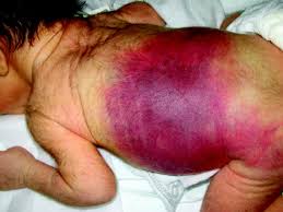
“In 1986, President Reagan recognized March as “National Hemophilia Month” which has since been changed to #BleedingDisordersAwarenessMonth to be inclusive of all #bleedingdisorders. We’ll be posting facts throughout the month to honor our history and raise awareness about all #bleedingdisorders. Hemophilia is usually an inherited bleeding disorder in which the blood does not clot properly. This can lead to spontaneous bleeding as well as bleeding following injuries or surgery.

World Federation of Hemophilia – WFH (https://www.hemophiliafed.org/resource/bleeding-disorders-awareness-month/)

April Awareness on Hemophilia.

hemophilia3hemophilia2

hemophilia1hemodialysis explained

What is this condition?

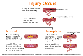
Hemophilia is a bleeding disorder characterized by low levels of clotting factor proteins. Correct diagnosis of Hemophilia is essential to providing effective treatment. Blood Center of Wisconsin offers one of the largest diagnostic menus to accurately and confidently diagnose Hemophilia.

The X and Y chromosomes are called sex chromosomes. The gene for hemophilia is carried on the X chromosome. Hemophilia is inherited in an X-linked recessive manner.  Females inherit two X chromosomes, one from their mother and one from their father (XX). Males inherit an X chromosome from their mother and a Y chromosome from their father (XY). That means if a son inherits an X chromosome carrying hemophilia from his mother, he will have hemophilia. It also means that fathers cannot pass hemophilia on to their sons.

But because daughters have two X chromosomes, even if they inherit the hemophilia gene from their mother, most likely they will inherit a healthy X chromosome from their father and not have hemophilia. A daughter who inherits an X chromosome that contains the gene for hemophilia is called a carrier. She can pass the gene on to her children. Hemophilia can occur in daughters, but is rare.

For a female carrier, there are four possible outcomes for each pregnancy:

  1. A girl who is not a carrier
  2. A girl who is a carrier
  3. A boy without hemophilia
  4. A boy with hemophilia

Hemophilia is an X-linked inherited bleeding disorder caused by mutation of the F8 gene that encodes for coagulation factor VIII or the F9 gene that encodes for coagulation factor IX. The degree of plasma factor deficiency correlates with both the clinical severity of disease and genetic findings. Severe hemophilia is characterized by plasma factor VIII or factor IX levels of under 1 IU/dl. Moderate and mild hemophilia are characterized by factor VIII or factor IX levels of 1-5 IU/dL or 6 – 40 IU/dL, respectively. Genetic analysis is useful for identification of the underlying genetic defect in males with severe, moderate or mild hemophilia and for determination of carrier status in the female individuals within their families. Additionally, data is emerging regarding the correlation between a patients mutation status and the risk of that patient developing an inhibitor.

People with hemophilia A often, bleed longer than other people. Bleeds can occur internally, into joints and muscles, or externally, from minor cuts, dental procedures or trauma. How frequently a person bleeds and the severity of those bleeds depends on how much FVIII is in the plasma, the straw-colored fluid portion of blood.

Normal plasma levels of FVIII range from 50% to 150%. Levels below 50%, or half of what is needed to form a clot, determine a person’s symptoms.

  • Mild hemophilia A-  6% up to 49% of FVIII in the blood. People with mild hemophilia Agenerally experience bleeding only after serious injury, trauma or surgery. In many cases, mild hemophilia is not diagnosed until an injury, surgery or tooth extraction results in prolonged bleeding. The first episode may not occur until adulthood. Women with mild hemophilia often experience menorrhagia, heavy menstrual periods, and can hemorrhage after childbirth.
  • Moderate hemophilia A. 1% up to 5% of FVIII in the blood. People with moderate hemophilia A  tend to have bleeding episodes after injuries. Bleeds that occur without obvious cause are called spontaneous bleeding episodes.
  • Severe hemophilia A.  <1% of FVIII in the blood. People with severe hemophilia A experience bleeding following an injury and may have frequent spontaneous bleeding episodes, often into their joints and musclesHemophilia A and B are diagnosed by measuring factor clotting activity. Individuals who have hemophilia A have low factor VIII clotting activity. Individuals who have hemophilia B have low factor IX clotting activity.Genetic testing is usually used to identify women who are carriers of a FVIII or FIX gene mutation, and to diagnose hemophilia in a fetus during a pregnancy (prenatal diagnosis). It is sometimes used to diagnose individuals who have mild symptoms of hemophilia A or B.There is currently no cure for hemophilia. Treatment depends on the severity of hemophilia.People who have moderate to severe hemophilia A or B may need to have an infusion of clotting factor taken from donated human blood or from genetically engineered products called recombinant clotting factors to stop the bleeding. If the potential for bleeding is serious, a doctor may give infusions of clotting factor to avoid bleeding (preventive infusions) before the bleeding begins. Repeated infusions may be necessary if the internal bleeding is serious. When a person who has hemophilia has a small cut or scrape, using pressure and a bandage will take care of the wound. An ice pack can be used when there are small areas of bleeding under the skin.
  • When bleeding has damaged joints, physical therapy is used to help them function better. Physical therapy helps to keep the joints moving and prevents the joints from becoming frozen or badly deformed. Sometimes the bleeding into joints damages them or destroys them. In this situation, the individual may be given an artificial joint.
  • Treatment may involve slow injection of a medicine called desmopressin (DDAVP) by the doctor into one of the veins. DDAVP helps to release more clotting factor to stop the bleeding. Sometimes, DDAVP is given as a medication that can be breathed in through the nose (nasal spray).
  • Diagnosing this condition:
  • Hemophilia is diagnosed with blood tests to determine if clotting factors are missing or at low levels, and which ones are causing the problem. If you have a family history of hemophilia, it is important that your doctors know the clotting factor your relatives are missing.
  • Treatment of the condition:
  • Genetic testing is also available for the factor VIII gene and the factor IX gene. Genetic testing of the FVIII gene finds a disease-causing mutation in up to 98 percent of individuals who have hemophilia A. Genetic testing of the FIX gene finds disease-causing mutations in more than 99 percent of individuals who have hemophilia B.
  • Researchers have been working to develop a gene replacement treatment (gene therapy) for Hemophilia A. Research of gene therapy for hemophilia A is now taking place. The results are encouraging. Researchers continue to evaluate the long-term safety of gene therapies. The hope is that there will be a genetic cure for hemophilia in the future.


QUOTE FOR TUESDAY:

  • Sarcoidosis is an inflammatory disease in which the immune system overreacts, causing groups of cells to form clusters of inflamed tissue called “granulomas” in one or more organs of the body.
  • Sarcoidosis most commonly affects the lungs and lymph nodes, but it can affect any organ including the eyes, skin, heart and nervous system.
  • Sarcoidosis is a rare disease with 150,000-200,000 cases in the United States per year and approximately 27,000 new cases per year.
  • Lofgren’s syndrome is an acute, milder form of sarcoidosis that usually goes away within six months to two years after diagnosis.
  • Black women are three times more likely to be diagnosed with sarcoidosis than White women, tend to have more severe disease, are more likely to be hospitalized and have a higher mortality rate.
  • In severe cases, sarcoidosis can be life-threatening if it progresses to heart or severe lung disease.
  • Severe fatigue can impact up to 70% of patients with sarcoidosis even when inflammation is controlled with medication. Fatigue can be disabling, and lead to loss of work.

Sarcoidosis in the lungs is often separated into the “Scadding stages” based on the chest X-ray. There are five Scadding stages, Stage 0 through Stage 4. The stages use the location of the granulomas in the lungs and lymph nodes, to predict the chance of the disease resolving on its own, with lower stages being more likely to resolve or not need treatment. At each stage you can have serious symptoms. You also can move between the stages of sarcoidosis. Patients of any Scadding stage can have involvement of other organs from sarcoidosis.”

American Lung Association (https://www.lung.org/lung-health-diseases/lung-disease-lookup/sarcoidosis/learn-about-sarcoidosis)

Part III Sarcoidosis Awareness Month – How it is diagnosed, the staging of it, & the treatments for it!


How Sarcoidosis can be diagnosed:

  • biopsy of the lungs, liver, skin, or other affected organs to check for granulomas
  • Blood tests, including complete blood counts, to check hormone levels and to test for other conditions that may cause sarcoidosis
  • Bronchoscopy, which may include rinsing an area of the lung to get cells or using a needle to take cells from the lymph nodes in the chest
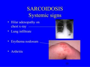
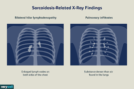
  • Chest X-ray to look for granulomas in the lungs and heart and determine the stage of the disease. Often, sarcoidosis is found because a chest X-ray is performed for another reason.
  • Neurological tests, such as electromyography, evoked potentials, spinal taps, or nerve conduction tests, to detect problems with the nervous system caused by sarcoidosis
  • Eye exam to look for eye damage, which can occur without symptoms in a person with sarcoidosis
  • Gallium scan external link , which uses a radioactive material called gallium to look for inflammation, usually in the eyes or lymph nodes.
  • High-resolution computed tomography (CT) scan to look for granulomas
  • Magnetic resonance imaging (MRI) to help find granulomas. Learn more in our Chest MRI Health Topic.
  • Positron electron tomography (PET) scan, a type of imaging that can help find granulomas
  • Pulmonary function tests to check whether you have breathing problems
  • Ultrasound to look for granulomas

There is a staging to Sarcoidosis:

Doctors use stages to describe the various imaging findings of sarcoidosis of the lung or lymph nodes of the chest. There are four stages of sarcoidosis, and they indicate where the granulomas are located. In each of the first three stages, sarcoidosis can range from mild to severe. This helps the doctor in deciding treatment for the patient.  Stage IV is the most severe and indicates permanent scarring in the lungs.

  • Stage I: Granulomas are located only in the lymph nodes.
  • Stage II: Granulomas are located in the lungs and lymph nodes.
  • Stage III: Granulomas are located in the lungs only.
  • Stage IV: Pulmonary fibrosis.

Treatment for Sarcoidosis:

Remember there is no cure.

The goal of treatment is remission, a state in which the condition is not causing problems. Not everyone who is diagnosed with sarcoidosis needs treatment. Sometimes the condition goes away on its own. Whether you need treatment—and what type you need—will depend on your signs and symptoms, which organs are affected, and whether those organs are working well; this is also where staging of the disease comes into play. Some people do not respond to treatment.


QUOTE FOR MONDAY:

“Sarcoidosis is considered a rare disease. There are usually fewer than 200,000 cases of sarcoidosis at any given time in the U.S.

Research suggests that a combination of genetics and environmental factors cause sarcoidosis, but its exact cause is unknown. We think certain people are more likely to have their immune system overreact to certain triggers (antigens), like bacteria and viruses. Despite the immune system’s involvement, sarcoidosis isn’t considered an autoimmune disorder.

Since we don’t know for sure which combination of genes and triggers causes sarcoidosis, it’s impossible to predict who will get it.”

Cleveland Clinic (https://my.clevelandclinic.org/health/diseases/11863-sarcoidosis#:~:text=Sarcoidosis%20is%20a%20condition%20that%20causes%20your%20immune%20system%20to,fibrosis%2C%20causing%20permanent%20lung%20scarring.)

Part II Sarcoidosis Awareness Month – Risk Factor, Complication and Treatment!

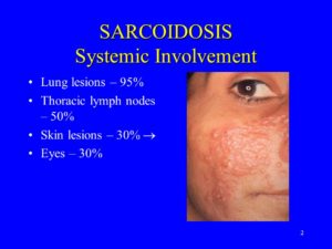
Lung lesions – 95% Thoracic lymph nodes – 50% Skin lesions – 30%  Eyes – 30%


Inflammatory Disease- Sarcoidosis or Sarcoid is a inflammatory disease that consists of granuloma.

Wide Spread Disease- Disease is wide spread in multiple organs.

Relapse-

  • The Sarcoidosis disease tends to come and go all of a sudden.
  • Disease may progressively develop as a serious illness.
  • Patient may experience several relapse throughout the life.

Granulomas-

  • Sarcoidosis or Sarcoid is a gradual progressive disease.
  • Microscopic lumps called granulomas start to appear in the affected organs.1
  • In most of the cases, these granulomas tend to clear with or without treatments.
  • There are few instances where granuloma grows in size and continues to be a part of the organ.
  • Granuloma eventually ends up as fibrotic lump but may cause several complications.

Causes

Doctors don’t know the exact cause of sarcoidosis. Some people appear to have a genetic predisposition to develop the disease, which may be triggered by bacteria, viruses, dust or chemicals.

This triggers an overreaction of your immune system and immune cells begin to collect in a pattern of inflammation called granulomas. As granulomas build up in an organ, the function of that organ can be affected.

Risk factors

While anyone can develop sarcoidosis, factors that may increase your risk include:

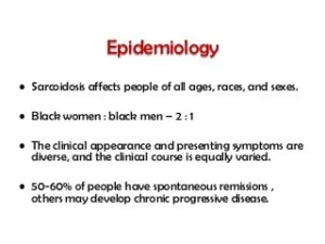
  • Age and sex. Sarcoidosis often occurs between the ages of 20 and 40. Women are slightly more likely to develop the disease.
  • Race. African-Americans have a higher incidence of sarcoidosis than do white Americans. Also, sarcoidosis may be more severe and may be more likely to recur and cause lung problems in African-Americans.
  • Family history. If someone in your family has had sarcoidosis, you’re more likely to develop the disease.

Complications

For most people, sarcoidosis resolves on its own with no lasting consequences. But sometimes it causes long-term problems.

  • Lungs. Untreated pulmonary sarcoidosis can lead to permanent scarring in your lungs, making it difficult to breathe.
  • Eyes. Inflammation can affect almost any part of your eye and can eventually cause blindness. Rarely, sarcoidosis also can cause cataracts and glaucoma.
  • Kidneys. Sarcoidosis can affect how your body handles calcium, which can lead to kidney failure.
  • Heart. Granulomas in your heart can cause abnormal heart rhythms and other heart problems. In rare instances, this may lead to death.
  • Nervous system. A small number of people with sarcoidosis develop problems related to the central nervous system when granulomas form in the brain and spinal cord. Inflammation in the facial nerves, for example, can cause facial paralysis.

QUOTE FOR THE WEEKEND:

“Sarcoidosis is a disease characterized by the growth of tiny collections of inflammatory cells (granulomas) in any part of your body — most commonly the lungs and lymph nodes. But it can also affect the eyes, skin, heart and other organs.

The cause of sarcoidosis is unknown, but experts think it results from the body’s immune system responding to an unknown substance. Some research suggests that infectious agents, chemicals, dust and a potential abnormal reaction to the body’s own proteins (self-proteins) could be responsible for the formation of granulomas in people who are genetically predisposed.”

MAYO CLINIC (https://www.mayoclinic.org/diseases-conditions/sarcoidosis/symptoms-causes/syc-20350358)

Part I Sarcoidosis Awareness Month – what it is, the areas of the body it can affect & symptoms!


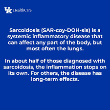
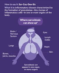
Sarcoidosis (pronounced SAR-COY-DOE-SIS) is an inflammatory disease characterized by the formation of granulomas, tiny clumps of inflammatory cells, in one or more organs of the body. When the immune system goes into overdrive and too many of these clumps form, they can interfere with an organ’s structure and function. When left unchecked, chronic inflammation can lead to fibrosis, which is permanent thickening or scarring of organ tissue.

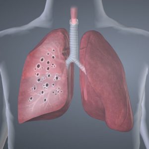
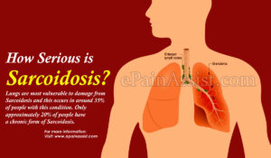
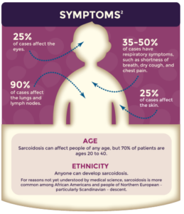
This disorder can affect almost any organ in the body, including the heart, skin, liver, kidneys, brain, sinuses, eyes, muscles, bones, and other areas. Sarcoidosis most commonly targets the lungs and the lymph nodes, which are an important part of the immune system. When it affects the lungs, it is called pulmonary sarcoidosis. Ninety percent or more of people diagnosed with the disease have lung involvement.

Doctors believe sarcoidosis results from the body’s immune system responding to an unknown substance, most likely something inhaled from the air.

There is no cure for sarcoidosis, but most people do very well with little or only modest treatment. In half of cases, sarcoidosis goes away on its own. In a few cases, however, sarcoidosis may last for years and may cause organ damage.

Symptoms in general to people affected by this disease sarcoidosis:

For many people, sarcoidosis begins with these symptoms in general:

  • Fatigue
  • Fever
  • Swollen lymph nodes

Symptoms caused by sarcoidosis at affected organ sites:

Lung symptoms

Many patients with sarcoidosis experience lung problems, which may include:

  • Persistent dry cough
  • Shortness of breath
  • Wheezing
  • Chest pain

Skin symptoms

Some people who have sarcoidosis develop skin problems, which may include:

  • A rash of red or reddish-purple bumps, usually located on the shins or ankles, which may be warm and tender to the touch
  • Disfiguring sores (lesions) on the nose, cheeks and ears
  • Areas of skin that are darker or lighter in color
  • Growths under the skin (nodules), particularly around scars or tattoos

Eye symptoms

Sarcoidosis can affect the eyes without causing any symptoms, so it’s important to have your eyes checked. When eye symptoms do occur, they may include:

  • Blurred vision
  • Eye pain
  • Severe redness
  • Sensitivity to light

Heart symptoms

Cardiac sarcoidosis: A heart under attack

Signs and symptoms related to cardiac sarcoidosis may include:

  • Chest pain
  • Shortness of breath (dyspnea)
  • Fainting (syncope)
  • Fatigue
  • Irregular heartbeats (arrhythmias)
  • Rapid or fluttering heart beat (palpitations)
  • Swelling caused by excess fluid (edema)

Stay tune and learn more about this illness in Part II tomorrow with Part III this weekend on Sarcoidosis!