Archive | September 2024

QUOTE FOR THE THURSDAY:

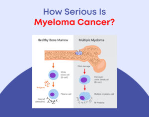
“Multiple myeloma is a cancer that forms in a type of white blood cell called a plasma cell. Healthy plasma cells help fight infections by making proteins called antibodies. Antibodies find and attack germs.

In multiple myeloma, cancerous plasma cells build up in bone marrow. The bone marrow is the soft matter inside bones where blood cells are made. In the bone marrow, the cancer cells crowd out healthy blood cells. Rather than make helpful antibodies, the cancer cells make proteins that don’t work right. This leads to complications of multiple myeloma.

Multiple myeloma treatment isn’t always needed right away. If the multiple myeloma is slow growing and isn’t causing symptoms, close watching might be the first step. For people with multiple myeloma who need treatment, there are a number of ways to help control the disease.”

MAYO CLINIC (https://www.mayoclinic.org/diseases-conditions/multiple-myeloma/symptoms-causes/syc-20353378)


Myeloma Cancer

What is Multiple Myeloma?

Multiple myeloma is a cancer of plasma cells (B and T cells).  Multiple myeloma is a cancer of plasma cells. Normal plasma cells are found in the bone marrow and are an important part of the immune system.The immune system is made up of several types of cells that work together to fight infections and other diseases.

We stated yesterday myeloma is most often found in bone marrow. Remember normal plasma cells are found in the bone marrow & an important part of the immune system.

The immune system is made up of several types of cells that work together to fight infections and other diseases. Lymphocytes (lymph cells) are one of the main types of white blood cells in the immune system and include T cells and B cells. Lymphocytes are in many areas of the body, such as lymph nodes, the bone marrow, the intestines, and the bloodstream.

When B cells respond to an infection, they mature and change into plasma cells. Plasma cells make the antibodies (also called immunoglobulins) that help the body attack and kill germs.

When plasma cells become cancerous=cancer cells (intially made in the bone marrow than released in the blood), they multiply and begin to crowd out healthy cells and produce abnormal antibodies called M proteins.  This continues to to replicate till the body dies or some treatment takes over decreasing them by destroying them (Ex. Chemo) or possibly putting the patient in remission.

What are the major types of myeloma?

There are four major sub-types of myeloma. Diagnosis depends on how localized or spread out the cancer has become and where it originated. The most common form of myeloma is multiple myeloma. Over 90% of people with myeloma have multiple myeloma.

Multiple myeloma -This is the one we will be discussing in this topic.
Plasmacytoma
Localized myeloma
Extramedullary myelom
Multiple Myeloma Complications:

In multiple myeloma, the overgrowth of plasma cells in the bone marrow can crowd out normal blood-forming cells:

1-Leading to low cell counts=RBCs-low in iron/anemia, level of platelets in the become low (called thrombocytopenia)=increased bleeding and bruising & WBCs low called leukopenia=problems fighting infections.

2-Myeloma cells make a substance that tells the osteoclasts to speed up dissolving the bone. So old bone is broken down without new bone to replace it, making the bones painful, weak, thinning the bones and easy to break.

3-Abnormal plasma cells cannot protect the body from infections. As mentioned before, normal plasma cells produce antibodies that attack germs. In multiple myeloma, the myeloma cells crowd out the normal plasma cells, so that antibodies to fight the infection can’t be made. The antibody made by the myeloma cells does not help fight infections. That’s because the myeloma cells are just many copies of the same plasma cell – all making copies of the same exact (or monoclonal) antibody.

A monoclonal gammopathy is when plasma cells make too many copies of the same antibody. It is usually found on a routine blood test when looking for other conditions.In monoclonal gammopathy of undetermined significance (MGUS), abnormal plasma cells make many copies of the same antibody (called a monoclonal protein). However, these plasma cells do not form an actual tumor or mass and do not cause any of the problems seen in multiple myeloma. MGUS usually does not affect a person’s health. It doesn’t cause weak bones, high calcium levels, kidney problems, or low blood counts.

4-Myeloma cells make an antibody that can harm the kidneys, leading to kidney damage and even kidney failure.

Factors that may increase your risk of multiple myeloma include:

Increasing age. Your risk of multiple myeloma increases as you age, with most people diagnosed in their mid-60s.
Male sex. Men are more likely to develop the disease than are women.
Black race. Black people are about twice as likely to develop multiple myeloma as are white people.
Family history of multiple myeloma. If a brother, sister or parent has multiple myeloma, you have an increased risk of the disease.
Personal history of a monoclonal gammopathy of undetermined significance (MGUS). Every year 1 percent of the people with MGUS in the United States develop multiple myeloma.
Diagnosing Multiple Myeloma:
Blood tests. Laboratory analysis of your blood may reveal the M proteins produced by myeloma cells. Another abnormal protein produced by myeloma cells — called beta-2-microglobulin — may be detected in your blood and give your doctor clues about the aggressiveness of your myeloma.Additionally, blood tests to examine your kidney function, blood cell counts, calcium levels and uric acid levels can give your doctor clues about your diagnosis.
Urine tests. Analysis of your urine may show M proteins, which are referred to as Bence Jones proteins when they’re detected in urine.
Examination of your bone marrow. Your doctor may remove a sample of bone marrow for laboratory testing. The sample is collected with a long needle inserted into a bone (bone marrow aspiration and biopsy).In the lab, the sample is examined for myeloma cells. Specialized tests, such as fluorescence in situ hybridization (FISH) can analyze myeloma cells to understand their genetic abnormalities. Tests are also done to measure the rate at which the myeloma cells are dividing.
Imaging tests. Imaging tests may be recommended to detect bone problems associated with multiple myeloma. Tests may include an X-ray, MRI, CT or positron emission tomography (PET).

After someone is diagnosed with cancer, doctors will try to figure out if it has spread, and if so, how far. This process is called staging (I,II,III,IV). The stage of a cancer describes how much cancer is in the body. It helps determine how serious the cancer is and how best to treat it.

Treatment:

1-Targeted therapy. Targeted drug treatment focuses on specific abnormalities within cancer cells that allow them to survive. Bortezomib (Velcade), carfilzomib (Kyprolis) and ixazomib (Ninlaro) are targeted drugs that block the action of a substance in myeloma cells that breaks down proteins. This action causes myeloma cells to die. Targeted-therapy drugs may be administered through a vein in your arm or in pill form.

Other targeted-therapy treatments include monoclonal antibody drugs that bind to the specific proteins present on myeloma cells, causing them to die.

2-Biological therapy. Biological therapy drugs use your body’s immune system to fight myeloma cells. The drugs thalidomide (Thalomid), lenalidomide (Revlimid) and pomalidomide (Pomalyst) enhance the immune system cells that identify and attack cancer cells. These medications are commonly taken in pill form.

3-Chemotherapy. Chemotherapy drugs kill fast-growing cells, including myeloma cells. Chemotherapy drugs can be given through a vein in your arm or taken in pill form. High doses of chemotherapy drugs are used before a bone marrow transplant.

4-Corticosteroids. Corticosteroids, such as prednisone and dexamethasone, regulate the immune system to control inflammation in the body. They are also active against myeloma cells. Corticosteroids can be taken in pill form or administered through a vein in your arm.

5-Bone marrow transplant. A bone marrow transplant, also known as a stem cell transplant, is a procedure to replace your diseased bone marrow with healthy bone marrow.

Before a bone marrow transplant, blood-forming stem cells are collected from your blood. You then receive high doses of chemotherapy to destroy your diseased bone marrow. Then your stem cells are infused into your body, where they travel to your bones and begin rebuilding your bone marrow.

6-Radiation therapy. This treatment uses beams of energy, such as X-rays and protons, to damage myeloma cells and stop their growth. Radiation therapy may be used to quickly shrink myeloma cells in a specific area — for instance, when a collection of abnormal plasma cells form a tumor (plasmacytoma) that’s causing pain or destroying a bone.

QUOTE FOR WEDNESDAY:

“An estimated 400,000 people were exposed to toxic contaminants, risk of physical injury, and physically and emotionally stressful conditions in the days, weeks, and months following the attacks. Responders who worked on the rescue, recovery, and relief efforts reported a range of illnesses soon after the attacks. Survivors who lived, worked, and attended school in Lower Manhattan, some who remained throughout and some who returned to their lives in the disaster area, started reporting illnesses as well. estimated 400,000 people were exposed to toxic contaminants, risk of physical injury, and physically and emotionally stressful conditions in the days, weeks, and months following the attacks. Responders who worked on the rescue, recovery, and relief efforts reported a range of illnesses soon after the attacks. Survivors who lived, worked, and attended school in Lower Manhattan, some who remained throughout and some who returned to their lives in the disaster area, started reporting illnesses as well.”

Centers for Disease Control and Prevention – CDC  (https://www.cdc.gov/wtc/exhibition/toxins-and-health-impacts.html)

Health Effects from 9/11 Disaster.


HEALTH EFFECTS FROM SEPTEMBER 11 DISASTER

The world trade center (WTC) terrorist attact and its aftermath exposed hundreds of thousands of people to dust, debris, smoke and fumes.  I was one that went down that day as a volunteer RN to help out my country.  It was a sight I will never forget and thank God nothing like it has happened since including developing anything from being exposed to the environment of this disaster.  September 11, 2001, among rescue and clean up workers, office workers, building evacuees, and residents of lower Manhattan living down their have shown increased respiratory and other physical health problems, like developing cancers after this terrible terrorist tragedy.  Following is a outline of the most common conditions experienced by individuals exposed to the WTC attacks and their aftermath.  The types of conditions are:

Upper Airway Cough Syndrome:  Formerly named postnasal drip syndrome which is commonly caused by continuous irritation or infection of the sinuses and the nose either due to allergies or from environmental irritants.  The signs or symptoms that arise from this are cough, nasal congestion, postnasal drip and frequent need to clear the throat.

Asthma/Reactive Airways Dysfunction Syndrome:  Some people exposed to the WTC disaster area have developed asthma related to exposure to irritants (also called reactive airways dysfunction syndrome [RADS]).  Signs and symptoms include:  Shortness of Breath (SOB); chest tightness, wheezing, coughing, phlegm, possible triggering of symptoms by colds or seasonal allergies or exercise or fragrances or extremes of temperature or humidity, recurrent episodes of respiratory infections requiring antibiotic treatment.

GASTROESOPHAGEAL REFLUX DISEASE OR LARYNGOPHARYNGEAL REFLUX DISEASE:  Some people exposed to the WTC disaster developed this condition.  GERD results from the flow back or return (reflux) of stomach contents into the esophagus. LPRD results from the reflux of stomach contents into the voice box or throat.

Symptoms of GERD: Heartburn, acid regurgitation, upset stomach, cough made worse with meals or at night

Symptoms of LPRD: Hoarseness or other vocal changes, sore throat, cough, sensation of having a lump in the throat

Long-Term Physical Health Concerns

There has been increased concern about sarcoidosis and cancer among individuals who were highly exposed during the WTC disaster. The NYC Department of Health and Mental Hygiene, in conjunction with other programs, is closely monitoring these conditions in order to detect any increases in these diseases.

At this time, there is no evidence of an increased rate of cancer among individuals highly exposed to the WTC disaster. Increased rates of sarcoidosis have been documented among fire department personnel ( Izbicki G, Chavko R, Banauch, GI, et al. World Trade Center “Sarcoid-Like” Granulomatous Pulmonary Disease in New York City Fire Department Workers. Chest. 2007;3:131;1414-1423). Rates of sarcoidosis among other groups are currently under investigation.

Sarcoidosis is difficult to verify because NYC providers are not required by law to report sarcoidosis cases to the Health Department.  Based on New York City death certificate records over the   past 10 years, there have been an average of 32 sarcoidosis deaths per year, with the annual number remaining the same since the WTC attacks. During the   same period, there have been between 362 and 439 sarcoidosis-related hospitalizations per year in NYC (five per 100,000 people), with the annual   rate remaining the same since the disaster.

* Breathing in beryllium, other metal dust or fumes or moldy materials can cause lung disease, which may resemble sarcoidosis.

.Sarcoidosis

Sarcoidosis* is an auto-immune disease that can attack any organ   of the body, although it often starts in the lungs or lymph nodes. It mainly   affects people between 20 and 40 years of age, with African-Americans three  times more likely to develop the condition than Caucasians and women twice as   likely to develop it as men. While most sarcoidosis patients recover without treatment, less than one-third develop chronic debilitating sarcoidosis and  fewer than 5% die from the disease. The exact cause of sarcoidosis is unknown.

Sarcoidosis and World Trade Center (WTC) Dust Exposure

While studies have not definitively linked dust exposure from   the WTC disaster to new-onset sarcoidosis among exposed workers, the data   does suggest elevated levels of sarcoidosis among firefighters.

There have been a few reported sarcoidosis cases among rescue   workers that may be related to Ground Zero dust exposure. In May 2007, the New York City Chief Medical Examiner determined that dust exposure from the disaster contributed to a sarcoidosis death, based on published epidemiologic findings among exposed firefighters.

Cancers

The collapse and burning of the WTC and neighboring buildings   released a complex mixture of irritant dust, smoke and gases. The dust cloud   also contained heavy metals, as well as asbestos and other carcinogens. In   addition, smoke released from the fires contained hazardous and potentially   cancer-causing substances.

Because of these exposures, there has been concern about the   possibility of increased cancer rates among WTC-exposed people. The NYC   Department of Health and Mental Hygiene, the Fire Department of New York and   other researchers are carefully monitoring cancer rates among highly exposed   people.

In 2007, the NYC Department of Health and Mental Hygiene brought together a panel of experts, including representatives from FDNY, the Mount Sinai School of Medicine, the New York State Department of Health and the National Institute for Occupational Safety and Health, to discuss the potential impact of the WTC disaster on cancer rates and mortality.

In 2010, after beginning the process of confirming cancer   diagnoses within their cohorts, the NYC Department of Health and Mental   Hygiene and FDNY co-chaired another conference of outside experts, including biostatisticians, environmental health scientists and cancer epidemiologists,   to help address the complex methodological questions associated with their   preliminary cancer investigations. The conference produced several   recommendations which are described in the 2010 WTC Medical Working group annual report.

Although cancer associated with specific exposures takes a long   time to develop, three early cancer studies based on verified diagnoses   within their cohorts through 2008 (the latest year for which data was   available when these analyses began) have been published:

  • FDNY found that nearly 9,000 firefighters with WTC exposure may be at greater risk for cancer than firefighters  who weren’t exposed.
  • The WTC Health Registry found small increases in rates of prostate cancer, thyroid cancer and multiple myeloma, a blood cancer, among nearly 34,000 WTC rescue and recovery workers in comparison to rates among New York State residents after adjusting for age, race/ethnicity and sex.
  • Prostate and thyroid cancer rates were higher than expected among nearly 21,000 rescue and recovery workers enrolled in the WTC Health Program in comparison to rates in New York, New Jersey, Connecticut and Pennsylvania where the majority  of workers lived.

Cancer analyses continue at FDNY, the WTC Health Registry and at   the Mount Sinai School of Medicine.

In 2012, the National Institute for Occupational Safety and Health  added many different types of cancers to the list of conditions that can be treated at the World Trade Center Health Program.

What the New York City  Health Department Is Doing:

Through the World Trade Center (WTC) Health Registry, the Health Department is conducting a cancer study that will help determine if cancer rates are higher among those exposed to the WTC disaster. Results of   the study will appear on this Web site and will be submitted to peer-reviewed scientific journals.

The Department is also conducting a broader public awareness campaign about cancer testing, and is working with its clinical partners to offer free cancer screenings. Identifying cancer early in the course of the disease reduces the risk of developing disability from cancer. Department efforts to reduce the rates of smoking among WTC disaster-exposed people also help reduce their risk for developing cancer and other diseases.

What You Can Do

Seek medical care.

If you were exposed to the WTC disaster and have developed symptoms or conditions you feel are related to this exposure, tell your doctor and bring the Clinical Guidelines for Physicians Treating Adults   Exposed to the WTC Disaster to   help your doctor diagnose and treat your symptoms. The Health Department also   released clinical guidelines  for   health care providers on how to treat children and adolescents exposed to the WTC disaster.

If you or your doctor believe that you require more specialized   care, you may be eligible for free treatment at a WTC   Center of Excellence or affiliated facility.

Practice preventive   health.

Whether or not you are currently experiencing symptoms, there   are things you can do to maintain your current health status, prevent   worsening of your health, and detect any new conditions. Suggested measures   include:

    1. If you smoke, quit! Tobacco is known to cause   cancer and make existing respiratory conditions worse. If you currently   smoke, there are programs in place to help you quit.
    2. Lead a healthy lifestyle by drinking alcohol   in moderation and avoiding more than moderate sun exposure. Increase physical   activity, keep your weight down and eat a healthful and nutritious diet.
    3. Have a physical exam every year that includes a   complete blood count (CBC). Be sure your exam includes all tests appropriate   for your sex and age, including cancer screenings.
    4. Avoid   risks at work and during leisure time. If you have any respiratory conditions  try to reduce your exposure to irritants such as dust, pollen, grass and   smoke both at home and at work. Consider giving up activities that expose you to irritants, and if activities cannot be avoided, wear personal protective   equipment to minimize exposure.
 

QUOTE FOR TUESDAY:

“If you’ve been diagnosed with cancer, and you aren’t sure about your diagnosis or treatment options, get a second opinion from Cedars-Sinai. Your Virtual Second Opinion will give you the information you need to make the best decisions about your care.  Our hodgkin or  non-hodgkin lymphoma specialists can provide a Virtual Second Opinion in the comfort of your home, and you can get the insights needed to make informed decisions about your care.   Treatments range from chemo, radiation and more.”

Cedars Sinai (https://secondopinion.cedars-sinai.org/department/cancer/?&cid=21046965596&agid=158830926683&tid=kwd-328662390414&kwt=p&adid=691743710360&ext=&dvc=c&loi=&lop=9004281&gclid=EAIaIQobChMI-PzYvvusiAMVFGpHAR3FHQ4aEAAYASAAEgKRwvD_BwE&gclsrc=aw.ds)


Part II Hodgkin’s versus Non=Hodgkin’s Lymphoma (Treatments)

Treatments for both Hodgkin’s Lymphoma & Non-Hodgkin’s Lymphoma:

First the doctor needs to know what stage of cancer your in ranging from I to IV.  This shows the doctor the following information to help the M.D. decide what treatment would be most effective to take.

  • Stage I. The cancer is limited to one lymph node region or a group of nearby nodes or limited to a single organ.
  • Stage II. In this stage, the cancer is in two lymph node regions, or the cancer has invaded one organ and the nearby lymph nodes as well. But the cancer is still limited to a section of the body either above or below the diaphragm.
  • Stage III. When the cancer moves to lymph nodes both above and below the diaphragm, it’s considered stage III. Cancer may also be found in the lymph nodes above the diaphragm and in the spleen in non-Hodgkins and may also be in one portion of tissue or an organ near the lymph node groups or in the spleen in Hodgkins.
  • Stage IV. This is the most advanced stage of non-Hodgkin’s and Hodgkin’s lymphoma. Cancer cells are in several portions of one or more organs and tissues.
  • Stage III. When the cancer moves to lymph nodes both above and below the diaphragm, it’s considered stage III. Cancer may also be in one portion of tissue or an organ near the lymph node groups or in the spleen.
  • Stage IV. This is the most advanced stage of Hodgkin’s lymphoma. Cancer cells are in several portions of one or more organs and tissues. Stage IV in both lymphomas affects not only the lymph nodes but also other parts of the body, such as the liver, lungs or bones.

Hodgkin Lymphoma Consultation

Your doctor will review your scans and discuss treatment options with you.

Which Hodgkin’s lymphoma treatments are right for you depends on the type and stage of your disease, your overall health, and your preferences. The goal of treatment is to destroy as many cancer cells as possible and bring the disease into remission.

Chemotherapy

Chemotherapy is a drug treatment that uses chemicals to kill lymphoma cells. Chemotherapy drugs travel through your bloodstream and can reach nearly all areas of your body.

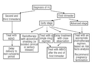
Chemotherapy is often combined with radiation therapy in people with early-stage classical type Hodgkin’s lymphoma. Radiation therapy is typically done after chemotherapy. In advanced Hodgkin’s lymphoma, chemotherapy may be used alone or combined with radiation therapy.

Chemotherapy drugs can be taken in pill form or through a vein in your arm, or sometimes both methods of administration are used. Several combinations of chemotherapy drugs are used to treat Hodgkin’s lymphoma.

Side effects of chemotherapy depend on the drugs you’re given. Common side effects are nausea and hair loss. Serious long-term complications can occur, such as heart damage, lung damage, fertility problems and other cancers, such as leukemia.

Radiation therapy

Radiation therapy uses high-energy beams, such as X-rays and protons, to kill cancer cells. For classical Hodgkin’s lymphoma, radiation therapy is often used after chemotherapy. People with early-stage nodular lymphocyte-predominant Hodgkin’s lymphoma may undergo radiation therapy alone.

During radiation therapy, you lie on a table and a large machine moves around you, directing the energy beams to specific points on your body. Radiation can be aimed at affected lymph nodes and the nearby area of nodes where the disease might progress. The length of radiation treatment varies, depending on the stage of the disease. A typical treatment plan might have you going to the hospital or clinic five days a week for several weeks. At each visit, you undergo a 30-minute radiation treatment.

Radiation therapy can cause skin redness and hair loss at the site where the radiation is aimed. Many people experience fatigue during radiation therapy. More-serious risks include heart disease, stroke, thyroid problems, infertility and other cancers, such as breast or lung cancer.

Bone marrow transplant

Bone marrow transplant, also known as stem cell transplant, is a treatment to replace your diseased bone marrow with healthy stem cells that help you grow new bone marrow. A bone marrow transplant may be an option if Hodgkin’s lymphoma returns despite treatment.

During a bone marrow transplant, your own blood stem cells are removed, frozen and stored for later use. Next you receive high-dose chemotherapy and radiation therapy to destroy cancerous cells in your body. Finally your stem cells are thawed and injected into your body through your veins. The stem cells help build healthy bone marrow.

People who undergo bone marrow transplant may be at increased risk of infection.

Other drug therapy

Other drugs used to treat Hodgkin’s lymphoma include targeted drugs that focus on specific vulnerabilities in your cancer cells and immunotherapy that works to activate your own immune system to kill the lymphoma cells. If other treatments haven’t helped or if your Hodgkin’s lymphoma returns, your lymphoma cells may be analyzed in a laboratory to look for genetic mutations. Your doctor may recommend treatment with a drug that targets the particular mutations present in your lymphoma cells.

Targeted therapy is an active area of cancer research. New targeted therapy drugs are being studied in clinical trials.

Now look at Non-Hodgkin’s Lymphoma Treatment and look at the similarities of Hodkin’s Lymphoma Rx; both are WBC’s Blood Cancers:

If your non-Hodgkin’s lymphoma is aggressive or causes signs and symptoms, your doctor may recommend treatment. Options may include:

Chemotherapy

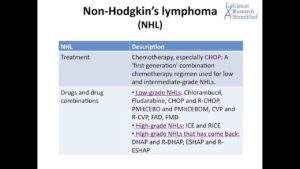
Chemotherapy is a drug treatment — given orally or by injection — that kills cancer cells. Chemotherapy drugs can be given alone, in combination with other chemotherapy drugs or combined with other treatments.

Side effects of chemotherapy depend on the drugs you’re given. Common side effects are nausea and hair loss. Serious long-term complications can occur, such as heart damage, lung damage, fertility problems and other cancers, such as leukemia.

Radiation therapy

Radiation therapy uses high-powered energy beams, such as X-rays and protons, to kill cancer cells. During radiation therapy, you’re positioned on a table and a large machine directs radiation at precise points on your body. Radiation therapy can be used alone or in combination with other cancer treatments.

During radiation therapy, you lie on a table and a large machine moves around you, directing the energy beams to specific points on your body. Radiation can be aimed at affected lymph nodes and the nearby area of nodes where the disease might progress. The length of radiation treatment varies, depending on the stage of the disease. A typical treatment plan might have you going to the hospital or clinic five days a week for several weeks, where you undergo a 30-minute radiation treatment at each visit.

Radiation therapy can cause skin redness and hair loss at the site where the radiation is aimed. Many people experience fatigue during radiation therapy. More-serious risks include heart disease, stroke, thyroid problems, infertility, and other cancers, such as breast or lung cancer.

Bone marrow transplant

Bone marrow transplant, also known as a stem cell transplant, involves using high doses of chemotherapy and radiation to suppress your bone marrow. Then healthy bone marrow stem cells from your body or from a donor are infused into your blood where they travel to your bones and rebuild your bone marrow.

People who undergo bone marrow transplant may be at increased risk of infection.

Other drug therapy

Biological therapy drugs help your body’s immune system fight cancer.

For example, one biological therapy called rituximab (Rituxan) is a type of monoclonal antibody that attaches to B cells and makes them more visible to the immune system, which can then attack. Rituximab lowers the number of B cells, including your healthy B cells, but your body produces new healthy B cells to replace these. The cancerous B cells are less likely to recur.

Also, a drug called ibrutinib (Imbruvica) has been approved by the Food and Drug Administration (FDA) for some people undergoing treatment for non-Hodgkin’s lymphoma.

Radioimmunotherapy drugs are made of monoclonal antibodies that carry radioactive isotopes. This allows the antibody to attach to cancer cells and deliver radiation directly to the cells. An example of a radioimmunotherapy drug used to treat non-Hodgkin’s lymphoma is ibritumomab tiuxetan (Zevalin).

Clinical trials

Clinical research studies (clinical trials) may be an option for people whose disease has not been controlled by other treatment options. Ask your doctor about possible clinical trials for your type of non-Hodgkin’s lymphoma.


QUOTE FOR MONDAY:

“The terms Hodgkin and non-Hodgkin lymphoma can be easily confused. Although they’re both named after the scientist who discovered them, they are different diseases that require different treatments to ensure the best results for patients.

Here, lymphoma specialist Paolo Strati, M.D., explains the differences between the two types of lymphoma and shares what newly diagnosed patients should know when seeking treatment.

Non-Hodgkin lymphoma is more common than Hodgkin lymphoma

Non-Hodgkin lymphoma is more common than Hodgkin lymphoma, and both types are slightly more common in men. Although both diseases can be diagnosed at any age, Hodgkin lymphoma is most common in young adults ages 15 to 40 and older adults over age 55. Non-Hodgkin lymphoma is typically diagnosed in adults over age 60.

Some viruses may make you more likely to get both Hodgkin and non-Hodgkin lymphoma. These include: Epstein-Barr virus, human immunodeficiency virus (HIV) and human T-cell lymphocytotropic virus. If you have had any of these viruses, talk with your doctor about whether you might be at increased risk for lymphoma.”

Dr. Anderson’s Cancer Center (https://www.mdanderson.org/cancerwise/hodgkin-vs–non-hodgkin-lymphoma–whats-the-difference.h00-159457689.html)


Part I What’s the difference between Hodgkin’s versus non Hodgkin’s Lymphoma?

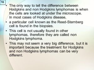
A particular cell known as the Reed-Sternberg cell is found in the biopsies. This cell is not usually found in other lymphomas, therefore they are called non Hodgkins lymphoma. This may not seem a very big difference, but it is important because the treatment for Hodgkins and non Hodgkins lymphomas can be very different.

Although the diseases may sound similar, there is a lot of difference between Hodgkin and non-Hodgkin lymphoma.

Both Hodgkin and non-Hodgkin lymphoma are malignancies of a family of white blood cells known as lymphocytes, which help the body fight off infections and other diseases.

Both Hodgkin’s lymphoma and non-Hodgkin’s lymphoma are lymphomas — a type of cancer that begins in a subset of white blood cells and these are called lymphocytes. Lymphocytes are an integral part of your immune system, which protects you from germs.

The main difference between Hodgkin’s lymphoma and non-Hodgkin’s lymphoma is in the specific lymphocyte each involves but also it includes:

Hodgkin lymphoma Non-Hodgkin lymphoma
Thirty-two percent of patients diagnosed with Hodgkin lymphoma are 20-34 years old. The median age of a patient diagnosed with the disease is 39. Seventy-five percent of patients diagnosed with Hodgkin lymphoma 55 or older. The median age of a patient diagnosed with the disease is 66.
Hodgkin lymphoma is rare, accounting for about .5 percent of all new cancers diagnosed. An estimated 8,500 cases were diagnosed in 2016. Non-Hodgkin lymphoma is the seventh most diagnosed cancer, accounting for an estimated 72,500 cases in 2016.
More than 86 percent of patients diagnosed with Hodgkin lymphoma survive five years or more. About 70 percent of patients diagnosed with non-Hodgkin lymphoma survive five years or more.
There are six varieties of Hodgkin lymphoma. The most common forms are nodular sclerosis classical Hodgkin lymphoma and mixed cellularity classical Hodgkin lymphoma. They account for about 90 percent of all cases. There are more than 61 types and subtypes of non-Hodgkin lymphoma. B-cell lymphomas account for 85 percent of all cases. Diffuse large B-cell lymphoma is the most common form on non-Hodgkin lymphoma.

Sources: National Cancer Institute and Lymphoma Research Foundation 2016

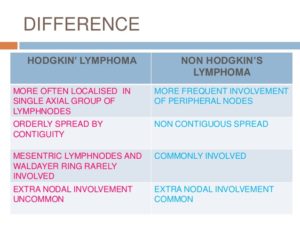
Hodgkin lymphoma is marked by the presence of Reed-Sternberg cells, which are mature B cells that have become malignant, are unusually large, and carry more than one nucleus. The first sign of the disease is often the appearance of enlarged lymph nodes. Non-Hodgkin lymphoma, by contrast, can be derived from B cells or T cells and can arise in the lymph nodes as well as other organs. (B cells and T cells play different roles in the body’s immune response to disease.)

The the Mayo Clinic it states both diseases are relatively rare, but non-Hodgkin lymphoma is more common in the United States, with more than 70,000 new cases diagnosed each year, compared to about 8,000 for Hodgkin lymphoma. The median age of patients with non-Hodgkin lymphoma is 60, but it occurs in all age groups. Hodgkin lymphoma most often occurs in people ages 15 to 24 and in people over 60. There are more than 60 distinct types of non-Hodgkin lymphoma, whereas Hodgkin lymphoma is a more homogeneous disease.

The two forms of lymphoma are marked by a painless swelling of the lymph nodes. Hodgkin lymphomas are more likely to arise in the upper portion of the body (the neck, underarms, or chest). Non-Hodgkin lymphoma can arise in lymph nodes throughout the body, but can also arise in normal organs. Patients with either type can have symptoms such as weight loss, fevers, and night sweats.

The diseases often follow different courses of progression. Hodgkin lymphoma tends to progress in an orderly fashion, moving from one group of lymph nodes to the next, and is often diagnosed before it reaches an advanced stage. Most patients with non-Hodgkin lymphoma are diagnosed at a more advanced stage.

Treatments for lymphoma vary depending on the type of disease, its aggressiveness, and location, along with the age and general health of the patient. As a general rule, however, Hodgkin lymphoma is considered one of the most treatable cancers, with more than 90 percent of patients surviving more than five years. Survival rates for patients with non-Hodgkin lymphoma tend to be lower, but for certain types of the disease, the survival rates are similar to those of patients with Hodgkin lymphoma.

A doctor can tell the difference between Hodgkin’s lymphoma and non-Hodgkin’s lymphoma by examining the cancer cells under a microscope. If in examining the cells, the doctor detects the presence of a specific type of abnormal cell called a Reed-Sternberg cell, the lymphoma is classified as Hodgkin’s. If the Reed-Sternberg cell is not present, the lymphoma is classified as non-Hodgkin’s.

Many subtypes of lymphoma exist, and your doctor will use laboratory tests to examine a sample of your lymphoma cells to determine your specific subtype. Expect to wait a few days to receive results from these specialized tests.

Your type of lymphoma helps your doctor determine your prognosis and your treatment options. The types of lymphoma have very different disease courses and treatment choices, so an accurate diagnosis is an integral part of getting the care you need.

How both Hodgkin’s and Non-Hodgkin’s Disease are diagnosed:

  • Physical exam. Your doctor checks for swollen lymph nodes, including in your neck, underarm and groin, as well as for a swollen spleen or liver.
  • Blood and urine tests. Blood and urine tests may help rule out an infection or other disease.  A sample of your blood is examined in a lab to see if anything in your blood indicates the possibility of cancer (in both of these diseases particularly the WBCs).
  • Imaging tests. Your doctor may recommend imaging tests to look for tumors in your body. Tests may include X-ray, CT, MRI and positron emission tomography (PET).
  • Lymph node test. Your doctor may recommend a lymph node biopsy procedure to remove all or part of a lymph node for laboratory testing. Analyzing lymph node tissue in a lab may reveal whether you have non-Hodgkin’s lymphoma and, if so, which type.  Your doctor may recommend a lymph node biopsy procedure to remove a lymph node for laboratory testing. He or she will diagnose classical Hodgkin’s lymphoma if abnormal cells called Reed-Sternberg cells are found within the lymph node.
  • Bone marrow test. A bone marrow biopsy and aspiration procedure involves inserting a needle into your hipbone to remove a sample of bone marrow. The sample is analyzed to look for non-Hodgkin’s lymphoma cells to look for non-Hodgkin’s lymphoma cells or to look for non-Hodgkin’s lymphoma cells.  This test is done on numerous types of cancer patients in helping to diagnose the cancer they have.


QUOTE FOR FRIDAY:

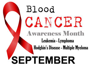
Blood cancer is a group of more than 100 cancers that most often begin in the bone marrow and can affect blood cells, lymph nodes, and other parts of the lymphatic system. Blood cancers occur when abnormal blood cells crowd out normal cells, interfering with the ability of normal blood cells to fight infection and reproduce.

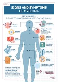
Many people discount their symptoms. Exhaustion or random aches and pains are often blamed on stress or age. Night sweats and recurring fevers sometimes get written off as a virus. Lots of itching… must be an allergy. Or a broken bone is usually just seen as just an accident.

They seem like innocent symptoms that can be associated with other common ailments, but any of them could point to blood cancer.

Many people aren’t familiar with blood cancer. In fact, some patients who have been diagnosed with leukemia or lymphoma, myeloma, myelodysplastic syndromes, or myeloproliferative neoplasms, may not realize they have a form of blood cancer.”

Leukemia and Lymphoma Society (https://www.lls.org/blog/defining-and-redefining-blood-cancer-diagnosis)

What is the difference between leukemia and lymphoma?


Today’s topics will be covering on leukemia and lymphoma than what is the difference between them actually.  It will also include the factors proning you to these cancers, the tests that the M.D. might do and the different treatments for all 3 cancers Leukemia, Lymphoma and Myeloma (discussed in Part II).

Leukemia

Leukemia and Lymphoma Society states leukemia begins in a cell in the bone marrow and in the blood.  Remember the bone marrow creates our cells releasing them into the blood stream.   The cell undergoes a change and becomes a type of leukemia cell. Once the marrow cell undergoes a leukemic change, the leukemia cells may grow and survive better than normal cells. Over time, the leukemia cells crowd out or suppress the development of normal cells. The rate at which leukemia progresses and how the cells replace the normal blood and marrow cells are different with each type of leukemia.

After diagnosis and treatment, many people with leukemia live many good, quality years.

Leukemia is a cancer of the early blood-forming cells, meaning just coming formed from the bone marrow. Most often, leukemia is a cancer of the white blood cells, but some leukemia (s) start in other blood cell types or effect the count of other cells in our bloodstream. Leukemia is often described as being either acute (fast growing) or chronic (slow growing). Different types of leukemia have different treatment options and outlooks.

The National Cancer Institute also says leukemia is a broad term for cancers of the blood cells. The type of leukemia depends on the type of blood cell that becomes cancer and whether it grows quickly or slowly. Leukemia occurs most often in adults older than 55, but it is also the most common cancer in children younger than 15.

Leukemia may affect red blood cells, white blood cells, and platelets.

In a healthy child, the bone marrow makes blood stem cells (immature cells) that become mature blood cells over time. A blood stem cell may become a myeloid stem cell or a lymphoid stem cell.

A myeloid stem cell becomes one of three types of mature blood cells:

  • Red blood cells that carry oxygen and other substances to all tissues of the body.
  • Platelets that form blood clots to stop bleeding.
  • White blood cells that fight infection and disease.

A lymphoid stem cell becomes a lymphoblast cell and then one of three types of lymphocytes (white blood cells):

  • B lymphocytes that make antibodies to help fight infection.
  • T lymphocytes that help B lymphocytes make the antibodies that help fight infection.
  • Natural killer cells that attack cancer cells and viruse

The major types of leukemia are:

  • Acute lymphocytic leukemia (ALL). This is the most common type of leukemia in young children. ALL can also occur in adults.
  • Acute myelogenous leukemia (AML). AML is a common type of leukemia. It occurs in children and adults. AML is the most common type of acute leukemia in adults.
  • Chronic lymphocytic leukemia (CLL). With CLL, the most common chronic adult leukemia, you may feel well for years without needing treatment.
  • Chronic myelogenous leukemia (CML). This type of leukemia mainly affects adults. A person with CML may have few or no symptoms for months or years before entering a phase in which the leukemia cells grow more quickly.
  • Other types. Other, rarer types of leukemia exist, including hairy cell leukemia, myelodysplastic syndromes and myeloproliferative disorders.

Statistics by the American Cancer Society:  Leukemia is the most common type of cancer in children and teens, accounting for 1 out of 3 cancers.   Most childhood leukemias are ALL Acute Lymphocytic Leukemia.  Most of the remaining cases for childhood are AML Acute Myeloid Leukemia.  Chronic Leukemia is rare in children.   However, because other types of leukemia become more common with age, most leukemia is found in among adults.

Decades of research have led to vastly improved outcomes for children diagnosed with ALL.

Lymphoma

Lymphoma starts in the immune system and affects the lymph nodes and lymphocytes, which are a type of white blood cell. There are two main types of lymphocyte, B cells and T cells.  Whereas Leukemia starts in the bone marrow affecting the white blood cells.  Doctors categorize leukemia based on which type of white blood— lymphocytes or myeloid cells — and whether the illness is developing very quickly (acute disease) or slowly over time (chronic disease).

There are numerous types of leukemia and lymphoma whose facts and figures are not nearly so rosy as, for instance, the promising 86% five-year-survival rate for Hodgkin lymphoma. Non-Hodgkin lymphoma is a far more lethal form of blood cancer, and while the survival rate has grown considerably since the 1990s, approximately 20,140 deaths — 11,450 men and 8,690 women — from this disease will still occur this year.

Cancer can affect any part of the body, including the blood. Leukemia and lymphoma are both forms of blood cancer. The main difference is that leukemia affects the blood and bone marrow, while lymphomas tend to affect the lymph nodes.  BUT remember leukemia can go into the lymph nodes since its in the blood stream the cancer cells and can effect the lymph nodes.  BOTH effect white blood cells. Myeloma is similar in that it effects plasma a white blood cell.

The origin of where Myeloma, Leukemia or Lymphoma.  Lets look at all 3 All start in the Bone Marrow but the effect is similar in general but the type with the stage of cancer the individual has is a major factor of ending results.

All 3 types of cancers this article in Part I to Part II are bad cancer cells that derive in all in the bone marrow and they all effect some type of White Blood Cell (WBC).  All White Blood Cells are to fight infection and prevent it.  

Myeloma effects the normal plasma cells originating in the bone marrow and are an important part of the immune system.  Lymphocytes (lymph cells) are one of the main types of white blood cells in the immune system and include T cells and B cells. Lymphocytes are in many areas of the body, such as lymph nodes, the bone marrow, the intestines, and the bloodstream.  These are the cells involved in this cancer being the problem.

Lymphoma is cancer that begins in infection-fighting cells of the immune system, called lymphocytes. Leukocytes are WBCs also that are affected in this cancer like Myeloma.  In Lymphoma the leukocytes become an abnormal amount (of WBCs) in the body.  Lymphocytes are a form of small leukocyte (white blood cell) with a single round nucleus (remember this is the brain for the cell), occurring especially in the lymphatic system.  Abnormal lymphocytes, a type of white blood cell that fights infection, become lymphoma cells, which multiply and collect in your lymph nodes. Over time, these cancerous cells impair your immune system. Lymphomas are divided into two categories: Hodgkin lymphoma and non-Hodgkin lymphoma (this is another topic in itself).  These cells are in the lymph nodes, spleen, thymus, bone marrow, and other parts of the body.

Leukemia is a cancer of the body’s blood-forming tissues, including the bone marrow and the lymphatic system.  In Leukemia many of the white blood cells produced in the bone marrow do not mature normally. These abnormal cells, called leukemic cells, are unable to fight infection the way healthy white cells can. As they grow in number, the leukemic cells also interfere with the production of other blood cells. Obviously the WBCs start in the bone marrow just like every cell does.

So these 3 cancers are similar in many ways with the ending results but where they effect initially or their primary area effected might be slightly different, as discussed already in this topic. 

Factors that may increase your risk of developing some types of any 3 cancers include:

  • Previous cancer treatment. People who’ve had certain types of chemotherapy and radiation therapy for other cancers have an increased risk of developing certain types of leukemia.
  • Genetic disorders. Genetic abnormalities seem to play a role in the development of leukemia. Certain genetic disorders, such as Down syndrome, are associated with an increased risk of leukemia.
  • Exposure to certain chemicals. Exposure to certain chemicals, such as benzene — which is found in gasoline and is used by the chemical industry — is linked to an increased risk of some kinds of leukemia.
  • Smoking. Smoking cigarettes increases the risk of acute myelogenous leukemia.
  • Family history of leukemia. If members of your family have been diagnosed with leukemia, your risk of the disease may be increased.

However, most people with known risk factors don’t get leukemia. And many people with leukemia have none of these risk factors.

Regarding Myeloma, Leukemia or Lymphoma:

Doctors may find in a routine blood test, before symptoms begin. If this happens, or if you have signs or symptoms that suggest any 3 of the cancers, you may undergo the following diagnostic exams:

  • Physical exam. Your doctor will look for physical signs of Myeloma or Leukemia or Lymphoma, such as pale skin from anemia, swelling of your lymph nodes, and enlargement of your liver and spleen.
  • Blood tests. By looking at a sample of your blood, your doctor can determine if you have abnormal levels of red or white blood cells or platelets — which may suggest leukemia.
  • Bone marrow test. Your doctor may recommend a procedure to remove a sample of bone marrow from your hipbone. The bone marrow is removed using a long, thin needle. The sample is sent to a laboratory to look for leukemia cells. Specialized tests of your cancer cells that may reveal certain characteristics that are used to determine your treatment options.

Treatment

Treatment for your Myeloma, Leukemia or Lymphoma depends on many factors. Your doctor determines your treatment options based on your age and overall health, the type of cancer of the 3 you have, and whether it has spread to other parts of your body, including the central nervous system.

Common treatments used to fight these 3 blood cancers include:

  • Chemotherapy. Chemotherapy is the major form of treatment for all 3. This drug treatment uses chemicals to kill cancer cells.Depending on the type of cancer you have, you may receive a single drug or a combination of drugs. These drugs may come in a pill form, or they may be injected directly into a vein.
  • Biological therapy. Biological therapy works by using treatments that help your immune system recognize and attack cancer cells.
  • Targeted therapy. Targeted therapy uses drugs that attack specific vulnerabilities within your cancer cells.For example, the drug imatinib (Gleevec) stops the action of a protein within the leukemia cells of people with chronic myelogenous leukemia. This can help control the disease.
  • Radiation therapy. Radiation therapy uses X-rays or other high-energy beams to damage cancer cells and stop their growth. During radiation therapy, you lie on a table while a large machine moves around you, directing the radiation to precise points on your body.You may receive radiation in one specific area of your body where there is a collection of cancer cells, or you may receive radiation over your whole body. Radiation therapy may be used to prepare for a stem cell transplant.
  • Stem cell transplant. A stem cell transplant is a procedure to replace your diseased bone marrow with healthy bone marrow.Before a stem cell transplant, you receive high doses of chemotherapy or radiation therapy to destroy your diseased bone marrow. Then you receive an infusion of blood-forming stem cells that help to rebuild your bone marrow.You may receive stem cells from a donor, or in some cases you may be able to use your own stem cells. A stem cell transplant is very similar to a bone marrow transplant.