QUOTE FOR MONDAY:

“Chronic obstructive pulmonary disease (COPD) is a chronic inflammatory lung disease that causes obstructed airflow from the lungs. Symptoms include breathing difficulty, cough, mucus (sputum) production and wheezing. It’s typically caused by long-term exposure to irritating gases or particulate matter, most often from cigarette smoke. People with COPD are at increased risk of developing heart disease, lung cancer and a variety of other conditions.

Emphysema and chronic bronchitis are the two most common conditions that contribute to COPD. These two conditions usually occur together and can vary in severity among individuals with COPD.

Chronic bronchitis is inflammation of the lining of the bronchial tubes, which carry air to and from the air sacs (alveoli) of the lungs. It’s characterized by daily cough and mucus (sputum) production.

Emphysema is a condition in which the alveoli at the end of the smallest air passages (bronchioles) of the lungs are destroyed as a result of damaging exposure to cigarette smoke and other irritating gases and particulate matter.

Although COPD is a progressive disease that gets worse over time, COPD is treatable.”

MAYO CLINIC

Part I What actually is Chronic Obstructive Pulmonary Disease (COPD)?

COPD2  COPD3 Usually due to smoking

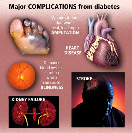
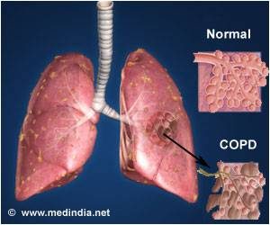
This is Healthy Lung Month covering COPD.

What is Chronic Obstructive Pulmonary Disease (COPD)?

Chronic obstructive pulmonary disease (COPD) is a term that applies to patients with chronic bronchitis, bronchiectasis, emphysema and, to a certain extent, asthma. A brief review of normal functional anatomy will provide a background for the discussion of pathology.

The airway down to the bronchioles normally is lined with ciliated pseudo-stratified columnar cells and goblet cells. Mucus derives from mucus glands that are freely distributed in the walls of the trachea and bronchi. The cilia sweep mucus and minor debris toward the upper airway. Low humidity, anesthesia gases, cigarette smoking and other chemical irritants paralyze the action of these cilia. The mucociliary action starts again after a matter of time. This is why people awaken to “smokers cough.”

“Chronic obstructive pulmonary disease (COPD) is a term that applies to patients with chronic bronchitis, bronchiectasis, emphysema and, to a certain extent, asthma.”

Bronchi run in septal connective tissue, but bronchioles are suspended in lung parenchyma by alveolar elastic tissue. The elastic tissue extends throughout alveolar walls, air passages, and vessels, connecting them in a delicate web. Bronchiolar epithelium is ciliated, single-layered and columnar or cuboidal. Beyond the bronchioles the epithelium is flat and lined with a film of phospholipid (surfactant), which lowers surface tension and thereby helps to keep these air spaces from collapsing. Remember that the phospholipid develops during later gestation in utero. This is the reason why premature infant’s lungs cannot stay inflated without the addition of surfactant therapy. Macrophages are found in alveolar lining. Smooth muscles surround the walls of all bronchi, bronchioles, and alveolar ducts and when stimulated they shorten and narrow the passages. Cartilage lends rigidity and lies in regular horse-shaped rings in the tracheal wall. Cartilage is absent in bronchi less than 1 mm in diameter.

The terminal bronchiole is lined with columnar epithelium and is the last purely conducting airway. An acinus includes a terminal bronchiole and its distal structures. Five to ten acini together constitute a secondary lobule, which is generally 1 to 2 cm in diameter and is partly surrounded by grossly visible fibrous septa. Passages distal to the terminal bronchiole include an average of three but as many as nine generations of respiratory bronchioles lined with both columnar and alveolar epithelium. Each of the last respiratory bronchioles gives rise to about six alveolar ducts, each of these to one or two alveolar sacs, and finally each of the sacs to perhaps seventy-five alveoli. Alveolar pores (pores of Kohn) may connect alveoli in adjacent lobules.

Two different circulations supply the lungs. The pulmonary arteries and veins are involved in gas exchange. The pulmonary arteries branch with the bronchi, dividing into capillaries at the level of the respiratory bronchiole, and supplying these as well as the alveolar ducts and alveoli. In the periphery of the lung, the pulmonary veins lie in the interlobular septa rather than accompanying the arteries and airways. The bronchial arteries are small and arise mostly from the aorta. They accompany the bronchi to supply their walls. In some cases of COPD, like bronchiectasis, extensive anastomoses develop between the pulmonary and bronchial circulations. This can allow major shunting and recirculation of blood, therefore contributing to cardiac overload and failure. Lymphatics run chiefly in bronchial walls and as a fine network in the pleural membrane. The lumina of the capillaries in the alveolar walls are separated from the alveolar lining surfaces by the alveolar-capillary membrane, consisting of thin endothelial and epithelial cells and a minute but expansile interstitial space. This interface between air and blood, only 2 microns in thickness, is the only place where gases may be exchanged effectively.

Disease Specific Review

Chronic Bronchitis

Chronic bronchitis is a clinical disorder characterized by excessive mucus secretion in the bronchi. It was traditionally defined by chronic or recurrent productive cough lasting for a minimum of three months per year and for at least two consecutive years, in which all other causes for the cough have been eliminated. Today’s definition remains more simplistic to include a productive cough progressing over a period of time and lasting longer and longer. Sometimes, chronic bronchitis is broken down into three types: simple, mucopurulent or obstructive. The pathologic changes consist of inflammation, primarily mononuclear, infiltrate in the bronchial wall, hypertrophy and hyperplasia of the mucus-secreting bronchial glands and mucosal goblet cells, metaplasia of bronchial and bronchiolar epithelium, and loss of cilia. Eventually, there may be distortion and scarring of the bronchial wall.

Asthma

Asthma is a disease characterized by increased responsiveness of the trachea and bronchi to various stimuli (intrinsic or extrinsic), causing difficulty in breathing due to narrowing airways. The narrowing is dynamic and changes in degree. It occurs either spontaneously or because of therapy. The basic defect appears to be an altered state of the host, which periodically produces a hyperirritable contraction of smooth muscle and hypersecretion of bronchial mucus. This mucus is abnormally sticky and therefore obstructive. In some instances, the illness seems related to an altered immunologic state.

Histological changes of asthma include an increase in the size and number of the mucosal goblet cells and submucosal mucus glands. There is marked thickening of the bronchial basement membrane and hypertrophy of bronchial and bronchiolar smooth muscle tissue. A submucosal infiltration of mononuclear inflammatory cells, eosinophils and plugs of mucus blocks small airways. Patients who have had asthma for many years may develop cor pulmonale and emphysema.
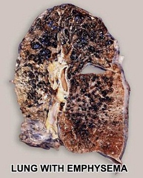
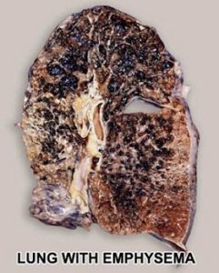
Emphysema

Pulmonary emphysema is described in clinical, radiological and physiologic terms, but the condition is best defined morphologically. It is an enlargement of the air spaces distal to the terminal non-respiratory bronchiole, with destruction of alveolar walls.

Although the normal lung has about 35,000 terminal bronchioles and their total internal cross-sectional area is at least 40 times as great as that of the lobar bronchi, the bronchioles are more delicate and vulnerable. Bronchioles may be obstructed partially or completely, temporarily or permanently, by thickening of their walls, by collapse due to loss of elasticity of the surrounding parenchyma, or by influx of exudate. In advanced emphysema, the lungs are large, pale, and relatively bloodless. They do not readily collapse. They many contain many superficial blebs or bullae, which occasionally are huge. The right ventricle of the heart is often enlarged (cor pulmonale), reflecting pulmonary arterial hypertension. Right ventricular enlargement is found in about 40% of autopsies of patients with severe emphysema. The distal air spaces are distended and disrupted, thus excessively confluent and reduced in number. There may be marked decrease in the number and size of the smaller vascular channels. The decrease in alveolar-capillary membrane surface area may be critical. Death may result from infection that obliterates the small bronchi and bronchioles. There is often organized pneumonia or scarring of the lung parenchyma due to previous infections.

Classification of emphysema relies on descriptive morphology, requiring the study of inflated lungs. The two principal types are centrilobular and panlobular emphysema. The two types may coexist in the same lung or lobe.

Centrilobular emphysema (CLE) or centriacinar emphysema affects respiratory bronchioles selectively. Fenestrations develop in the walls, enlarge, become confluent, and tend to form a single space as the walls disintegrate. There is often bronchiolitis with narrowing of lumina. The more distal parenchyma (alveolar ducts and sacs and alveoli) is initially preserved, then similarly destroyed as fenestrations develop and progress.

The disease commonly affects the upper portions of the lung more severely, but it tends to be unevenly distributed. The walls of the emphysematous spaces may be deeply pigmented. This discoloration may represent failure of clearance mechanisms to remove dust particles, or perhaps the pigment plays an active role in lung destruction. CLE is much more prevalent in males than in females. It is usually associated with chronic bronchitis and is seldom found in nonsmokers.

Panlobular emphysema (PLE) or panacinar emphysema is a nearly uniform enlargement and destruction of the alveoli in the pulmonary acinus. As the disease progresses, there is gradual loss of all components of the acinus until only a few strands of tissue, which are usually blood vessels, remain. PLE is usually diffuse, but is more severe in the lower lung areas. It is often found to some degree in older people, who do not have chronic bronchitis or clinical impairment of lung function. The term senile emphysema was formerly applied to this condition. PLE occurs as commonly in women and men, but is less frequent than CLE. It is a characteristic finding in those with homozygous deficiency of serum alpha-1 antitrypsin. It has also been found that certain populations of IV Ritalin abusers show PLE.

Bullae are common in both CLE and PLE, but may exist in the absence of either. Air-filled spaces in the visceral pleura are commonly termed blebs, and those in the parenchyma greater than 1 cm in diameter are called bullae. A valve mechanism in the bronchial communication of a bulla permits air trapping and enlargement of the air space. This scenario may compress the surrounding normal lung. Blebs may rupture into the pleural cavity causing a pneumothorax, and through a valve mechanism in the bronchopleural fistula a tension pneumothorax may develop.

Paracicatricial emphysema occurring adjacent to pulmonary scars represents another type of localized emphysema. When the air spaces distal to terminal bronchioles are increased beyond the normal size but do not show destructive changes of the alveolar walls, the condition is called pulmonary overinflation. This condition may be obstructive, because of air trapping beyond an incomplete bronchial obstruction due to a foreign body or a neoplasm. Many lung lobules may be simultaneously affected as a result of many check-valve obstructions, as in bronchial asthma. Pulmonary overinflation may also be nonobstructive, less properly called “compensatory emphysema”, when associated with atelectasis or resection of other areas of the lung.

Bronchiectasis

Bronchiectasis means irreversible dilation and distortion of the bronchi and bronchioles. Saccular bronchiectasis is the classic advanced form characterized by irregular dilatations and narrowing. The term cystic is used when the dilatations are especially large and numerous. Cystic bronchiectasis can be further classified as fusiform or varicose.

Tubular bronchiectasis is simply the absence of normal bronchial tapering and is usually a manifestation of severe chronic bronchitis rather than of true bronchial wall destruction.

Repeated or prolonged episodes of pneumonitis, inhaled foreign objects or neoplasms have been known to cause bronchiectasis. When the bronchiectatic process involves most or all of the bronchial tree, whether in one or both lungs, it is believed to be genetic or developmental in origin.

Mucoviscidosis, Kartagener’s syndrome (bronchiectasis with dextrocardia and paranasal sinusitis), and agammaglobulinemia are all examples of inherited or developmental diseases associated with bronchiectasis. The term pseudobronchiectasis is applied to cylindrical bronchial widening, which may complicate a pneumonitis but which disappears after a few months. Bronchiectasis is true saccular bronchiectasis but without cough or expectoration. It is located especially in the upper lobes where good dependent drainage is available. A proximal form of bronchiectasis (with normal distal airways) complicates aspergillus mucus plugging.

Advanced bronchiectasis is often accompanied by anastomoses between the bronchial and pulmonary vessels. These cause right-to-left shunts, with resulting hypoxemia, pulmonary hypertension and cor pulmonale.

Keeping a healthy lung prevents emphysema.  So for starters don’t smoke and exercise; which includes don’t be exposed to smoke frequently!

QUOTE FOR WEEKEND:

“Regarding newly diagnosed diabetics 1.5 million people will be diagnosed with DM this year. $237 billion is spent each year on direct medical costs and another $90 billion on reduced productivity. Some people can manage it with healthy eating and exercise, or with oral medications, while others may also need to use insulin.  Sometimes one medication will be enough, but in other cases, your doctor may prescribe a combination of medications.  It’s common for your medication needs to change over time. And that’s a good thing. The most important thing is to get to feeling your best.”

American Diabetes Association (https://diabetes.org/)


QUOTE FOR FRIDAY:

“Research shows that people with prediabetes or type 2 diabetes have a higher risk of getting Alzheimer’s disease and other types of dementia later in life.

Scientists think there are a few ways that problems with blood sugar control can lead to problems with your memory and thinking.

Insulin Resistance-When your cells don’t use insulin the way they should, that affects the mechanics of your brain.

  • 1-Your cells don’t get the fuel they need, so your brain can’t work right.
  • 2-Your blood sugar goes up, and over time, that can cause harmful fatty deposits in your blood vessels.
  • 3-Too much insulin can throw off the balance of chemicals in your brain.

These effects on the brain are so strong that some scientists feel that Alzheimer’s related to insulin resistance should be called “type 3 diabetes.”

WEB MD (https://www.webmd.com/)


Part IV November Diabetes Awareness – Diabetes increases your risk of Alzheimer’s dramatically.

If You Have Diabetes, Your Risk of Alzheimer’s Increases Dramatically

Diabetes is linked to a 65 percent increased risk of developing Alzheimer’s, which may be due, in part, because insulin resistance and/or diabetes appear to accelerate the development of plaque in your brain, which is a hallmark of Alzheimer’s. Separate research has found that impaired insulin response was associated with a 30 percent higher risk of Alzheimer’s disease, and overall dementia and cognitive risks were associated with high fasting serum insulin, insulin resistance, impaired insulin secretion and glucose intolerance.

A drop in insulin production in your brain may contribute to the degeneration of your brain cells, mainly by depriving them of glucose, and studies have found that people with lower levels of insulin and insulin receptors in their brain often have Alzheimer’s disease (people with type 2 diabetes often wind up with low levels of insulin in their brains as well). As explained in New Scientist, which highlighted this latest research:

What’s more, it encourages the process through which neurons change shape, make new connections and strengthen others. And it is important for the function and growth of blood vessels, which supply the brain with oxygen and glucose.

As a result, reducing the level of insulin in the brain can immediately impair cognition. Spatial memory, in particular, seems to suffer when you block insulin uptake in the hippocampus… Conversely, a boost of insulin seems to improve its functioning.

When people frequently gorge on fatty, sugary food, their insulin spikes repeatedly until it sticks at a high level. Muscle, liver and fat cells then stop responding to the hormone, meaning they don’t mop up glucose and fat in the blood. As a result, the pancreas desperately works overtime to make more insulin to control the glucose – and levels of the two molecules skyrocket.

The pancreas can’t keep up with the demand indefinitely, however, and as time passes people with type 2 diabetes often end up with abnormally low levels of insulin.”

Alzheimer’s Might be “Brain Diabetes”

BBA – Molecular Basis of Disease, Accepted manuscript. doi:10.1016/j.bbadis.2016.04.017

It’s becoming increasingly clear that the same pathological process that leads to insulin resistance and type 2 diabetes may also hold true for your brain. As you over-indulge on sugar and grains, your brain becomes overwhelmed by the consistently high levels of insulin and eventually shuts down its insulin signaling, leading to impairments in your thinking and memory abilities, and eventually causing permanent brain damage.

Regularly consuming more than 25 grams of fructose per day will dramatically increase your risk of dementia and Alzheimer’s disease. Consuming too much fructose will inevitably wreak havoc on your body’s ability to regulate proper insulin levels.

Although fructose is relatively “low glycemic” on the front end, it reduces the affinity for insulin for its receptor leading to chronic insulin resistance and elevated blood sugar on the back end. So, while you may not notice a steep increase in blood sugar immediately following fructose consumption, it is likely changing your entire endocrine system’s ability to function properly behind the scenes.

Additionally, fructose has other modes of neurotoxicity, including causing damage to the circulatory system upon which the health of your nervous system depends, as well as profoundly changing your brain’s craving mechanism, often resulting in excessive hunger and subsequent consumption of additional empty carbohydrate-based calories.

In one study from UCLA, researchers found that rats fed a fructose-rich and omega-3 fat deficient diet (similar to what is consumed by many Americans) developed both insulin resistance and impaired brain function in just six weeks.

Plus, when your liver is busy processing fructose (which your liver turns into fat), it severely hampers its ability to make cholesterol , an essential building block of your brain crucial to its health. This is yet another important facet that explains how and why excessive fructose consumption is so detrimental to your health.  Decreasing fructose intake is one of the most important moves you can take in decreasing the risk of Alzheimer’s disease in your lifetime.


QUOTE FOR THURSDAY:

“It’s important to keep your blood sugar levels in your target range as much as possible to help prevent or delay long-term, serious health problems, such as heart disease, vision loss, and kidney disease. Staying in your target range of blood glucose (Before a meal glucose in the blood should range the following: 80 to 130 mg/dL. Two hours after the start of a meal: Less than 180 mg/dL.).  Keeping the glucose in therapeutic range can also help improve your energy and mood.”

Center for Disease Control and Prevention (CDC)

Find answers to common questions about blood sugar for people with diabetes

Part III November Diabetes Awareness. How to take control of your Diabetes and decrease complications!


Here is a fast review of Part I and Part II:

Diabetes occurs when the pancreas, a gland behind the stomach, does not produce enough of the hormone insulin, or the body cannot use insulin properly. Insulin helps carry sugar from the bloodstream into the cells. Once inside the cells, sugar is converted into energy for immediate use or stored for the future. That energy fuels many of our bodily functions.

The body produces glucose from the foods you eat. The liver also releases sugar when you are not eating. The pancreas produces the hormone insulin, which allows glucose from the bloodstream to enter the body’s cells where it is used for energy. In type 2 diabetes, too little insulin is produced, or the body cannot use insulin properly, or both. This results in a build-up of glucose in the blood.

People with diabetes are at risk of developing signs and symptoms of hyperglycemia to serious health problems (complications).

HOW we can decrease the risk of complications and decrease the chance of diabetes worsening = KEEP IT UNDER CONTROL = PRACTICING VERY GOOD MANAGEMENT IN CARING FOR YOUR DIABETES

This is how you can reach this goal:

-Controlling your blood glucose, blood pressure, and cholesterol can make a huge difference in staying healthy. Talk with your doctor about what your goals should be and how to reach them but make sure you are given information on paper or write down what it is you have discussed in the doctor’s office based on your care for diabetes and what to do. Usually diabetic information on paper is available and given to you.

To reach controlling your glucose and treatment for Diabetes:

-Your healthy eating plan that you and your doctor with a dietician have discussed.

-Overweight? Than diet down to your therapeutic weight range for your height after discussed with by you with your doctor.

-Be physically active for 30 to 60 minutes most days but if this is new get your doctor to clear this activity for you with what kind of activity you are allowed and not allowed.

-Take your medicines as directed and keep taking them even after you’ve reached your goals; or you will be at high risk of ending up the way you were earlier=Diabetes badly controlled with running into the problems you had earlier.It’s very important to take your diabetes medications as recommended by your doctor. Left untreated, diabetes can lead to serious, even life-threatening complications.

-If you smoke=QUIT.

-Check your skin daily in particular the FEET and LOWER LEGS to check for redness, swelling to blisters, sores and sore toenails

-Ask your doctor if you should be taking aspirin to prevent a heart attack or stroke by making the blood less thick to thinner making it easier for the heart to pump and less stress to the organ.

The key is to controlling your DIABETES is to be living a healthy life! This consists of diet, exercise or activity and healthy habits learned and practiced routinely in your life that will help prevent or assist in treating diabetic disease. The better we treat ourselves regarding health the higher the odds we will live a longer and healthier life. There is not just one food to eat or one type of exercise to do or one healthy habit to practice in order to keep you healthy, there’s choices. To be a part learn what healthier habits or changes you want for a healthier way of living; learn how to eat out of the 4 food groups to prevent Diabetes or eating out of the 4 food groups that are following your diabetic diet as ordered by your MD. It allows you to make all the decisions in what you want to do regarding what to eat (diet). Now with diet you must include exercise/activity, and what healthy habits you want to add in your life that are not so healthy; you know what that is and if not read a book on how to get heathier-including how to prevent diabetes where the library and book stores have many options for you. Provide yourself with the information and healthy foods in your diet, if you decide you want it. You make all the choices.

The ending line of all problems resulting from Diabetes is due to the thick high glucose blood in the blood stream filtering throughout the different organs in our body causing from peripheral neuropathy to necrotic skin to amputations for LE’s usually or same effect elsewhere causing macular degeneration to blindness or increase of cancers, heart disease, Diabetes Alzheimer’s and we could go on about the effects of diabetes.  Get it now its control your blood glucose keeping it in therapeutic range  decreasing the odds of developing these conditions or the severity of these conditions.

If you don’t have diabetes than take the steps to prevent being diagnosed with it later in life.  WHAT are those steps? Eat Right (Healthy), Keep your weight in therapeutic range, Exercise the body balancing it with rest, decrease stress, and take care of yourself.  BUT if there is heredity in the family, especially your nuclear family, when you see your primary care doctor every 6 months or yearly have your glucose checked to see if it is high or not.  Simply get a BMP or CMP blood test that looks at blood electrolyte levels that includes glucose.  If its high the next step is getting the doctor to check your hemoglobin A1C another blood test done with no eating for 12 hrs prior to see what your real glucose level is prior to your first meal in the morning (done on a empty stomach).  For if you eat prior to the test it won’t accurate on your true glucose level.  2 Easy blood tests.

It is all up to you!

Wouldn’t you want less disease/illness for yourself, for your family, others significant to you and even throughout the nation including our future generations. Wouldn’t it be great to see Diabetes decrease in America for future years and giving us an ending result of higher probability that we would overall a healthier country with less diseases. If that included Diabetes decreased significantly what an impact it would play in decreasing other diseases, that occurred due to the diabetes alone  (That would decrease cardiac disease, renal disease, blurred vision, neuropathy, I could go on).  Besides how much it would decrease in this country to take care of patients with diabetes.

I’m not a diabetic but eating overall healthy and in my diet range (barely) but there and trying to increase my activity. Do yourself and maybe others a favor by making yourself and America a healthier country for less Diabetes and the diseases it can cause from cardiac to vision to renal to brain, etc…

Again its all up to you!

REFERENCES for Part I, Part II & III this week on diabetes:

1.)  Center for Disease (CDC) – “National Diabetes Fact Sheet”

2.)  NYS Dept. of Health –Diabetes

3.)  Diabetic Neuropathy.org “All about diabetic neuropathy and nerve damage caused by Diabetes.”

4.)  NIDDK “National Institute of Diabetes and Digestive and Kidney Diseases.

5.)  National Diabetes Information Clearinghouse (NIDC) – U.S. Department of Health and Human Services.       “Preventing Diabetes Problems: What you need to know”


QUOTE FOR WEDNESDAY:

” No matter what type of diabetes you have, it can lead to excess sugar in the blood that does not absorb into the cell like normally causing hyperglycemia. Too much sugar in the blood can lead to serious health problems.  Diabetes symptoms depend on how high your blood sugar is. Type I known as juvenile diabetes.  Some people, especially if they have prediabetes or type 2 diabetes, may not have symptoms. In type 1 diabetes, symptoms tend to come on quickly and be more severe .  Type 2 diabetes used to be known as adult-onset diabetes, but both type 1 and type 2 diabetes can begin during childhood and adulthood.  Still Type II DM most of the times you get it 40 yrs old and up; not as soon as Type 1 (usually in childhood or teen years you get it).   Type I exposes the person to high sugar in the intensity &/or length of free sugar in the blood outside the cells due to the age they got the disease, that causes the S/S & complications to arise sooner than Type II patients.”.

MAYO CLINIC

QUOTE FOR TUESDAY:

“Diabetes is a chronic (long-lasting) health condition that affects how your body turns food into energy.
Your body breaks down most of the food you eat into sugar (glucose) and releases it into your bloodstream. When your blood sugar goes up, it signals your pancreas to release insulin. Insulin acts like a key to let the blood sugar into your body’s cells for use as energy.
With diabetes, your body doesn’t make enough insulin or can’t use it as well as it should. When there isn’t enough insulin or cells stop responding to insulin, too much blood sugar s tays in your bloodstream. Over time, that can cause serious health problems.”


Centers for Disease Control and Prevention (CDC)

QUOTE FOR MONDAY:

“Everyone loves a good scare on Halloween, but not when it comes to the safety of those little trick-or-treaters. Fortunately, there are lots of easy things parents and kids can do to stay safe on the spookiest of holidays.

Hard Facts about Halloween Safety

On average, children are more than twice as likely to be hit by a car and killed on Halloween than on any other day of the year.”

Safe Kids Worldwide (https://www.safekids.org/halloween)