High Blood Pressure and Kidney Disease!

What is high blood pressure?

Blood pressure is the force of blood pushing against blood vessel walls as your heart pumps out blood. High blood pressure NIH external link, also called hypertension, is an increase in the amount of force that blood places on blood vessels as it moves through the body.

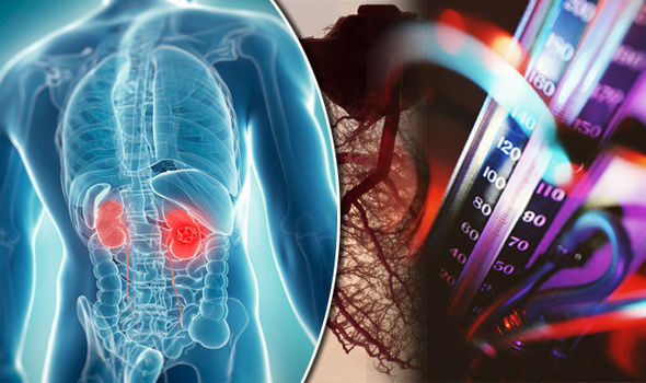
What are the kidneys and what do they do?

Healthy kidneys filter about a half cup of blood every minute, removing wastes and extra water to make urine. The urine flows from each kidney to the bladder through a pair of thin tubes called ureters, one on each side of your bladder. Your bladder stores urine. Your kidneys, ureters, and bladder are part of your urinary tract system.

How does high blood pressure affect the kidneys?

High blood pressure can constrict and narrow the blood vessels, which eventually damages and weakens them throughout the body, including in the kidneys. The narrowing reduces blood flow.

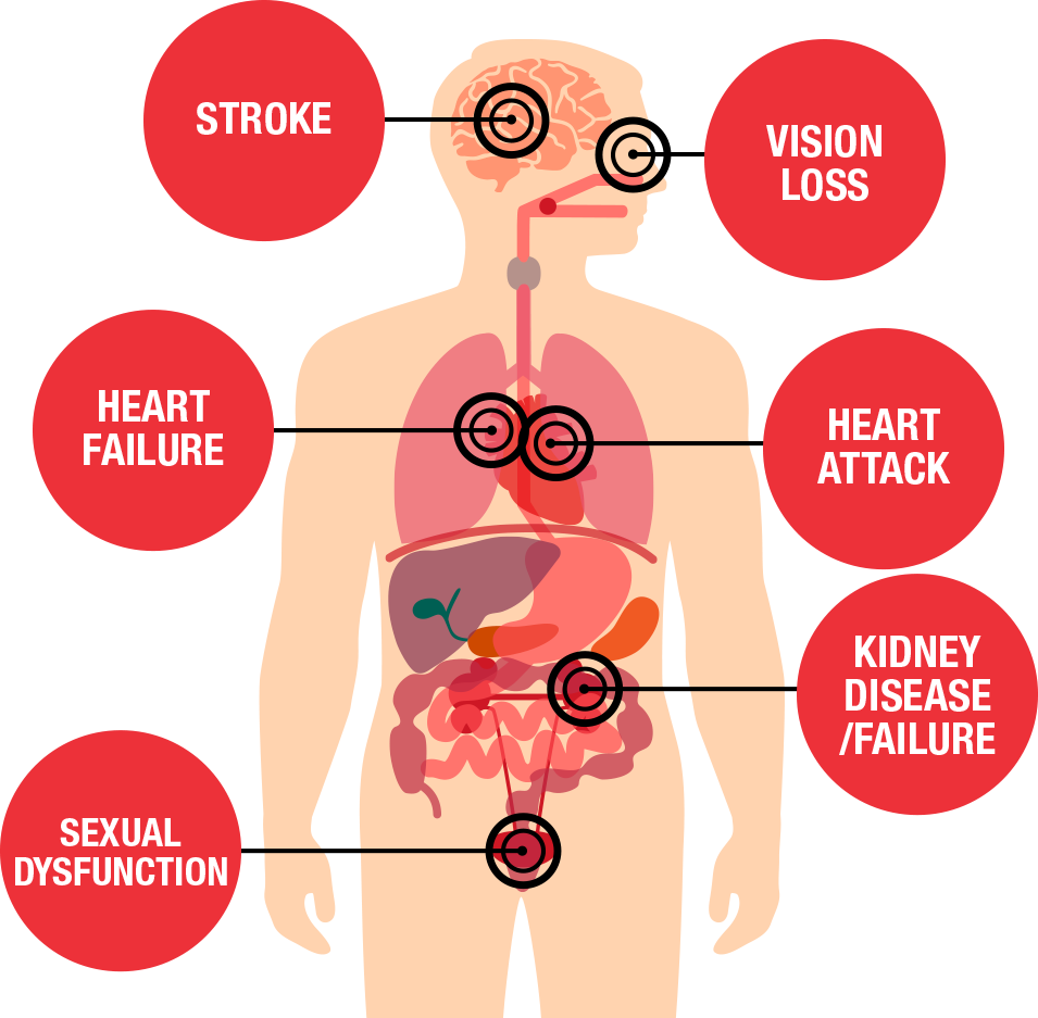
If your kidneys’ blood vessels are damaged, they may no longer work properly. When this happens, the kidneys are not able to remove all wastes and extra fluid from your body. Extra fluid in the blood vessels can raise your blood pressure even more, creating a dangerous cycle, and cause more damage leading to kidney failure.

How common are high blood pressure and kidney disease?

Almost 1 in 2 U.S. adults—or about 108 million people—have high blood pressure.1

More than 1 in 7 U.S. adults—or about 37 million people—may have chronic kidney disease (CKD).

High blood pressure is the second leading cause of kidney failure in the United States after diabetes, as illustrated in Figure 1.A pie chart showing the causes of kidney failure in the United States, with diabetes at 38%, high blood pressure at 26%, glomerulonephritis at 16%, other causes at 15%, and unknown causes at 5%.

Figure 1. Causes of kidney failure in the United States

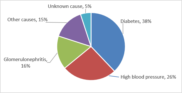
Who is more likely to have high blood pressure or kidney disease?

High blood pressure

You are more likely to have high blood pressure if you

  • are older. Blood pressures tends to increase with age. Our blood vessels naturally thicken and stiffen over time.
  • have family members with high blood pressure. High blood pressure tends to run in families.
  • have unhealthy lifestyle habits. Unhealthy habits such as eating too much sodium (salt), drinking too many alcoholic beverages, or not being physically active can increase your risk of high blood pressure.
  • are African American. High blood pressure is more common in African American adults than in Caucasian, Hispanic, or Asian adults.
  • are male. Men are more likely to develop high blood pressure before age 55; women are more likely to develop it after age 55.

Kidney disease

In addition to high blood pressure, other factors that increase your risk of kidney disease are

  • diabetes
  • a family history of kidney failure
  • race or ethnicity—African Americans, Hispanics, and American Indians tend to have a greater risk for CKD

High blood pressure can be both a cause and a result of kidney disease.

What are the symptoms of high blood pressure and kidney disease?

Most people with high blood pressure do not have symptoms. In rare cases, high blood pressure can cause headaches.

Early CKD also may not have symptoms. As kidney disease gets worse, some people may have swelling, called edema. Edema happens when the kidneys cannot get rid of extra fluid and salt. Edema can occur in the legs, feet, ankles, or—less often—in the hands or face.

Symptoms of advanced kidney disease can include

  • loss of appetite, nausea, or vomiting
  • drowsiness, feeling tired, or sleep problems
  • headaches or trouble concentrating
  • increased or decreased urination
  • generalized itching or numbness, dry skin, or darkened skin
  • weight loss
  • muscle cramps
  • chest pain or shortness of breath

How do health care professionals diagnose high blood pressure and kidney disease?

High blood pressure

Blood pressure test results are written with the two numbers separated by a slash. The top number is called the systolic pressure and represents the pressure as the heart beats and pushes blood through the blood vessels. The bottom number is called the diastolic pressure and represents the pressure as blood vessels relax between heartbeats.

Your health care professional will diagnose you with high blood pressure if your blood pressure readings are consistently higher than 130/80 when tested repeatedly in a health care office.

Health care professionals measure blood pressure NIH external link with a blood pressure cuff. You can also buy a blood pressure cuff to monitor your blood pressure at home.

A health care professional measures the blood pressure of an older patient using a blood pressure cuff.
Health care professionals measure blood pressure with a blood pressure cuff.

Kidney disease

To check for kidney disease, health care professionals use

  • a blood test that checks how well your kidneys are filtering your blood, called GFR, which stands for glomerular filtration rate.
  • a urine test to check for albumin. Albumin is a protein that can pass into the urine when the kidneys are damaged.

If you have kidney disease, your health care professional will use the same two tests to monitor your kidney disease.

How can I prevent or slow the progression of kidney disease from high blood pressure?

The best way to slow or prevent kidney disease from high blood pressure is to take steps to lower your blood pressure. These steps include a combination of medicines and lifestyle changes, such as

  • being physically active
  • maintaining a healthy weight
  • quitting smoking
  • managing stress
  • following a healthy diet, including less sodium (salt) intake

No matter what the cause of your kidney disease, high blood pressure can make your kidneys worse. If you have kidney disease, you should talk with your health care professional about your individual blood pressure goals and how often you should have your blood pressure checked.

Medicines

Medicines that lower blood pressure can also significantly slow the progression of kidney disease. Two types of blood pressure-lowering medications, angiotensin-converting enzyme (ACE) inhibitors and angiotensin receptor blockers (ARBs), may be effective in slowing the progression of kidney disease.

Many people require two or more medications to control their blood pressure. In addition to an ACE inhibitor or an ARB, a health care professional may prescribe a diuretic—a medication that helps the kidneys remove fluid from the blood—or other blood pressure medications NIH external link.

Physical activity

Regular physical activity can lower your blood pressure and reduce your chances of other health problems.

Aim for at least 150 minutes per week of moderate-intensity aerobic activity. These activities make your heart beat faster and may cause you to breathe harder. Start by trying to be active for at least 10 minutes at a time without breaks. You can count each 10-minute segment of activity toward your physical activity goal. Aerobic activities include

  • biking (Don’t forget the helmet.)
  • swimming
  • brisk walking
  • wheeling yourself in a wheelchair or engaging in activities that will support you such as chair aerobics
An older couple biking in the countryside, wearing helmets.
Biking is one type of moderate-intensity exercise that can help lower your blood pressure and weight.

If you have concerns, a health care professional can provide information about how much and what kinds of activity are safe for you.

Body weight

If you are overweight or have obesity, aim to reduce your weight by 7 to 10 percent during the first year of treatment for high blood pressure. This amount of weight loss can lower your chance of developing health problems related to high blood pressure.

Body Mass Index (BMI) is the tool most commonly used to estimate and screen for overweight and obesity in adults. BMI is a measure based on your weight in relation to your height. Your BMI can tell if you are at a normal or healthy weight, are overweight, or have obesity.

  • Normal or healthy weight. A person with a BMI of 18.5 to 24.9 is in the normal or healthy range.
  • Overweight. A person with a BMI of 25 to 29.9 is considered overweight.
  • Obesity. A person with a BMI of 30 to 39.9 is considered to have obesity.
  • Extreme obesity. A person with a BMI of 40 or greater is considered to have extreme obesity.

Your goal should be a BMI lower than 25 to help keep your blood pressure under control.3

Smoking

If you smoke, you should quit. Smoking can damage blood vessels, raise the chance of developing high blood pressure, and worsen health problems related to high blood pressure.

If you have high blood pressure, talk with your health care professional about programs and products to help you quit smoking.

Stress

Learning how to manage stress, relax, and cope with problems can improve your emotional and physical health. Some activities that may help you reduce stress include

  • being physically active
  • practicing yoga NIH external link or tai chi NIH external link
  • listening to music
  • focusing on something calm or peaceful
  • meditating NIH external link
Older man and woman in exercise clothes stretching in a park.
Physical activity can reduce stress and lower blood pressure.

How does eating, diet, and nutrition affect high blood pressure and kidney disease?

Following a healthy eating plan can help lower your blood pressure. Reducing the amount of sodium in your diet is an important part of any healthy eating plan. Your health care professional may recommend the Dietary Approaches to Stop Hypertension (DASH) eating plan NIH external link. DASH focuses on fruits, vegetables, whole grains, and other foods that are healthy for your heart and lower in sodium, which often comes from salt. The DASH eating plan

  • is low in fat and cholesterol
  • features fat-free or low-fat milk and dairy products, fish, poultry, and nuts
  • suggests less red meat, sweets, added sugars, and sugar-containing beverages
  • is rich in nutrients, protein, and fiber
A variety of healthy, nutritious foods including vegetables, fruits, whole grains, cheese, eggs, milk, and chicken.
A healthy eating plan includes a variety of healthy, nutritious foods.

A registered dietitian can help tailor your diet to your kidney disease. If you have congestive heart failure or edema, a diet low in sodium intake can help reduce edema and lower blood pressure. Reducing saturated fat and cholesterol can help control high levels of lipids, or fats, in the blood.

People with advanced kidney disease should speak with their health care professional about their diet.

What should I avoid eating if I have high blood pressure or kidney disease?

If you have kidney disease, avoid foods and beverages that are high in sodium NIH external link.

Additional steps you can take to meet your blood pressure goals may include eating heart-healthy and low-sodium meals, quitting smoking, being active, getting enough sleep, and taking your medicines as prescribed. You should also limit alcoholic drinks—no more than two per day for men and one per day for women—because consuming too many alcoholic beverages raises blood pressure.

In addition, a health care professional may recommend that you eat moderate or reduced amounts of protein.

Proteins break down into waste products that the kidneys filter from the blood. Eating more protein than your body needs may burden your kidneys and cause kidney function to decline faster. However, eating too little protein may lead to malnutrition, a condition that occurs when the body does not get enough nutrients.

If you have kidney disease and are on a restricted protein diet, a health care professional will use blood tests to monitor your nutrient levels.

QUOTE FOR THURSDAY:

“There’s no cure, so the goal of treatment is to ease your symptoms and slow the disease. Your doctor will also want to prevent or treat any complications and improve your overall quality of life.

One of the best things you can do to stop your COPD from getting worse is to stop smoking. Talk to your doctor about different things you can try.”

WEB MD

Part IV COPD Awareness – Treatment for COPD Meds and Surgery


If you have COPD, you may have symptoms such as:

  • trouble breathing
  • cough
  • wheezing
  • tightness in your chest

Smoking often causes COPD, but in some cases, breathing in toxins from the environment is the cause.

There’s currently no cure for COPD, and the damage to the lungs and airways is permanent.

However, several medications can help reduce inflammation and open your airways to help you breathe easier with COPD.

Short Acting Bronchodilators:

Bronchodilators help open your airways to make breathing easier. Your doctor may prescribe short-acting bronchodilators for an emergency situation or for quick relief as needed.

You take them using an inhaler or nebulizer.

Examples of short-acting bronchodilators include:

  • albuterol (Proair HFA, Ventolin HFA)
  • levalbuterol (Xopenex)
  • ipratropium (Atrovent HFA)
  • albuterol/ipratropium (Combivent Respimat)

Short-acting bronchodilators can cause side effects such as:

  • dry mouth
  • headache
  • cough

These effects should go away over time.

Other side effects include:

  • tremors (shaking)
  • nervousness
  • a fast heartbeat

If you have a heart condition, tell your doctor before taking a short-acting bronchodilator.

Corticosteroids:

With COPD, your airways can be inflamed, causing them to become swollen and irritated. Inflammation makes it harder to breathe.

Corticosteroids are a type of medication that reduces inflammation in the body, making air flow easier in the lungs.

Several types of corticosteroids are available. Some are inhalable and should be used every day as directed. They’re usually prescribed in combination with a long-acting COPD drug.

Other corticosteroids are injected or taken by mouth. These forms are used on a short-term basis when your COPD suddenly gets worse.

The corticosteroids that doctors most often prescribe for COPD are:

  • Fluticasone (Flovent). This comes as an inhaler you use twice daily. Side effects can include headache, sore throat, voice changes, nausea, cold-like symptoms, and thrush.
  • Budesonide (Pulmicort). This comes as a handheld inhaler or for use in a nebulizer. Side effects can include colds and thrush.
  • Prednisolone. This comes as a pill, liquid, or shot. It’s usually given for emergency rescue treatment. Side effects can include headache, muscle weakness, upset stomach, and weight gain.

Methylxanthines:

For some people with severe COPD, the typical first-line treatments, such as fast-acting bronchodilators and corticosteroids, don’t seem to help when used on their own.

When this happens, some doctors prescribe a drug called theophylline along with a bronchodilator.

Theophylline works as an anti-inflammatory drug and relaxes the muscles in the airways. It comes as a pill or liquid you take daily.

Side effects of theophylline can include:

  • nausea or vomiting
  • tremors
  • headache
  • trouble sleeping

Long Acting Bronchodilators:

Long-acting bronchodilators are medications that are used to treat COPD over a longer period of time. They’re usually taken once or twice daily using inhalers or nebulizers.

Because these drugs work gradually to help ease breathing, they don’t act as quickly as rescue medication. They’re not meant to be used in an emergency situation.

The long-acting bronchodilators currently available are:

  • aclidinium (Tudorza)
  • arformoterol (Brovana)
  • formoterol (Foradil, Perforomist)
  • glycopyrrolate (Seebri Neohaler, Lonhala Magnair)
  • indacaterol (Arcapta)
  • olodaterol (Striverdi Respimat)
  • revefenacin (Yupelri)
  • salmeterol (Serevent)
  • tiotropium (Spiriva)
  • umeclidinium (Incruse Ellipta)

Side effects of long-acting bronchodilators can include:

  • dry mouth
  • dizziness
  • tremors
  • runny nose
  • irritated or scratchy throat
  • upset stomach

More serious side effects include:

  • blurry vision
  • rapid or irregular heart rate
  • an allergic reaction with rash or swelling

Combination Drugs:

Several COPD drugs come as combination medications. These are mainly combinations of either two long-acting bronchodilators or an inhaled corticosteroid and a long-acting bronchodilator.

For people with COPD who experience shortness of breath or trouble breathing during exercise, the American Thoracic Society strongly recommends a long-acting beta agonist (LABA) combined with a long- acting muscarinic antagonist (LAMA).

Triple therapy, a combination of an inhaled corticosteroid and two long-acting bronchodilators, is recommended for those who continue to have shortness of breath or trouble breathing and are currently using LABA and LAMA combination therapy.

Recommended LABA/LAMA combination bronchodilator therapies include:

  • aclidinium/formoterol (Duaklir)
  • glycopyrrolate/formoterol (Bevespi Aerosphere)
  • tiotropium/olodaterol (Stiolto Respimat)
  • umeclidinium/vilanterol (Anoro Ellipta)

Combinations of an inhaled corticosteroid and a long-acting bronchodilator include:

  • budesonide/formoterol (Symbicort)
  • fluticasone/salmeterol (Advair)
  • fluticasone/vilanterol (Breo Ellipta)

Combinations of an inhaled corticosteroid and two long-acting bronchodilators, called triple therapy, include fluticasone/vilanterol/umeclidinium (Trelegy Ellipta).

A 2018 research reviewTrusted Source found that triple therapy reduced flare-ups and improved lung function in people with advanced COPD.

According to current guidelines, the inhaled corticosteroid may be withdrawn if you have not had a flare-up in the past year.

However, it also indicated that pneumonia was more likely to develop with triple therapy than with a combination of two medications.

Antibiotics:

Antibiotics

Regular treatment with antibiotics like azithromycin and erythromycin may help manage COPD.

Long term antibiotic therapy needs further research studies.

Cancer Medications for COPD:

Several cancer drugs could possibly help reduce inflammation and limit damage from COPD.

A 2019 study found that the drug tyrphostin AG825 helped lower inflammation levels in zebrafish.

The medication also sped up the rate of death of neutrophils, which are cells that promote inflammation, in mice with inflamed lungs similar to COPD.

Research is still limited on using tyrphostin AG825 and similar drugs for COPD and other inflammatory conditions. Eventually, they may become a treatment option for COPD.

Different types of medications treat different aspects and symptoms of COPD. Your doctor will prescribe medications that will best treat your particular condition.

Types of surgery for COPD:

Some considerations for surgery candidates include:

  • You must be strong enough to have the surgery.
  • You must participate in a pulmonary rehabilitation program.
  • You cannot be a current smoker.

Some lung surgeries require that the lung damage must be in an area that is localized (a specific area) and can be removed. The decision for surgery is based on the results of many tests. Talk to your doctor to find out if lung surgery is right for you.

There are two types of lung surgery performed to address COPD:

  • Bullectomy is a procedure where doctors remove one or more of the very large bullae or blebs from the lungs. Bullae are large air sacs that form from hundreds of destroyed alveoli. These air spaces can become so large that they crowd out the better functioning lung and interfere with breathing. For those people, removing the destroyed air sacs improves breathing.
  • Lung Volume Reduction Surgery (LVRS) is a procedure to help people with severe emphysema affecting the upper lung lobes. LVRS is not a cure for COPD but can improve one’s exercise capacity and quality of life. The goal of the surgery is to reduce the size of the lungs by removing about 30 percent of the most diseased lung tissues so that the remaining healthier portion can perform better. LVRS also can allow the diaphragm to return to its normal shape, helping you breathe more efficiently. The surgery has been shown to help improve breathing ability, lung capacity and overall quality of life among those who qualify for it.

Surgery Transplantation for patients who are candidates:

Lung transplantation can prolong and dramatically improve quality of life for patients with advanced lung diseases. The Center for Advanced Lung Disease and Lung Transplantation at NewYork-Presbyterian/Columbia University Irving Medical Center is one of the oldest in the United States, having performed more than 1,300 lung and heart-lung transplants since 1988. Between 2001 and 2019, with the launch of new program leadership, they performed over 1,000 lung transplants.

Their patient survival rates are much higher than the national average — even though they treat sicker patients than most U.S. centers. We’ve also worked to expand the pool of donor lungs through innovative technologies. Over the years, they have earned a reputation for our clinical expertise and rigorous commitment to excellence.


QUOTE FOR WEDNESDAY:

“To diagnose chronic obstructive pulmonary disease (COPD), which includes chronic bronchitis and emphysema, your doctor will evaluate your symptoms, ask for your complete health history, conduct a health exam and examine test results.  The pt is tested through spirometry. Spirometry is a simple test of how well your lungs work. For this test, you blow air into a mouthpiece and tubing attached to a small machine. The machine measures the amount of air you blow out and how fast you can blow it.   Also a chest X-ray and/or other tests, such as an arterial blood gas test, which measures the oxygen level in your blood. When you are diagnosed with chronic obstructive pulmonary disease (COPD), which includes chronic bronchitis and emphysema, you likely will have many questions and the answers may not always be clear at first. Not all people with COPD have the same symptoms and treatment may differ from person to person. It is important to talk to your doctor about your treatment options and to get answers to all of your questions.”

American Lung Association (https://www.lung.org/lung-health-diseases/lung-disease-lookup/copd/treating)

Part III COPD – Applied Abnormalities in Cardiopulmonary physiology with COPD

copd3copd-part-iii copd-part-iiibcopd-part-iiic


The normal lung is capable of receiving and distributing a large flow of air and blood to its alveoli. In emphysema, the elastic recoil of the lung decreases with loss of alveolar septa, presumably because the reduced alveolar surface area exerts a lower surface tension. Inspiration lowers alveolar pressure, allowing air to flow into the lungs; the bronchiole dilates when the pressure in the surrounding alveoli is less than that within the lumen of the bronchiole. Conversely, in expiration, the airways are compressed because the alveolar pressure surrounding the bronchiole exceeds that within the bronchiolar lumen. There is a greater tendency for airflow obstruction during expiration. In emphysema, bronchiolar obstruction due to loss of alveolar structure is irreversible.

The bronchial glands and goblet cells may be hypertrophied, producing excessive amounts of mucus, which frequently obstructs bronchiolar lumina. One aspect of therapy focuses on increasing the fluidity and mobility of mucus. Submucosal edema and cellular infiltration cause a thickening of the bronchiolar wall and narrowing of the lumen. Because vasodilatation often leads to edema, another aspect of treatment is to cause vasoconstriction by means of alpha-adrenergics. The smooth muscle may be hypertrophied in bronchitis or asthma, narrowing the lumen. Adrenergic drugs are used to smooth the muscle. COPD is usually insidious, existing in an asymptomatic unrecognized form for years prior to the appearance of noticeable dyspnea on exertion. With mild to moderate COPD, bronchiolar obstruction is found in a patchy distribution throughout the lungs. This results in uneven ventilation/perfusion ratios, which will be discussed at the end of this section. The less involved, better-ventilated lung units become insufficient to compensate for the more involved, poorly ventilated units in cases of advanced COPD or superimposed viral or bacterial infections.

Severe arterial hypoxemia is likely to increase production of erythropoietin, which stimulates the bone marrow causing erythrocytosis. This erythrocytosis may be either useful or harmful. The higher hemoglobin associated with increased O2 capacity is good; but the increased blood volume in the presence of a failing heart is not. Increased blood viscosity causes a harmful resistance to blood flow through the lungs and coronary vessels. Early medicine utilized phlebotomies to treat hypoxia instead of O2. This resulted in a stimulus for increased erythropoiesis causing a snowball effect.

Patients with severe bronchitis have mismatched ventilation/­perfusion. This leads to arterial hypoxemia, secondary erythrocytosis, and cor pulmonale with congestive heart failure. They are called blue bloaters due to their cyanosis and edema, or anasarca. A patient with severe emphysema may have decreased cardiac output and a relatively small heart, but as long as he/she can effectively hyperventilate and match ventilation/perfusion, he/she will not develop hypoxemia. They are called pink puffers because they maintain a near normal PaO2 and are hyperpneic.

Auscultation

Auscultation of the lungs provides information about the airflow through the tracheobronchial tree and the presence of fluid, mucus or obstruction of the airway. Vesicular breath sounds are normally heard over the chest. They are soft and low in pitch. Bronchovesicular breath sounds are medium in intensity and pitch and heard over the large, main stem bronchi. Bronchial breath sounds are loud and high in pitch and normally heard over the trachea. One type of bronchial breath sound rarely heard is the amphoric breath sound heard over a thick walled cavity that communicates freely with a large sized bronchus. The sound resembles blowing over the top of a wine bottle. Vesicular breath sounds last longest on inspiration and when airflow to an area is diminished, they may be decreased or absent. Bronchial breath sounds are longest on expiration. Consolidation of lung tissue, as occurs in pneumonia, blocks the passage of air through the affected area and prevents the exchange of sound quality.

Remember that a patient with particularly severe asthma may have a rather quiet chest on auscultation. This is probably because airflow is so slow that it can no longer generate much sound. Breath sounds will also be absent or decreased in COPD. This is caused by lung distention and poor transmission of sound to the chest wall.

Abnormal breath sounds (adventitious or “added”) include rales, rhonchi, wheezes and pleural friction rubs. Rales are noisy murmurs caused by passage of air through liquid. Moisture causes a sound like soda fizzing, cellophane crinkling, or the sound you hear when you roll your hair between your fingers near your ears. Rales are usually heard on inspiration. Coarse rales may clear after a cough but fine rales near the bases of long fields rarely do. Rales are sometimes called “crackles.” The crackles of interstitial lung disease, such as fibrosing alveolitis, are typically heard on late inspiration as opposed to crackles from secretions.

Rhonchi are rumbling, snoring or rattling sounds caused by obstruction of a large bronchus or the collection of secretions in a large bronchus. They are most prominent on expiration. Another name for rhonchus is a “wheeze.” Snoring sounds are called sonorous rhonchi, and high-pitched musical sounds are called sibilant rhonchi. Wheezes may be audible without a stethoscope.

Pleural friction rubs occur when the pleural fluid that normally lubricates the pleura is decreased or absent. The membranes rub together causing a loud creak or a soft click that resembles a grating sound. They are heard on inspiration and expiration and are associated with pain and splinting.

Ventilation/Perfusion (V/Q) Ratio

Effective gas exchange depends on uniform distribution of function throughout the lung. Ventilation must be distributed to 300 million alveoli through 23 generations of branching airways along with blood distribution through a myriad of capillaries. Even in normal lung function, distribution is not uniform. There is a gravity-dependent gradient of pleural pressure in the upright lung of about 0.3 cm H2O pressure/cm vertical distance. The pleural pressure over a normal adult lung 30 cm in height is about 9 cm H2O more negative at the apex than at the base. Lung units near the lung apex are distended by a greater trans­pulmonary pressure and are more fully inflated than those at the base.

Blood flow, like ventilation, is least at the apex and increases down the lung. However, alveolar ventilation and perfusion are not evenly matched, so the gradient of perfusion is steeper than that of ventilation. The average V/Q (Ventilation-Perfusion Ratio) is 0.8.

In regions of the lung where the V/Q ratio is increased above normal, wasted ventilation occurs. This has the effect of adding a space that is ventilated but does not participate adequately in gas exchange. An extreme example can occur when perfusion is virtually eliminated, by a blood clot or following ligation of a pulmonary artery.

Ventilation of regions of the lung with high V/Q ratios is partly wasted and contributes to alveolar dead space ventilation. In decreased states, this is not uncommon. It results in hyperventilation and increased work of breathing.

When ventilation is impaired without decreased blood flow or when perfusion continues to non-ventilated regions of the lung, as in atelectasis, there is a decreased V/Q. Gas exchange is extremely impaired or absent and perfusing blood is poorly oxygenated. Hyperventilation can help hypercapnia, but not hypoxemia. The addition of poorly oxygenated blood from areas of low V/Q to normally oxygenated blood acts like a shunt. This “physiologic shunting” must be differentiated from true venous admixture produced by an “anatomic” shunt.

A shunt study can be performed by having the patient breathe 100% O2 for 20 minutes and then obtaining arterial blood gases. True venous admixture will not be changed by breathing 100% O2. Use extreme caution in some patients, however, making sure hypoxic drive is what is keeping them ventilated.

Clinical Features of COPD:

History & Physical Findings

Patients with COPD have at least one symptom in common: undue breathlessness on exertion. Chronic bronchitis is unusual in nonsmokers and is more common in men than in women. Cough is often worse on arising due to accumulation of secretions while sleeping. Wheezing and exercise intolerance are often present and tend to worsen during acute infections of the lower respiratory tract. The sputum may become mucopurulent or purulent. Unless the patient has a hobby or job that requires strenuous exertion, the disease may go unnoticed until quite extensive.

In general, the COPDer appears anxious and malnourished, and complains of lost appetite, use of accessory muscles, muscle atrophy, jugular engorgement, cyanosis, and digital clubbing.

The COPDer’s chest will have increased AP diameter, barrel chest, or hyper-resonant chest, with decreased breath sounds and adventitious breath sounds. Their ventilatory pattern may include paradoxical movement of the abdomen, prolonged expiratory time, active exhalation and pursed lip breathing. In advanced disease, peripheral edema may be present.

Asthmatics who show some degree of persistent airway obstruction and exertional dyspnea are classified as COPD. The accompanying cough is often paroxysmal, and wheezing is severe. Asthma can be brought on by intrinsic or extrinsic factors. An example of an intrinsic factor would be an emotional upset that brings on an attack; extrinsic factors would include specific allergens, etc. Usually by the time an emphysema patient reaches the fifth decade, dyspnea is the primary complaint. Hyperventilation may be present if the patient becomes anxious, but true orthopnea is uncommon unless heart failure is present.

The history may be helpful to distinguish other conditions like chronic pulmonary fibrosis, recurrent pulmonary thromboembolism, polycythemia vera, the diseases of hypoventilation, and myxedema. Aerophagia with gastric distension causes early satiety. Patients often complain of upper abdominal soreness, distention, and fullness, or even epigastric pain. It is important to note that 20 to 25% of emphysema patients develop ulcers at some stage of their disease.

With deteriorating blood gases, there will be gradual impairment of mental acuity, memory, and judgment, along with headache and insomnia. Patients with cor pulmonale complain of easy fatigability, and may have anterior chest pain and palpitation on exertion. With right heart failure, ankle edema appears and liver enlargement with or without ascites develops.

Clinical features of bronchiectasis principally include a chronic, loose cough with mucopurulent, foul-smelling sputum. In advanced cases, the mucus settles out into three layers: cloudy on top, clear saliva in the middle, and cloudy, purulent material on the bottom. It is frequently associated with chronic paranasal sinusitis. Hemoptysis, occasionally severe, occurs in at least a half of all cases. Advanced cases result in chronic malnutrition, sinusitis, clubbing, cor pulmonale and right heart failure. Physical signs are variable; rales may be present at times. A plain chest film may not be helpful if dilatations of air fluid levels are not present.

Often the diagnosis of the disease can be made from history alone. It is confirmed by bronchography after vigorous treatment for at least one week. A lung resection may be indicated. Iodized oil and iodine in water have been the standard contrast media for many years. Powdered tantalum appears to offer a reliable substitute without the risk of iodine sensitivity. (We will be learning more about roentgenologic features in the next section.) Bronchoscopy in bronchiectasis often reveals a deep velvety red mucosa with pus swelling up from areas of involvement. Gram stains may show fusospirochetal organisms and cultures will reveal common mouth flora and anaerobic streptococci or others. Microscopic exam of sputum may show necrotic tissue, muscle fibers and epithelial debris.

Roentgenologic Features

Correlation among symptoms, physical findings, and the appearance of chest x-rays is often poor in COPD. Films of moderately advanced disease can be read “essentially normal,” but at least they can be used to rule out other complications. In acute asthma, hyperlucency may mask emphysema, but will clear after attack. Emphysema patients will show attenuation of the peripheral pulmonary vasculature. Those with alpha-1-antitrypsin will have scarcity of vascular markings in bases, and hilar shadows present.

“By far the best ways to treat COPD are to catch it early and to stop smoking.”

Increased prominence of the basal vascular markings is often seen in patients with severe chronic bronchitis or bronchiectasis, with or without emphysema. In patients with pulmonary hypertension and right ventricular enlargement, classically there is prominence of the main pulmonary artery segment, bulging of the anterior cardiac contour into the retrosternal space, and enlargement of the right and left pulmonary artery shadows. In combined right and left ventricular failure, the transverse diameter of the heart is widened, and the basal vascular markings show increased prominence. Comparison with x-rays previously taken may show progressive flattening of the diaphragm, increased radiolucency of the lung fields, increased size of bullous areas, and increased heart size.

The best radiologic criteria for the presence of emphysema is a flattened diaphragm, as seen in lateral view, and an increased depth of the retrosternal space of more than 3 cm between the anterior wall of the origin of the ascending aorta and the sternum. Fluoroscopy in COPD may be helpful because radiolucency of the lung bases tend to persist during forced expiration, in contrast to the increased density seen in normal subjects. Expiratory films should be obtained four or five seconds after the command to exhale is given, to allow time for the full effects of airway obstruction to be registered. CT Scans and modern MRI’s have replaced most need for older lung laminagrams to demonstrate size and location of bullae. Lung photoscans following intravenous injection of macroaggregated particles of serum albumin tagged with iodine are helpful in demonstrating areas of non-perfused or under-perfused areas. Occasionally, Xenon scans are used for this purpose. Pulmonary arteriograms may be indicated to rule out embolism.

EKG Aspects

The electrocardiogram is often normal in early or moderate emphysema. One of the most frequent changes in COPD is a shift of the P wave axis toward the right, often greater than +80 degrees in the frontal plane. Observing the P wave in a VL easily assesses this; it is isoelectric at the +60 degree axis and becomes increasingly negative as its axis moves further to the right, greater than +60 degrees. The P waves frequently are symmetrically peaked in leads II, III, and a VF; and when their height is 2.5 mm or more they are classified as “P pulmonale.”

The QRS complexes often show low voltage in both the limb leads and the precordial leads, especially leads V5- 6. The mean QRS axis is displaced posteriorly and superiorly and shifted toward the left (clockwise rotation). The frontal electrical axis is often vertical, frequently more than +70 degrees. Superior rotation of the electrical vector manifested by a late R wave in a VR ABG gives rise to a SI, SII, SIII pattern with an indeterminate mean axis. With more severe rotation, axes greater than -30 degrees (left axis deviation) may be seen.

When right ventricular hypertrophy develops as a result of increased pulmonary vascular resistance and pulmonary hypertension, the QRS vector shift anteriorly and to the right. R waves then appear in the right precordial leads. Complete right bundle branch block is occasionally observed.

The QRS abnormalities may sometimes simulate those of myocardial infarction, particularly of the inferior portion of the heart. The presence of abnormal pulmonale-type P Ò26 waves suggests that emphysema is the sole cause of the EKG abnormality.

QUOTE FOR TUESDAY:

“Chronic obstructive pulmonary disease (COPD) is characterised by poorly reversible airflow obstruction and an abnormal inflammatory response in the lungs.  The latter represents the innate and adaptive immune responses to long term exposure to noxious particles and gases, particularly cigarette smoke. People with COPD are at increased risk of developing heart disease, lung cancer and a variety of other conditions especially if its due to smoking.  Inflammation is present in the lungs, particularly the small airways, of all people who smoke. This normal protective response to the inhaled toxins is amplified in COPD, leading to tissue destruction, impairment of the defence mechanisms that limit such destruction, and disruption of the repair mechanisms. In general, the inflammatory and structural changes in the airways increase with disease severity and persist even after smoking cessation.”

National Library of Medicine NIH

Part II Etiology and Pathogenesis of Chronic Obstructive Pulmonary Disease (COPD)

copd-facts  copd-facts2


Etiology

By far the most common etiological cause of COPD remains smoking. Even after the client quits smoking, the disease process continues to worsen. Air pollution and occupation also play an important role in COPD. Smog and second-hand smoke contribute to worsening of the disease.

Occupational exposure to irritating fumes and dusts may aggravate COPD. Silicosis and other pneumonoconioses may bring about lung fibrosis and focal emphysema. Exposure to certain vegetable dusts, such as cotton fiber, molds and fungi in grain dust, may increase airway resistance and sometimes produce permanent respiratory impairment. Exposures to irritating gases, such as chlorine and oxides of nitrogen and sulfur, produce pulmonary edema, bronchiolitis and at times permanent parenchymal damage.

Repeated bronchopulmonary infections can also intensify the existing pathological changes, playing a role in destruction of lung parenchyma and the progression of COPD.

Heredity or biological factors can determine the reactions of pulmonary tissue to noxious agents. For example, a genetic familial form of emphysema involves a deficiency of the major normal serum alpha-1 globulin (alpha-1 antitrypsin). A single autosomal recessive gene transmits this deficiency. The homozygotes may develop severe panlobular emphysema (PLE) early in adult life. The heterozygotes appear to be predisposed to the development of centrilobular emphysema related to cigarette smoking. The other better-known cause of chronic lung disease is mucoviscidosis or cystic fibrosis, which produces thickened secretions via the endocrine system and throughout the body.

Aging by itself is not a primary cause of COPD, but some degree of panlobular emphysema is commonly discovered on histopathologic examination. Age related dorsal kyphosis with the barrel-shaped thorax has often been called senile emphysema, even though there is little destruction of interalveolar septa. The morphologic changes consist of dilated air spaces and pores of Kohn.

Pathogenesis

The pathogenesis of COPD is not fully understood despite attempts to correlate the morphologic appearance of lungs at necropsy to the clinical measurements of functioning during life. Chronic bronchitis and centrilobular emphysema do seem to develop after prolonged exposure to cigarette smoke and/or other air pollutants. Whatever the causes, bronchiolar obstruction by itself does not result in focal atelectasis, provided there is collateral ventilation from adjacent pulmonary parenchyma via the pores of Kohn.

It has been proposed that airway obstruction at times may result in a check-valve mechanism leading to overdistension and rupture of alveolar septa, especially if the latter are inflamed and exposed to high positive pressure (i.e. barotrauma). This concept of pathogenesis of emphysema is entirely speculative. Airflow obstruction alone does not necessarily result in tissue destruction. Moreover, both centrilobular and panlobular emphysema may exist in lungs of asymptomatic individuals. It has been reported that up to 30% of lung tissue can be destroyed by emphysema without resulting in demonstrable airflow obstruction. Normally, radial traction forces of the attached alveolar septa support the bronchiolar walls. With loss of alveolar surface in emphysema, there is a decrease in surface tension, resulting in expiratory airway collapse. Additional investigative work continues in an effort to link disease states to pathogenesis.

Control of Ventilation

A brief description of respiratory control mechanisms will help the you with COPD or family members or the nurse better understand how the progression of COPD results in pathophysiologic changes. The respiratory centers impart rhythmicity to breathing. The sensory-motor mechanisms provide fine regulation of respiratory muscle tension and the chemical or humoral regulation that maintains normal arterial blood gases. This will help the nurse to understand why hypercapnia (increased PaCO2) results in the COPDers’ extreme reliance on the hypoxic drive.

The reticular formation of the medulla oblongata constitutes the medullar control center responsible for respiratory rhythmicity. The mechanism whereby rhythmicity is established is not clear, but it may be the end result of the interaction of two oscillating circuits, one for inspiration and one for expiration, which inhibit each other. Although medullar centers are inherently rhythmic, medullar breathing without pontine influence is not well coordinated; therefore, pontine as well as medullar centers participate in producing normal respiratory rhythm.

In the pons, a neural mechanism has been identified as the pneumotaxic center. Stimulation of this center leads to an increase in respiratory frequency with an inspiratory shift, whereas ablation of the center leads to a slowing of respiration. The pneumotaxic center has no intrinsic rhythmicity but appears to serve by modulation of the tonic activity of the apneustic center. The latter is located in the middle and caudal pons. Stimulation of the apneustic center results in respiratory arrest in the maximal inspiratory position, or apneusis.

Respiratory muscles, like other skeletal muscles, possess muscle spindles, which, by sensing length, form a part of a reflex loop that assures that the muscle contraction is appropriate to the anticipated respiratory load and required effort. This servo-­mechanism facilitates fine regulation of respiratory movements and may stabilize the normal respiration in spite of changes in mechanical loading. Breathing is automatic when the respiratory load is constant or when changes in load are subconsciously anticipated. Thus, because it is anticipated, we are not consciously aware of the increase in expiratory resistance during phonation. Under such circumstances the increase in effort is not sensed because it is appropriate to the expected load.

It has been suggested that signals from respiratory muscle and joint mechano-receptors are integrated to produce a sensation that may reach consciousness when there is this “length tension appropriateness.”

Humoral regulation of the medullar centers is mediated by chemosensitive areas in the medulla and through peripheral chemoreceptors. Peripheral chemoreceptors are primarily responsible for the hypoxic drive. These receptors are highly vascular structures located at the carotid bifurcation and arch of the aorta. A diminution of oxygen supply results in anaerobic metabolism in cells of these carotid and aortic bodies. The resulting locally produced metabolites stimulate receptor nerve endings and, through signals conveyed to medullar control centers, lead to increased ventilation. The extremely high blood flow of the chemoreceptors and their almost immeasurable arterial-venous difference make them sensitive to reduced arterial oxygen tension (PaO2) but not to a reduction in oxygen content alone. However, a decrease in blood flow to these chemoreceptor organs, by permitting accumulation of metabolites, results in their stimulation and an increase in ventilation. Very high PaCO2 minimizes receptor stimulation regardless of blood flow.

A decrease in arterial pH also stimulates these peripheral chemoreceptors. The stimulation resulting from an increase in arterial carbon dioxide tension (PaCO2) is probably secondary to the increase in pH. The effect of pH has been attributed to dilatation of arteriovenous anastomoses in the periphery of the chemoreceptor bodies, with resulting reduction in blood flow to the chemosensitive cells. However, the effect of carbon dioxide and pH on respiration is mediated only to a limited extent by peripheral chemoreceptors. Denervation of these receptor organs abolishes the hypoxic drive to respiration but has little effect on the influence on ventilation of carbon dioxide or pH.

Changes in PaCO2 have a profound effect on central chemoreceptors located in the medulla. These are primarily responsible for mediating the hypercapnic respiratory drive. The precise location and characteristics of these central chemoreceptor sites nor their neural connections with the medullar respiratory control centers have been established. The chemosensitive areas appear to be directly responsive to hydrogen ions rather than to carbon dioxide.

Central chemoreceptors are sensitive to changes in pH, and through this mechanism they appear to be specifically responsive to PaCO2. Hydrogen ions themselves do not readily traverse the blood-brain barrier. Under normal circumstances, CO2 plays the primary role in chemical control of ventilation while PaO2 and extracellular pH have lesser roles. Normal subjects increase their ventilation more than two-fold while breathing 5% CO2 gas mixture.

Chronic elevation of PaCO2 (hypercapnia) is found in patients having COPD. The respiratory response to CO2 is markedly diminished in these clients and they become markedly sensitive to their diminished PaO2 (hypoxemia). An exuberant use of oxygen for hours may have dire consequences by removing the dominant respiratory stimulus in these clients.  If a patient has Emphysema whose brain is use to high carbon diozide levels in their blood secondary to bad breathing and getting low 02 blood levels in their body so their brain gets use to being messaged to tell the patient to breath on low levels of carbon dioxide blood levels when reaching the brain.  If this emphysema pt is given high doses of O2 for hours it turns the brain off making it think it doesn’t need to send messages to the person to breath.  A normal person with no emphysema COPD is use to breathing due to hypoxia but a emphysema is use to breathing when they have hypocapnia.  That is why when a emphysema pt who is no respiratory arrest is given 2L or less daily.  When is distress high 02 levels temporarily unlikely to hurt the pt, since the high 02 is given for a short period.

QUOTE FOR MONDAY:

“Chronic obstructive pulmonary disease (COPD) is a chronic inflammatory lung disease that causes obstructed airflow from the lungs. Symptoms include breathing difficulty, cough, mucus (sputum) production and wheezing. It’s typically caused by long-term exposure to irritating gases or particulate matter, most often from cigarette smoke. People with COPD are at increased risk of developing heart disease, lung cancer and a variety of other conditions.

Emphysema and chronic bronchitis are the two most common conditions that contribute to COPD. These two conditions usually occur together and can vary in severity among individuals with COPD.

Chronic bronchitis is inflammation of the lining of the bronchial tubes, which carry air to and from the air sacs (alveoli) of the lungs. It’s characterized by daily cough and mucus (sputum) production.

Emphysema is a condition in which the alveoli at the end of the smallest air passages (bronchioles) of the lungs are destroyed as a result of damaging exposure to cigarette smoke and other irritating gases and particulate matter.

Although COPD is a progressive disease that gets worse over time, COPD is treatable.”

MAYO CLINIC

Part I What actually is Chronic Obstructive Pulmonary Disease (COPD)?

COPD2  COPD3 Usually due to smoking

This is Healthy Lung Month covering COPD.

What is Chronic Obstructive Pulmonary Disease (COPD)?

Chronic obstructive pulmonary disease (COPD) is a term that applies to patients with chronic bronchitis, bronchiectasis, emphysema and, to a certain extent, asthma. A brief review of normal functional anatomy will provide a background for the discussion of pathology.

The airway down to the bronchioles normally is lined with ciliated pseudo-stratified columnar cells and goblet cells. Mucus derives from mucus glands that are freely distributed in the walls of the trachea and bronchi. The cilia sweep mucus and minor debris toward the upper airway. Low humidity, anesthesia gases, cigarette smoking and other chemical irritants paralyze the action of these cilia. The mucociliary action starts again after a matter of time. This is why people awaken to “smokers cough.”

“Chronic obstructive pulmonary disease (COPD) is a term that applies to patients with chronic bronchitis, bronchiectasis, emphysema and, to a certain extent, asthma.”

Bronchi run in septal connective tissue, but bronchioles are suspended in lung parenchyma by alveolar elastic tissue. The elastic tissue extends throughout alveolar walls, air passages, and vessels, connecting them in a delicate web. Bronchiolar epithelium is ciliated, single-layered and columnar or cuboidal. Beyond the bronchioles the epithelium is flat and lined with a film of phospholipid (surfactant), which lowers surface tension and thereby helps to keep these air spaces from collapsing. Remember that the phospholipid develops during later gestation in utero. This is the reason why premature infant’s lungs cannot stay inflated without the addition of surfactant therapy. Macrophages are found in alveolar lining. Smooth muscles surround the walls of all bronchi, bronchioles, and alveolar ducts and when stimulated they shorten and narrow the passages. Cartilage lends rigidity and lies in regular horse-shaped rings in the tracheal wall. Cartilage is absent in bronchi less than 1 mm in diameter.

The terminal bronchiole is lined with columnar epithelium and is the last purely conducting airway. An acinus includes a terminal bronchiole and its distal structures. Five to ten acini together constitute a secondary lobule, which is generally 1 to 2 cm in diameter and is partly surrounded by grossly visible fibrous septa. Passages distal to the terminal bronchiole include an average of three but as many as nine generations of respiratory bronchioles lined with both columnar and alveolar epithelium. Each of the last respiratory bronchioles gives rise to about six alveolar ducts, each of these to one or two alveolar sacs, and finally each of the sacs to perhaps seventy-five alveoli. Alveolar pores (pores of Kohn) may connect alveoli in adjacent lobules.

Two different circulations supply the lungs. The pulmonary arteries and veins are involved in gas exchange. The pulmonary arteries branch with the bronchi, dividing into capillaries at the level of the respiratory bronchiole, and supplying these as well as the alveolar ducts and alveoli. In the periphery of the lung, the pulmonary veins lie in the interlobular septa rather than accompanying the arteries and airways. The bronchial arteries are small and arise mostly from the aorta. They accompany the bronchi to supply their walls. In some cases of COPD, like bronchiectasis, extensive anastomoses develop between the pulmonary and bronchial circulations. This can allow major shunting and recirculation of blood, therefore contributing to cardiac overload and failure. Lymphatics run chiefly in bronchial walls and as a fine network in the pleural membrane. The lumina of the capillaries in the alveolar walls are separated from the alveolar lining surfaces by the alveolar-capillary membrane, consisting of thin endothelial and epithelial cells and a minute but expansile interstitial space. This interface between air and blood, only 2 microns in thickness, is the only place where gases may be exchanged effectively.

Disease Specific Review

Chronic Bronchitis

Chronic bronchitis is a clinical disorder characterized by excessive mucus secretion in the bronchi. It was traditionally defined by chronic or recurrent productive cough lasting for a minimum of three months per year and for at least two consecutive years, in which all other causes for the cough have been eliminated. Today’s definition remains more simplistic to include a productive cough progressing over a period of time and lasting longer and longer. Sometimes, chronic bronchitis is broken down into three types: simple, mucopurulent or obstructive. The pathologic changes consist of inflammation, primarily mononuclear, infiltrate in the bronchial wall, hypertrophy and hyperplasia of the mucus-secreting bronchial glands and mucosal goblet cells, metaplasia of bronchial and bronchiolar epithelium, and loss of cilia. Eventually, there may be distortion and scarring of the bronchial wall.

Asthma

Asthma is a disease characterized by increased responsiveness of the trachea and bronchi to various stimuli (intrinsic or extrinsic), causing difficulty in breathing due to narrowing airways. The narrowing is dynamic and changes in degree. It occurs either spontaneously or because of therapy. The basic defect appears to be an altered state of the host, which periodically produces a hyperirritable contraction of smooth muscle and hypersecretion of bronchial mucus. This mucus is abnormally sticky and therefore obstructive. In some instances, the illness seems related to an altered immunologic state.

Histological changes of asthma include an increase in the size and number of the mucosal goblet cells and submucosal mucus glands. There is marked thickening of the bronchial basement membrane and hypertrophy of bronchial and bronchiolar smooth muscle tissue. A submucosal infiltration of mononuclear inflammatory cells, eosinophils and plugs of mucus blocks small airways. Patients who have had asthma for many years may develop cor pulmonale and emphysema.

Emphysema

Pulmonary emphysema is described in clinical, radiological and physiologic terms, but the condition is best defined morphologically. It is an enlargement of the air spaces distal to the terminal non-respiratory bronchiole, with destruction of alveolar walls.

Although the normal lung has about 35,000 terminal bronchioles and their total internal cross-sectional area is at least 40 times as great as that of the lobar bronchi, the bronchioles are more delicate and vulnerable. Bronchioles may be obstructed partially or completely, temporarily or permanently, by thickening of their walls, by collapse due to loss of elasticity of the surrounding parenchyma, or by influx of exudate. In advanced emphysema, the lungs are large, pale, and relatively bloodless. They do not readily collapse. They many contain many superficial blebs or bullae, which occasionally are huge. The right ventricle of the heart is often enlarged (cor pulmonale), reflecting pulmonary arterial hypertension. Right ventricular enlargement is found in about 40% of autopsies of patients with severe emphysema. The distal air spaces are distended and disrupted, thus excessively confluent and reduced in number. There may be marked decrease in the number and size of the smaller vascular channels. The decrease in alveolar-capillary membrane surface area may be critical. Death may result from infection that obliterates the small bronchi and bronchioles. There is often organized pneumonia or scarring of the lung parenchyma due to previous infections.

Classification of emphysema relies on descriptive morphology, requiring the study of inflated lungs. The two principal types are centrilobular and panlobular emphysema. The two types may coexist in the same lung or lobe.

Centrilobular emphysema (CLE) or centriacinar emphysema affects respiratory bronchioles selectively. Fenestrations develop in the walls, enlarge, become confluent, and tend to form a single space as the walls disintegrate. There is often bronchiolitis with narrowing of lumina. The more distal parenchyma (alveolar ducts and sacs and alveoli) is initially preserved, then similarly destroyed as fenestrations develop and progress.

The disease commonly affects the upper portions of the lung more severely, but it tends to be unevenly distributed. The walls of the emphysematous spaces may be deeply pigmented. This discoloration may represent failure of clearance mechanisms to remove dust particles, or perhaps the pigment plays an active role in lung destruction. CLE is much more prevalent in males than in females. It is usually associated with chronic bronchitis and is seldom found in nonsmokers.

Panlobular emphysema (PLE) or panacinar emphysema is a nearly uniform enlargement and destruction of the alveoli in the pulmonary acinus. As the disease progresses, there is gradual loss of all components of the acinus until only a few strands of tissue, which are usually blood vessels, remain. PLE is usually diffuse, but is more severe in the lower lung areas. It is often found to some degree in older people, who do not have chronic bronchitis or clinical impairment of lung function. The term senile emphysema was formerly applied to this condition. PLE occurs as commonly in women and men, but is less frequent than CLE. It is a characteristic finding in those with homozygous deficiency of serum alpha-1 antitrypsin. It has also been found that certain populations of IV Ritalin abusers show PLE.

Bullae are common in both CLE and PLE, but may exist in the absence of either. Air-filled spaces in the visceral pleura are commonly termed blebs, and those in the parenchyma greater than 1 cm in diameter are called bullae. A valve mechanism in the bronchial communication of a bulla permits air trapping and enlargement of the air space. This scenario may compress the surrounding normal lung. Blebs may rupture into the pleural cavity causing a pneumothorax, and through a valve mechanism in the bronchopleural fistula a tension pneumothorax may develop.

Paracicatricial emphysema occurring adjacent to pulmonary scars represents another type of localized emphysema. When the air spaces distal to terminal bronchioles are increased beyond the normal size but do not show destructive changes of the alveolar walls, the condition is called pulmonary overinflation. This condition may be obstructive, because of air trapping beyond an incomplete bronchial obstruction due to a foreign body or a neoplasm. Many lung lobules may be simultaneously affected as a result of many check-valve obstructions, as in bronchial asthma. Pulmonary overinflation may also be nonobstructive, less properly called “compensatory emphysema”, when associated with atelectasis or resection of other areas of the lung.

Bronchiectasis

Bronchiectasis means irreversible dilation and distortion of the bronchi and bronchioles. Saccular bronchiectasis is the classic advanced form characterized by irregular dilatations and narrowing. The term cystic is used when the dilatations are especially large and numerous. Cystic bronchiectasis can be further classified as fusiform or varicose.

Tubular bronchiectasis is simply the absence of normal bronchial tapering and is usually a manifestation of severe chronic bronchitis rather than of true bronchial wall destruction.

Repeated or prolonged episodes of pneumonitis, inhaled foreign objects or neoplasms have been known to cause bronchiectasis. When the bronchiectatic process involves most or all of the bronchial tree, whether in one or both lungs, it is believed to be genetic or developmental in origin.

Mucoviscidosis, Kartagener’s syndrome (bronchiectasis with dextrocardia and paranasal sinusitis), and agammaglobulinemia are all examples of inherited or developmental diseases associated with bronchiectasis. The term pseudobronchiectasis is applied to cylindrical bronchial widening, which may complicate a pneumonitis but which disappears after a few months. Bronchiectasis is true saccular bronchiectasis but without cough or expectoration. It is located especially in the upper lobes where good dependent drainage is available. A proximal form of bronchiectasis (with normal distal airways) complicates aspergillus mucus plugging.

Advanced bronchiectasis is often accompanied by anastomoses between the bronchial and pulmonary vessels. These cause right-to-left shunts, with resulting hypoxemia, pulmonary hypertension and cor pulmonale.

Keeping a healthy lung prevents emphysema.  So for starters don’t smoke and exercise; which includes don’t be exposed to smoke frequently!

QUOTE FOR WEEKEND:

“Regarding newly diagnosed diabetics 1.5 million people will be diagnosed with DM this year. $237 billion is spent each year on direct medical costs and another $90 billion on reduced productivity. Some people can manage it with healthy eating and exercise, or with oral medications, while others may also need to use insulin.  Sometimes one medication will be enough, but in other cases, your doctor may prescribe a combination of medications.  It’s common for your medication needs to change over time. And that’s a good thing. The most important thing is to get to feeling your best.”

American Diabetes Association (https://diabetes.org/)