Month for LIver Cancer!

liver awareness

liver awareness2

The liver is a large, meaty organ that sits on the right side of the belly. Weighing about 3 pounds, the liver is reddish-brown in color and feels rubbery to the touch. Normally you can’t feel the liver, because it’s protected by the rib cage.

The liver has two large sections, called the right and the left lobes. The gallbladder sits under the liver, along with parts of the pancreas and intestines. The liver and these organs work together to digest, absorb, and process food.

The liver’s main job is to filter the blood coming from the digestive tract, before passing it to the rest of the body. The liver also detoxifies chemicals and metabolizes drugs. As it does so, the liver secretes bile that ends up back in the intestines. The liver also makes proteins important for blood clotting and other functions.

The liver is a vital organ of vertebrates and some other animals. In the human it is located in the upper right quadrant of the abdomen, below the diaphragm. The liver has a wide range of functions, including detoxification of various metabolites, protein synthesis, and the production of biochemicals necessary for digestion.

The liver is a gland and plays a major role in metabolism with numerous functions in the human body, including regulation of glycogen storage, decomposition of red blood cells, plasma protein synthesis, hormone production, and detoxification.[3] It is an accessory digestive gland and produces bile, an alkaline compound which aids in digestion via the emulsification of lipids. The gallbladder, a small pouch that sits just under the liver, stores bile produced by the liver. The liver’s highly specialized tissue consisting of mostly hepatocytes regulates a wide variety of high-volume biochemical reactions, including the synthesis and breakdown of small and complex molecules, many of which are necessary for normal vital functions Estimates regarding the organ’s total number of functions vary, but textbooks generally cite it being around 500.

Several types of cancer can form in the liver. The most common type of liver cancer is hepatocellular carcinoma, which begins in the main type of liver cell (hepatocyte). Other types of liver cancer, such as intrahepatic cholangiocarcinoma and hepatoblastoma, are much less common.

Not all cancers that affect the liver are considered liver cancer. Cancer that begins in another area of the body — such as the colon, lung or breast — and then spreads to the liver is called metastatic cancer rather than liver cancer.

Most people don’t have signs and symptoms in the early stages of primary liver cancer. When signs and symptoms do appear, they may include:

  • Losing weight without trying
  • Loss of appetite
  • Upper abdominal pain
  • Nausea and vomiting
  • General weakness and fatigue
  • Abdominal swelling
  • Yellow discoloration of your skin and the whites of your eyes (jaundice)
  • White, chalky stools

It’s not clear what causes most cases of liver cancer. But in some cases, the cause is known. For instance, chronic infection with certain hepatitis viruses can cause liver cancer.

Liver cancer occurs when liver cells develop changes (mutations) in their DNA — the material that provides instructions for every chemical process in your body. DNA mutations cause changes in these instructions. One result is that cells may begin to grow out of control and eventually form a tumor — a mass of cancerous cells.

Factors that increase the risk of primary liver cancer include:

  • Chronic infection with HBV or HCV. Chronic infection with the hepatitis B virus (HBV) or hepatitis C virus (HCV) increases your risk of liver cancer.
  • Cirrhosis. This progressive and irreversible condition causes scar tissue to form in your liver and increases your chances of developing liver cancer.
  • Certain inherited liver diseases. Liver diseases that can increase the risk of liver cancer include hemochromatosis and Wilson’s disease.
  • Diabetes. People with this blood sugar disorder have a greater risk of liver cancer than those who don’t have diabetes.
  • Nonalcoholic fatty liver disease. An accumulation of fat in the liver increases the risk of liver cancer.
  • Exposure to aflatoxins. Aflatoxins are poisons produced by molds that grow on crops that are stored poorly. Crops such as corn and peanuts can become contaminated with aflatoxins, which can end up in foods made of these products. In the United States, safety regulations limit aflatoxin contamination. Aflatoxin contamination is more common in certain parts of Africa and Asia.
  • Excessive alcohol consumption. Consuming more than a moderate amount of alcohol daily over many years can lead to irreversible liver damage and increase your risk of liver cancer.

MOST IMPORTANT is PREVENTION:

Reduce your risk of cirrhosis

Cirrhosis is scarring of the liver, and it increases the risk of liver cancer. You can reduce your risk of cirrhosis if you:

  • Drink alcohol in moderation, if at all. If you choose to drink alcohol, limit the amount you drink. For women, this means no more than one drink a day. For men, this means no more than two drinks a day.
  • Maintain a healthy weight. If your current weight is healthy, work to maintain it by choosing a healthy diet and exercising most days of the week. If you need to lose weight, reduce the number of calories you eat each day and increase the amount of exercise you do. Aim to lose weight slowly — 1 or 2 pounds (0.5 to 1 kilograms) each week.
  • Use caution with chemicals. Follow instructions on chemicals you use at home or at work.

Get vaccinated against hepatitis B

You can reduce your risk of hepatitis B by receiving the hepatitis B vaccine, which provides more than 90 percent protection for both adults and children. The vaccine can be given to almost anyone, including infants, older adults and those with compromised immune systems.

Take measures to prevent hepatitis C

No vaccine for hepatitis C exists, but you can reduce your risk of infection.

  • Know the health status of any sexual partner. Don’t engage in unprotected sex unless you’re certain your partner isn’t infected with HBV, HCV or any other sexually transmitted infection. If you don’t know the health status of your partner, use a condom every time you have sexual intercourse.
  • Don’t use intravenous (IV) drugs, but if you do, use a clean needle. Reduce your risk of HCV by not injecting illegal drugs. But if that isn’t an option for you, make sure any needle you use is sterile, and don’t share it. Contaminated drug paraphernalia is a common cause of hepatitis C infection. Take advantage of needle-exchange programs in your community and consider seeking help for your drug use.
  • Seek safe, clean shops when getting a piercing or tattoo. Needles that may not be properly sterilized can spread the hepatitis C virus. Before getting a piercing or tattoo, check out the shops in your area and ask staff members about their safety practices. If employees at a shop refuse to answer your questions or don’t take your questions seriously, take that as a sign that the facility isn’t right for you.

Ask your doctor about liver cancer screening

For the general population, screening for liver cancer hasn’t been proved to reduce the risk of dying of liver cancer, so it isn’t generally recommended. The American Association for the Study of Liver Diseases recommends liver cancer screening for those thought to have a high risk, including people who have:

  • Hepatitis B and one or more of the following apply: are Asian or African, have liver cirrhosis, or have a family history of liver cancer
  • Hepatitis C infection and liver cirrhosis
  • Liver cirrhosis from other causes, such as an autoimmune disease, excessive alcohol use, nonalcoholic fatty liver disease and inherited hemochromatosis
  • Primary biliary cirrhosis

Discuss the pros and cons of screening with your doctor. Together you can decide whether screening is right for you based on your risk. Screening typically involves an ultrasound exam every six months.

Diagnosing liver cancer

Tests and procedures used to diagnose liver cancer include:

  • Blood tests. Blood tests may reveal liver function abnormalities.
  • Imaging tests. Your doctor may recommend imaging tests, such as an ultrasound, computerized tomography (CT) scan and magnetic resonance imaging (MRI).
  • Removing a sample of liver tissue for testing. Your doctor may recommend removing a piece of liver tissue for laboratory testing in order to make a definitive diagnosis of liver cancer.

    During a liver biopsy, your doctor inserts a thin needle through your skin and into your liver to obtain a tissue sample. In the lab, doctors examine the tissue under a microscope to look for cancer cells. Liver biopsy carries a risk of bleeding, bruising and infection.

Determining the extent of the liver cancer

Once liver cancer is diagnosed, your doctor will work to determine the extent (stage) of the cancer. Staging tests help determine the size and location of cancer and whether it has spread. Imaging tests used to stage liver cancer include CTs, MRIs and bone scans.

There are different methods of staging liver cancer. One method uses Roman numerals I through IV, and another uses letters A through D. Your doctor uses your cancer’s stage to determine your treatment options and your prognosis. Stage IV and stage D indicate the most advanced liver cancer with the worst prognosis.

Treatment

Treatments for primary liver cancer depend on the extent (stage) of the disease as well as your age, overall health and personal preferences.

Surgery

Operations used to treat liver cancer include:

  • Surgery to remove the tumor. In certain situations, your doctor may recommend an operation to remove the liver cancer and a small portion of healthy liver tissue that surrounds it if your tumor is small and your liver function is good.Whether this is an option for you also depends on the location of your cancer within the liver, how well your liver functions and your overall health.
  • Liver transplant surgery. During liver transplant surgery, your diseased liver is removed and replaced with a healthy liver from a donor. Liver transplant surgery is only an option for a small percentage of people with early-stage liver cancer.

Localized treatments

Localized treatments for liver cancer are those that are administered directly to the cancer cells or the area surrounding the cancer cells. Localized treatment options for liver cancer include:

  • Heating cancer cells. In a procedure called radiofrequency ablation, electric current is used to heat and destroy cancer cells. Using an ultrasound or CT scan as a guide, your surgeon inserts one or more thin needles into small incisions in your abdomen. When the needles reach the tumor, they’re heated with an electric current, destroying the cancer cells.
  • Freezing cancer cells. Cryoablation uses extreme cold to destroy cancer cells. During the procedure, your doctor places an instrument (cryoprobe) containing liquid nitrogen directly onto liver tumors. Ultrasound images are used to guide the cryoprobe and monitor the freezing of the cells.
  • Injecting alcohol into the tumor. During alcohol injection, pure alcohol is injected directly into tumors, either through the skin or during an operation. Alcohol causes the tumor cells to die.
  • Injecting chemotherapy drugs into the liver. Chemoembolization is a type of chemotherapy treatment that supplies strong anti-cancer drugs directly to the liver.
  • Placing beads filled with radiation in the liver. Tiny spheres that contain radiation may be placed directly in the liver where they can deliver radiation directly to the tumor.

Radiation therapy

This treatment uses high-powered energy from sources such as X-rays and protons to destroy cancer cells and shrink tumors. Doctors carefully direct the energy to the liver, while sparing the surrounding healthy tissue.

During external beam radiation therapy treatment, you lie on a table and a machine directs the energy beams at a precise point on your body.

A specialized type of radiation therapy, called stereotactic radiosurgery, involves focusing many beams of radiation simultaneously at one point in your body.

Targeted drug therapy

Targeted drugs work by interfering with specific abnormalities within a tumor. They have been shown to slow or stop advanced hepatocellular carcinoma from progressing for a few months longer than with no treatment.

More studies are needed to understand how targeted therapies, such as the drug sorafenib (Nexavar), may be used to control advanced liver cancer.

Supportive (palliative) care

Palliative care is specialized medical care that focuses on providing relief from pain and other symptoms of a serious illness. Palliative care specialists work with you, your family and your other doctors to provide an extra layer of support that complements your ongoing care. Palliative care can be used while undergoing other aggressive treatments, such as surgery, chemotherapy or radiation therapy.

When palliative care is used along with all of the other appropriate treatments, people with cancer may feel better and live longer.

Palliative care is provided by a team of doctors, nurses and other specially trained professionals. Palliative care teams aim to improve the quality of life for people with cancer and their families. This form of care is offered alongside curative or other treatments you may be receiving.

What you can do in being preparred to see your doctor:

  • Be aware of any pre-appointment restrictions. At the time you make the appointment, be sure to ask if there’s anything you need to do in advance, such as restrict your diet.
  • Write down any symptoms you’re experiencing, including any that may seem unrelated to the reason for which you scheduled the appointment.
  • Write down key personal information, including any major stresses or recent life changes.
  • Make a list of all medications, as well as any vitamins or supplements, that you’re taking.
  • Consider taking a family member or friend along. Sometimes it can be difficult to remember all the information provided during an appointment. Someone who accompanies you may remember something that you missed or forgot.
  • Write down questions to ask your doctor.


QUOTE FOR FRIDAY:

“Your liver is a powerhouse, performing over 500 life-sustaining functions. This 3-pound organ and is located in the upper-right part of your abdomen.  Can you live without a liver? No. The liver is so crucial to existence that while you can live with only part of a liver, you can’t live without any liver at all. Without a liver:

  • your blood won’t properly clot, causing uncontrolled bleeding
  • toxins and chemical and digestive byproducts will build up in the blood
  • you’ll have fewer defenses against bacterial and fungal infections
  • you can have swelling, including deadly swelling of the brain

Without a liver, death would occur in a matter of days.”

Healthline (https://www.healthline.com/health/can-you-live-without-liver)

Why the liver is so vital to our human body!

healthy liver

The liver is like our transmission to the human body—it cleans out end products of what enters our body keeping the essentials we need inside. This is what this organ does for us:

Your liver is your very own chemical processing plant. It receives 30% of the blood circulating in your system every minute – performing chemical reactions to remove harmful toxins and distribute and store essential nutrients. This vital process is called ‘metabolism’ and cells in the liver, known as hepatocytes, are put to work to keep your body working at its best. Essentially, your liver loves and cares for you.

Once carbs have been broken down into glucose in your gastrointestinal tract, the glucose enters the blood stream and is taken straight to your liver to regulate and maintain healthy levels. Your liver also stores excess glucose in the form of glycogen (inactive glucose) and the liver will fill up with this glycogenl (like a gas tank). When the tank of the liver gets full the excess of the sugar floating in your bloodstream is used by our tissues that need it right than.  The sugar that couldn’t go in the liver is extra glucose that now needs a place to store (if the liver is full) than it gets dumped in our fat tissue next. This is how we get obese. This is the logic of eating small meals not 3 large meals a day. The small meals are utilized mostly where there is no extra floating glucose that needs to be stored anywhere like in our case the fatty tissue. The average American doesn’t realize this with knowing that glycogen (inactive glucose) when needed by our body is ready for converting back into glucose when levels drop between meals which usually occurs during exercise or when you’re fasting; which most of us in America don’t do by overall population. For our liver to do these processes of breakdown of our foods (including medications), to convert active glucose to inactive glucose (glycogen), store glycogen in the liver and to do much more the liver has to be working meaning healthy.

And here’s the really clever thing. Your liver can also convert non-sugars, such as amino acids, into glucose to keep levels healthy. It does some pretty impressive things with fats too.

Every time you eat either through food or protein fluid drinks in place of your food, your liver feeds you. Once food is digested, nutrients enters the blood from the stomach after digestion in that organ takes place including the small intestines, which are taken straight to your liver for processing. Depending on how low or plentiful these nutrients are in your body, the liver cells will either release the goodness of these processed broken down nutrient end products to where it’s needed (regarding our tissues of the body) or store they will be stored away for when your body tissues needs a boost later.

And here’s the really clever thing. Your liver can also convert non-sugars, such as amino acids, into glucose to keep levels healthy. It does some pretty impressive things with fats too.

Your liver is your fat processing factory – it breaks down fat and compounds such as lipoproteins, cholesterol and phospholipids. If fat is in excess, the liver combines fatty acids and glycerol to form a storage molecule and transports it to your body’s storage depots, such as the subcutaneous tissue (tissue just under the skin). Then, at times when energy levels are low, between meals and during exercise, this stored fat is converted back into glycerol and the liver turns the remaining fatty acids into an alternative energy supply. To aid absorption of fat and fat-soluble vitamins and flush out unwanted substances from your body, your liver produces bile. It stores the bile in your gall bladder, where it can be emptied into your intestines when needed.

Proteins are also vital for a healthy body, and your liver takes charge of these too. Once proteins are broken down into amino acids in your intestines, they enter the blood stream and flow direct to the liver. Here, the liver cells (hepatocytes) go to work on removing nitrogen from the proteins which rapidly changes into ammonia – a highly toxic substance. Your liver then acts fast to convert this into urea to be excreted into the urine and eliminated from your body. With excess amino acids, your liver converts them into fat for storage or, if your body needs an energy boost, it will use them to create glucose.

Ending line the liver breaks down our Calories & CHOs, Fats, and Proteins that enter our body.

Our liver watches out for us. When harmful toxins and substances enter your blood stream, your liver acts fast to detoxify and destroy them. Some may simply be a by-product of a normal metabolism, others may be ingested or inhaled substances such as drugs and alcohol. Filtering the blood, your liver removes dead cells and invading bacteria, processes nitrogen and cholesterol and neutralises harmful hormones.

The problem comes when the body liver can’t do this function anymore, meaning it can’t break down or do the processing of out nutrients we eat causing toxins to build up in our body. Soon this break down of the liver with no reversal or without treatment will go into liver failure. Liver failure can put a big hold up in your life but if you can reverse it your smart since you addressed the problem in getting cared for by a doctor. You can even be better than this in being healthy to your body which is you taking PREVENTION in allowing yourself never to deal with this headache.

Liver failure or hepatic insufficiency is the inability of the liver to perform its normal synthetic and metabolic function as part of normal physiology. Two forms are recognized, acute and chronic.

Acute liver failure defined as “the rapid development of hepatocellular dysfunction, specifically coagulopathy and mental status changes (encephalopathy) in a patient without known prior liver disease.”.

Chronic liver failure usually occurs in the context of cirrhosis, itself potentially the result of many possible causes, such as excessive alcohol intake, hepatitis B or C, autoimmune, hereditary and metabolic causes (such as iron or copper overload, Steatohepatitis or non-alcoholic fatty liver disease).

CHRONIC can be prevented. How important is your health?

QUOTE FOR THURSDAY:

“October is Healthy Lung  Awareness.  Tips to keeping a healthy lung are the following:

1-Cigarette smoking is the major cause of lung cancer and chronic obstructive pulmonary disease (COPD), which includes chronic bronchitis and emphysema. Cigarette smoke can narrow the air passages and make breathing more difficult. It causes chronic inflammation, or swelling in the lung, which can lead to chronic bronchitis. Over time cigarette smoke destroys lung tissue and may trigger changes that grow into cancer.

2-Whether you are young or old, slender or large, able-bodied or living with a chronic illness or disability, being physically active can help keep your lungs healthy.

3-Secondhand smoke, chemicals in the home and workplace, mold and radon all can cause or worsen lung disease.”

American Lung Association (https://www.lung.org/lung-health-diseases/wellness/protecting-your-lungs)

Healthy Lung Month

October is a month set aside for lung awareness. It’s National Healthy Lung Month, and Lung Health Day is October 28. National Respiratory Care Week is October 25-30. It may seem arbitrary to set aside a particular month for health awareness, but it’s useful to remind ourselves of the things in our environment that can harm us and to act with compassion toward those who have already been harmed.

Lung disease affects a staggering number of Americans. About 10 million adults are diagnosed with chronic bronchitis each year, and about 4.7 million others have ever been diagnosed with emphysema. Chronic bronchitis and emphysema constitute the diseases that make up Chronic Obstructive Pulmonary Disease (COPD). About 25 million people live with asthma as well.

Occupational lung diseases, which include asbestosis and mesothelioma, also affect many Americans. Occupational illnesses are estimated to cost $150 billion annually. Chronic Obstructive Pulmonary Disease (COPD), typically caused by long-term exposure to lung irritants that damage the lungs and airways, is attributed to occupational exposure 19.2% of the time.

In support of Healthy Lung Month, let’s raise awareness about the rapid escalation of lung disease in the United States.

The notion that harmful air pollutants exist only outdoors is false. Some pollutants occurring in the home can be more harmful than those commonly encountered outdoors. Modern homes harbor many sources of respiratory irritation, but it’s not terribly difficult to lung-proof your home. Some common lung irritants found indoors are lead, formaldehyde, radon, cleaning agent vapors, and fire-retardants. Natural pollutants also make their way into the home, including pet dander, dust mites, and mold. Here are a few simple ways the people, especially those with lung disease, can avoid these lung damaging agents.

  • Clean the Air:Buy a vacuum with a HEPA filter to reduce lead, chemical build-up, and allergens such as pet dander in the home. Follow this step by mopping with a microfiber mop to soak up any leftover particles.
  • Avoid exposure to indoor pollutants that can damage your lungs.
  • Minimize exposure to outdoor air pollution.
  • Green Your Space:Fill your place with plants! Indoor plants help purify the air, removing toxins such as formaldehyde, benzene, toluene, and xylene. Spider plants and aloe vera plants are good choices for the home.
  • Go Natural: Fragrances in cleaning products, laundry detergents, and air fresheners can all damage the lungs opt for fragrance-free whenever possible.
  • Test for Radon: Make sure your home has a radon detector. This colorless, odorless gas is the second leading cause of lung cancer in the United States today.
  • Don’t Vape or Smoke

Your entire body depends on your lungs to keep breathing and distributing oxygen-rich blood throughout your body and to get rid of the body’s gaseous waste, carbon dioxide. Our bodies do have a natural defense system designed to protect the lungs, keeping dirt, germs and other irritants at bay. But there are some important things you can do on a regular basis to keep your lungs healthy and reduce the risk of disease.

Sometimes we take our health for granted. Lungs keep us alive and well and for the most part, we don’t need to think about them.  Remember without lungs we couldn’t survive.  Love your lungs this October and work to make any necessary changes to keep yours healthy!

QUOTE FOR WEDNESDAY:

“Spina bifida is a type of birth defect affecting the development of the baby’s spine. It occurs when a developing baby’s spine does not develop or close properly, leaving a section of the spinal cord and nerves exposed.

In the first month of pregnancy, a special set of cells form the neural tube. The top of the neural tube forms the baby’s brain and skull, while the rest of the neural tube becomes the baby’s spinal cord and other structures surrounding the spinal cord. Normally, the neural tube completely closes 28 days after conception. In people with spina bifida, though, parts of the neural tube stay open. An open neural tube leaves the brain or spinal cord exposed and at risk for damage, potentially causing physical and intellectual disabilities.”

Radiology Affiliates Imaging-RAI  (https://4rai.com/2022/09/15/october-is-spina-bifida-awareness-month/)

Part III Spina BIfida Awareness Month – How its diagnosed and treated!


How is Spina Bifida Diagnosed:

In most cases, spina bifida is diagnosed before birth (prenatal). However, some mild cases may go unnoticed until after birth (postnatal). Very mild forms of spinal bifida are found when doing tests for other conditions or may never be detected.

PRENATAL DIAGNOSED:

The most common screening methods used to look for spina bifida during pregnancy are maternal serum alpha fetoprotein (MSAFP) screening and fetal ultrasound.  A doctor can also perform an amniocentesis test.

  • Maternal serum alpha fetoprotein (MSAFP) screen.  At 16 to 18 weeks of pregnancy, a sample of the mother’s blood is taken to measure the level of a protein called alpha-fetoprotein (AFP), which is made naturally by the fetus and placenta.  During pregnancy, a small amount of AFP normally crosses the placenta and enters the mother’s bloodstream.  Abnormally high levels of AFP may indicate that the fetus has spina bifida or other neural tube defect.  This test is not specific for spina bifida and cannot definitively determine that there is a problem with the fetus.  This means that a high AFP level alone is not enough to be sure the fetus has a neural tube defect.  If a high level of AFP is detected, the doctor may request additional testing, such as an ultrasound or amniocentesis.

The second trimester MSAFP screen may be performed alone or as part of a larger, multiple-marker screen.  Multiple-marker screens can look for neural tube defects and other birth defects, including Down syndrome and other chromosomal abnormalities.  First trimester screens for chromosomal abnormalities also exist but signs of spina bifida are not evident until the second trimester when the MSAFP screening is performed.

  • Ultrasound.  A fetal ultrasound uses high-frequency sound waves to create a picture of the developing baby inside the womb.  It is highly accurate in diagnosing some birth defects during pregnancy, including spina bifida.  Fetal ultrasound can be performed during the first trimester (usually between 11-14 weeks) and the second trimester (usually at 18-22 weeks), and diagnosis is more accurate during the second trimester.
  • Amniocentesis.  In this test, a doctor removes a sample of the amniotic fluid that surrounds the fetus and tests it for protein levels that may indicate a neural tube defect and genetic disorders.

POSTNATAL DIAGNOSED:

Closed neural tube defects are often recognized at birth due to an abnormal fatty mass, tuft or clump of hair, or a small dimple or birthmark on the skin at the site of the spinal malformation.  Spina bifida occulta is usually found when x-rays are done for another reason.

In rare cases, myelomeningocele and meningocele are not diagnosed during routine prenatal tests.  The baby will be diagnosed when they are born with a bubble on their back.  Babies with myelomeningocele and closed neural tube defects may have muscle weakness in their feet, hips, and legs that result in joint deformities first noticed at birth.  Mild cases of spina bifida (occulta, closed neural tube defects) not diagnosed during prenatal testing may be detected postnatally using ultrasound or X-ray imaging to look at the spine.

Doctors may use magnetic resonance imaging (MRI) or a computed tomography (CT) scan to get a clearer view of the spinal cord and vertebrae.  To evaluate for hydrocephalus, the doctor will request a head ultrasound, CT or MRI to look for extra cerebrospinal fluid inside the brain.

HOW SPINA BIFIDA CAN BE PREVENTED, THE BEST TREATMENT TO GIVE YOUR BABY; being FOLIC ACID!

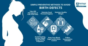
Women of childbearing age can reduce their risk of having a child with spina bifida by taking 400 micrograms (mcg) of folic acid every day pre-conception. Because it is water soluble, folic acid does not stay in the body for very long and needs to be taken every day to be effective against neural defects. Since half of all pregnancies in the United States are unplanned, folic acid must be taken whether a woman is planning a pregnancy or not. Research has shown that if all women of childbearing age took a multivitamin with the B-vitamin folic acid, the risk of neural tube defects could be reduced by up to 70%.  If you can prevent this the quality life is sure to be the best!

TREATMENTS FOR SPINA BIFIDA:

Some children with myelomeningocele and closed neural tube defects will need surgery to improve the alignment of their feet, legs, or spine.  Children with myelomeningocele usually have hydrocephalus and may require surgery to help drain fluid in the brain, such as the placement of a shunt or ETV.  Multiple surgeries may be required to replace the shunt, which may become clogged, infected, or disconnected.

Some individuals with myelomeningocele or closed neural tube defects require assistive devices for mobility such as braces, walkers, crutches, or wheelchairs.  The location of the defect on the spine often determines the type of assistive devices needed.  Children with a defect high on the spine will have little movement of the legs and will use a wheelchair for mobility.  Children with a defect lower on the spine usually have more strength in the legs.  They may be able to walk independently, or they may use crutches, leg braces, walkers, and wheelchairs depending on the activity.  Children with myelomeningocele usually have some degree of delayed mobility, so they are referred to physical therapists early on to maximize their strength and function.

Treatment for bladder and bowel dysfunction typically begins soon after birth.  Children with myelomeningocele and some closed neural tube defects have damage to the lowest spinal nerves which control typical bowel and bladder function.  Some children may be able to urinate typically, but most will need to drain their bladders with a catheter or thin tube 4-6 times a day to remain dry in between and to prevent kidney damage.  Kidneys are monitored closely so that medications or surgeries can be performed to prevent renal failure.  To prevent bowel accidents many people with myelomeningocele and closed neural type defects will use rectal medications or large volume enemas to have planned bowel movements.  Close follow-up with a spina bifida specialty clinic is recommended to develop a safe bowel and bladder program.

Treatment for progressive tethering of the spinal cord (called tethered cord syndrome) can be treated with surgery to help prevent further neurological deterioration.


QUOTE FOR TUESDAY:

“The cause of spina bifida is still unknown. Researchers believe it may be a combination of genetics and the environment that causes the condition. There are some things women who are pregnant or could get pregnant can do to help prevent spina bifida, including:

  • Take at least 400 micrograms of folic acid each day. Spina bifida happens early in a pregnancy, often before a woman even knows she is pregnant. All women who are sexually active should take folic acid supplements. While taking folic acid and getting the best prenatal care cannot completely prevent spina bifida, it can reduce the risk.
  • Talk with your primary care physician or pharmacist. If you’re planning to get pregnant or are sexually active, understanding how any prescriptions, over-the-counter medicines or supplements could affect a potential pregnancy can help you make decisions about your care.
  • Work with your provider to monitor and manage your medical conditions. If you have a condition such as Type 1 or Type 2 diabetes or are overweight, you will want to work with your OB/GYN or primary care provider before getting pregnant. Once you are pregnant, your OB/GYN may refer you to a maternal-fetal medicine specialist to get extra care during your pregnancy.
  • Avoid overheating your body (such as in a sauna or hot tub), and treat any fever you may have.”

NORTON Children’s (https://nortonchildrens.com/news/news-what-is-spina-bifida/)

Part II Spina Bifida Awareness-The complications that can arise from Spina Bifida!

  SPINA BIFIDA


TETHERED SPINALCORD (shown above)


FETUS WITH HYDROCEPHALUS (shown above)

  HYDROCEPHALUS

Spina Bifida is one of the causes of Tethered Spinal Cord and Hydrocephalus.

Tethered Spinal Cord is a stretch-induced functional disorder associated with the fixation (tethering) effect of inelastic tissue on the caudal spinal cord, limiting its movement. This abnormal attachment is associated with progressive stretching and increased tension of the spinal cord as a child ages, potentially resulting in a variety of neurological and other symptoms.

Hydrocephalus is caused by either increased production of CSF or impaired circulation and absorption

Spina bifida is a birth defect that affects the spine. Here are the latest national statistics on spina bifida in the United States:

1.)Each year, about 1,400 babies are born with spina bifida, or 1 in every 2,758 births, according to the U.S. Centers for Disease Control and Prevention. The exact cause of spina bifida is unknown. There is no cure but most people with spina bifida lead long and productive lives.

2.)Who it effects the most: Hispanic women have the highest rate of having a child affected by spina bifida, when compared with non-Hispanic white and non-Hispanic black women. Data from 12 state-based birth defects tracking programs were used to estimate the total number of pregnancies affected by spina bifida compared to the total number of live births (also called the prevalence of spina bifida) for each racial/ethnic group:

  • Hispanic: 3.80 per 10,000 live births
  • Non-Hispanic black or African-American: 2.73 per 10,000 live births
  • Non-Hispanic white: 3.09 per 10,000 live births

The symptoms listed below are some of the ways in which tethered spinal cord syndrome may be exhibited in children:

  • Lesion on the lower back
  • Fatty tumor or deep dimple on the lower back
  • Skin discoloration on the lower back
  • Hairy patch on the lower back
  • Back pain, worsened by activity and relieved with rest
  • Leg pain, especially in the back of legs
  • Leg numbness or tingling
  • Changes in leg strength
  • Progressive or repeated muscle contractions
  • Bowel and bladder problems

Spina bifida consequences that may occur, know the type of the spina bifida gives an idea of how many problems the patient will have:

  • Abnormal sensation or paralysis, which mostly occurs with closed neural tube defects and myelomeningocele.  People with these conditions typically have some degree of leg and core muscle weakness and loss of feeling in the groin and feet or legs.  The sensation can be more significant on one side of the body.
  • You may see deterioration of the gait in the patient’s walking.  Typically, the lower in the spine where the condition occurs results in less weakness and loss of feeling.  The strength and feeling do not improve with age due to nerve damage.  People with these types of spina bifida may lose strength and sensation as they grow and mobility can become more difficult with age.  People with these conditions may walk independently or use some combination of leg braces, walkers, crutches, or wheelchairs.  As they age, they may require more of these supports.
  • Chiari II malformation, in which parts of the brain called the brain stem and the cerebellum (hindbrain) protrude downward into the spinal canal or neck area.  It is almost always seen on advanced imagining of the brain in people with myelomeningocele, but it rarely causes symptoms.  When it does, this condition can press on the spinal cord and cause a variety of symptoms including difficulty breathing, swallowing, and arm weakness.  Surgery is sometimes required to reduce pressure in this area.
  • Blockage of cerebrospinal fluid, causing a condition called hydrocephalus.  Hydrocephalus is the abnormal buildup of the fluid that surrounds the brain.  Most people with myelomeningocele have this condition, which is not seen in the other types of spina bifida.  This buildup can put damaging pressure on the brain.  Hydrocephalus is commonly treated by surgically implanting a shunt—a hollow tube—in the brain which allows drainage of the excess fluid into the abdomen where it is absorbed by the body.  The tube is tunneled under the skin and not very noticeable to others.  Another treatment option is an endoscopic third ventriculostomy or ETV, a procedure that creates a new path for the fluid to flow.
  • Meningitis, an infection in the meninges covering the brain.  It can sometimes be associated with shunts.  Meningitis may cause brain injury and can be life-threatening.
  • Tethered cord syndrome can occur with all forms of spina bifida, although it is very rare in individuals with spina bifida occulta.  Usually the spinal cord and nerves float freely.  A tethered cord means that there is some type of tissue attached to and pulling the cord down.  This can cause damage to the nerves and decrease feeling and strength, as well as problems with bowel and bladder control.  It is surgically treated if a person has symptoms.
  • Bowel and bladder incontinence affect most individuals with myelomeningocele and closed neural tube defects.  The nerves at the very bottom of the spine control bowel and bladder function and don’t usually work properly in people with these types of spina bifida.  Most people with myelomeningocele and some types of closed neural tube defects need a regimen or other assistance to drain their bladders periodically or to have regularly scheduled bowel movements.
  • Learning disabilities, including difficulty paying attention, understanding concepts, impaired motor skills, impaired memory, and difficulty with organization and problem solving are commonly seen in children with myelomeningocele.  People with strength lower down in their legs tend to have less difficulty than those with more leg weakness.  Evaluation for an individualized education plan is recommended for all children with myelomeningocele.
  • Other complications such as skin ulcers, low bone mineral density, impaired male fertility, obesity, and kidney failure can be seen in people with myelomeningocele and neural tube defects as they age.  Additionally, people with myelomeningocele are at risk for precocious puberty (when changes to that of an adult occur too soon), sleep apnea, and depression.

The estimated lifetime cost of care for a person with spina bifida, with caregiving costs, is $791,900.

Learn tomorrow how you can prevent Spina Bifida with much more!


QUOTE FOR MONDAY:

“October is Spina Bifida Awareness Month—a time to celebrate the hundreds of thousands of people living with Spina Bifida. Every October, we highlight community stories that challenge us to raise more awareness and support for those living with Spina Bifida.  As a community, we can do this by raising awareness about Spina Bifida either in our own circles or larger networks. It’s also a great time for us to challenge ourselves to find new ways to get involved in raising awareness and advocacy activities.

Spina Bifida is a birth defect that is something you are born with, its not something you develop over time.  There is no cure to Spina Bifida. ”

Spina Bifida Association (https://www.spinabifidaassociation.org/awareness-month/)