Part II National Brain Injury Awareness Month!

What are the causes?

Common causes include falls, car or motorcycle crashes, vehicular accidents involving pedestrians, athletics, and assaults with or without a weapon.

Who is affected?

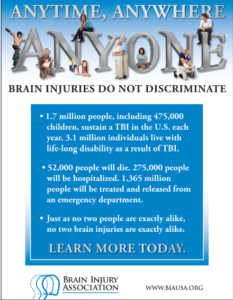
Approximately 1.5 to 2 million adults and children suffer a traumatic brain injury (TBI) each year in the United States. Most people who experience a head injury, about 1.1 million, will have a mild injury that does not require an admission to the hospital. Another 235,000 individuals will be hospitalized with a moderate to severe head injury, and approximately 50,000 will die.

How is a diagnosis made?

When a person is brought to the emergency room with a head injury, doctors will learn as much as possible about his or her symptoms and how the injury occurred. The person’s condition is assessed quickly to determine the extent of injury.

The Glasgow Coma Score (GCS) is a 15-point test used to grade a patient’s level of consciousness. Doctors assess the patient’s ability to 1) open his or her eyes, 2) ability to respond appropriately to orientation questions, (“What is your name? What is the date today?”), and 3) ability to follow commands (“Hold up two fingers, or give a thumbs up”). If unconscious or unable to follow commands, his or her response to painful stimulation is checked. A number is taken from each category and added together to get the total GCS score. The score ranges from 3 to 15 and helps doctors classify an injury as mild, moderate, or severe. Mild TBI has a score of 13-15. Moderate TBI has a score of 9-12, and severe TBI has a score of 8 and below.

Diagnostic imaging tests will be performed:

CT of TBI

Figure 3. CT scan shows a blood clot (hematoma) collecting under the bone (red arrows) and displacing brain (yellow arrow) to the other side of the skull.

  • Computed Tomography (CT) is a noninvasive X-ray that provides detailed images of anatomical structures within the brain. A CT scan of the head is taken at the time of injury to quickly identify fractures, bleeding in the brain, blood clots (hematomas) and the extent of injury (Fig. 3). CT scans are used throughout recovery to evaluate the evolution of the injury and to help guide decision-making about the patient’s care.
  • Magnetic Resonance Imaging (MRI) is a noninvasive test that uses a magnetic field and radiofrequency waves to give a detailed view of the soft tissues of the brain. A dye (contrast agent) may be injected into the patient’s bloodstream. MRI can detect subtle changes in the brain that cannot be seen on a CT scan.
  • Magnetic Resonance Spectroscopy (MRS) gives information about the metabolism of the brain. The numbers generated from this scan provide a general prognosis about the patient’s ability to recover from the injury.

What treatments are available?

Mild TBI usually requires rest and medication to relieve headache. Moderate to severe TBI require intensive care in a hospital. Bleeding and swelling in the brain can become an emergency that requires surgery. However, there are times when a patient does not require surgery and can be safely monitored by nurses and physicians in the neuroscience intensive care unit (NSICU).

The goals of treatment are to resuscitate and support the critically ill patient, minimize secondary brain injury and complications, and facilitate the patient’s transition to a recovery environment. Despite significant research, doctors only have measures to control brain swelling, but do not have a way to eliminate swelling from occurring.

Neurocritical care
Neurocritical care is the intensive care of patients who have suffered a life-threatening brain injury. Many patients with severe TBI are comatose or paralyzed; they also may have suffered injuries in other parts of the body. Their care is overseen by a neurointensivist, a specialty-trained physician who coordinates the patient’s complex neurological and medical care. Patients are monitored and awakened every hour for nursing assessments of their mental status or brain function.

Click to view larger image with labels

Figure 4. In the NSICU, the patient is connected to numerous machines, tubes, and monitors. The monitoring equipment provides information about body functions and helps guide care. Some equipment may take over certain functions, such as breathing, nutrition, and urination, until the patient’s body is able to do these things on its own.

Seeing a patient who has suffered a severe TBI can be shocking. It is possible that your loved one’s appearance will be altered because of facial injury and equipment that is used for monitoring. Numerous tubes, lines, and equipment may be used to closely monitor his or her heart rate, blood pressure, and other critical body functions. (Fig. 4)

ICP monitor

Figure 5. A brain oxygen and cerebral blood flow monitor is inserted into the brain tissue and secured to the skull with a bolt. A catheter is inserted into the ventricle of the brain to monitor intracranial pressure (ICP). If pressure is too high, the CSF fluid can be drained from the ventricles.

  • Intracranial pressure (ICP) monitor. A catheter is placed through a small hole in the skull and positioned inside the ventricle (fluid-filled area deep within the brain) to measure pressure inside the head (Fig. 5). The ICP monitor allows the NSICU team to intervene quickly if the pressure becomes too high. Typical intracranial pressure is less than 20 mmHg. However, there are times when a higher number is safe and acceptable.
  • Brain oxygen monitor (Licox). A catheter is placed through a small hole in the skull and positioned within the brain tissue. The Licox measures the oxygen level and temperature within the brain. Adjustments in the amount of oxygen given to the patient are often made to maximize the brain’s oxygen level. A cerebral blood flow monitor, called a Hemedex, is a newer monitor that is placed with the Licox and helps the NSICU team evaluate blood flow through the brain.
  • Ventilator. Some patients may require a ventilator, a machine that helps them breathe. The ventilator is connected to the patient by the endotracheal tube, or ET tube. The tube is placed into the patient’s mouth and down into the trachea, or windpipe. The tube allows the machine to push air into and out of the lungs, thereby helping the patient breathe.
  • Feeding tube. When patients are on a ventilator or have a decreased level of alertness, they may not be able to eat or get sufficient nutrition to meet their needs. A nasal-gastric feeding tube may be inserted through the patient’s nose and passed down the throat into the stomach. It delivers liquid nutrition as well as any medication that is required.
  • Seizures and EEG monitoring. A seizure is an abnormal electrical discharge from the brain. Approximately 24% of patients who suffer a TBI will have a seizure that is undetected unless they are monitored by an electroencephalogram (EEG). Seizures that are not visible to the human eye are referred to as non-convulsive seizures. Because these seizures are serious, all patients with a severe TBI are monitored with continuous EEG for 24 to 72 hours after injury.

Medication

  • Sedation and pain. After a head injury it may be necessary to keep the patient sedated with medications. These medications can be turned off quickly in order to awaken the patient and check their mental status. Because patients often have other injuries, pain medication is given to keep them comfortable.
  • Controlling intracranial pressure. Hypertonic saline is a medication used to control pressure within the brain. It works by drawing the extra water out of the brain cells into the blood vessels and allowing the kidneys to filter it out of the blood.
  • Preventing seizures. Patients who’ve had a moderate to severe traumatic brain injury are at higher risk of having seizures during the first week after their injury. Patients are given an anti-seizure medication (levetiracetam or phenytoin) to prevent seizures from occurring.
  • Preventing infection. Although every attempt is made to prevent infection, the risk is always present. Any device placed within the patient has the potential to introduce a microbe. If an infection is suspected, a test will be sent to a laboratory for analysis. If an infection is present, it will be treated with antibiotics.

Surgery

Surgery is sometimes necessary to repair skull fractures, repair bleeding vessels, or remove large blood clots (hematomas). It is also performed to relieve extremely high intracranial pressure.

  • Craniotomy involves cutting a hole in the skull to remove a bone flap so that the surgeon can access the brain. The surgeon then repairs the damage (e.g., skull fracture, bleeding vessel, remove large blood clots). The bone flap is replaced in its normal position and secured to the skull with plates and screws.

Figure 6. A large decompressive craniectomy is removed and the dura is opened to allow the brain to expand. Blood clots are removed and bleeding vessels are repaired. The bone flap is frozen and replaced about 6 weeks later.

  • Decompressive craniectomy involves removing a large section of bone so that the brain can swell and expand. This is typically performed when extremely high intracranial pressure becomes life threatening. At that time the patient is taken to the operating room where a large portion of the skull is removed to give the brain more room to swell (Fig. 6). A special biologic tissue is placed on top of the exposed brain and the skin is closed. The bone flap is stored in a freezer. One to 3 months after the swelling has resolved and the patient has stabilized from the injury, the bone flap is replaced in another surgery, called cranioplasty.

Other surgical procedures may be performed to aid in the patient’s recovery:

  • Tracheotomy involves making a small incision in the neck to insert the breathing tube directly into the windpipe. The ventilator will then be connected to this new location on the neck and the old tube is removed from the mouth.
  • Percutaneous Endoscopic Gastrostomy Tube (PEG) is a feeding tube inserted directly into the stomach through the abdominal wall. A small camera is placed down the patient’s throat into the stomach to aid with the procedure and to ensure correct placement of the PEG tube (see Surgical Procedures for Accelerated Recovery).

Clinical trials

Clinical trials are research studies in which new treatments—drugs, diagnostics, procedures, and other therapies—are tested in people to see if they are safe and effective. Research is always being conducted to improve the standard of medical care. Information about current clinical trials, including eligibility, protocol, and locations, are found on the Web. Studies can be sponsored by the National Institutes of Health (see clinicaltrials.gov) as well as private industry and pharmaceutical companies (see www.centerwatch.com).

Recovery & prevention

The recovery process varies depending on the severity of the injury, but typically progresses through stages: coma, confusion / amnesia, and recovery.

  • When a patient is in a coma, his or her eyes are closed and they show minimal reaction when spoken to or stimulated. Movements that may be seen at this time are basic reflexes or automatic responses to a stimulus. The brain wave activity in a comatose person is very different from that of a sleeping person.
  • When a patient begins to awaken, the first natural response is that of bodily protection. Patients at this stage will move away from any stimulus or tend to pull at items attached to them in an attempt to remove anything that is uncomfortable or irritating. His or her eyes may be open more often, but they may not be aware of their behavior or be able to interact in a meaningful way. It is common for a patient to respond to each stimulus (hearing, seeing, or touching) in the same way. Responses may include increased rate of breathing, moaning, moving, sweating, or a rise in blood pressure.
  • As the patient continues to wake up, their interactions may become more purposeful. They may look at a person and follow them around the room with their eyes, or follow simple commands such as “Hold up your thumb.” Patients tend to be confused and may have inappropriate or agitated behaviors.

Not all head injuries are the same. Patients recover at different rates and to varying degrees. It is difficult to determine at what point a patient will start understanding and interacting with their caregivers or family in a meaningful way. It is important to have patience; recovery from a brain injury can take weeks, months, or even years.

The Family’s Role
Many family members express feelings of helplessness when their loved one is in the NSICU. You are not alone. Please take care of yourself and use your energy wisely.

Visiting hours are limited in the NSICU. Too much stimulation can agitate the patient and raise his or her blood pressure. You can most effectively convey your concern by sitting quietly and holding your loved one’s hand. Be aware that the patient, though silent, may hear anything you say. Never speak as if the patient were not there.

As patients recover, they need help understanding what has happened to them during this “lost period of time.” Keep in mind that the recovery of consciousness is a gradual process – not just a matter of waking up. Progress is usually tracked in three areas: movement, thinking, and interacting. You can help by keeping a diary of their progress. Family photos may help with regaining memory.

Rehabilitation
Most patients are discharged from the hospital when their condition has stabilized and they no longer require intensive care. A social worker will work closely with the family as preparations are made for a return home or for transfer to a long-term care or rehabilitation center.

  • A long-term acute care (LTAC) facility is a place for patients who have stabilized from their initial injury but who still require a ventilator or frequent nursing care. Many patients are discharged to an LTAC to continue being weaned from the ventilator. Once off the ventilator, they can be moved to a rehabilitation or skilled nursing facility.
  • A rehabilitation facility is a place for patients who do not require a ventilator but who still require help with basic daily activities. Physical and occupational therapists work with patients to help them achieve their maximum potential for recovery. Rehab facilities are either Acute Inpatient Rehab that require patients to participate in 3 hours or more of rehab a day or a Skilled Nursing Facility (SNF) that provide 1-3 hours of rehab a day depending on what the patient can tolerate.

Recovering from a brain injury relies on the brain’s plasticity—the ability for undamaged areas of the brain to take over functions of the damaged areas. It also relies on regeneration and repair of nerve cells. And most importantly, on the patient’s hard work to relearn and compensate for lost abilities.

  • A physical therapist helps patients rebuild and maintain strength, balance, and coordination. They can work with the patient in any facility.
  • An occupational therapist helps patients to perform activities of daily living, such as dressing, feeding, bathing, toileting, and transferring themselves from one place to another. They also provide adaptive equipment if a patient has difficultly performing a task.
  • A speech therapist helps patients by monitoring their ability to safely swallow food and helping with communication and cognition.
  • A neuropsychologist helps patients relearn cognitive functions and develop compensation skills to cope with memory, thinking, and emotional needs.

Prevention

Tips to reduce the risk for a head injury:

  • Always wear your helmet when riding a bicycle, motorcycle, skateboard, or all-terrain vehicle.
  • Never drive under the influence of alcohol or drugs.
  • Always wear your seat belt and ensure that children are secured in the appropriate child safety seats.
  • Avoid falls in the home by keeping unsecured items off the floor, installing safety features such as non-slip mats in the bathtub, handrails on stairways, and keeping items off of stairs.
  • Avoid falls by exercising to increase strength, balance, and coordination.
  • Store firearms in a locked cabinet with bullets in a separate location.
  • Wear protective headgear while playing sports.

Reference for Part I and Part II: Mayfield Brain & Spine at 513-221-1100.
QUOTE FOR TUESDAY:

“National Brain Injury Awareness Month! Every year the Brain Injury Association of America (BIAA) proudly leads Americans throughout Brain Injury Awareness Month. The theme for this year is #ChangeYourMind. And its goal is to:

  • Help de-stigmatize brain injury in the community,
  • Share the stories of those who are living with brain injuries, and those that care for them,
  • And spread awareness of the ways people living with brain injuries can receive support.”

BrainForest (brainforest.com)

Part I National Brain Injury Awareness Month!

Overview

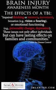
Traumatic brain injury (TBI) is sudden damage to the brain caused by a blow or jolt to the head. Common causes include car or motorcycle crashes, falls, sports injuries, and assaults. Injuries can range from mild concussions to severe permanent brain damage. While treatment for mild TBI may include rest and medication, severe TBI may require intensive care and life-saving surgery. Those who survive a brain injury can face lasting effects in their physical and mental abilities as well as emotions and personality. Most people who suffer moderate to severe TBI will need rehabilitation to recover and relearn skills.

What is a traumatic brain injury?

TBI is an injury to the brain caused by a blow or jolt to the head from blunt or penetrating trauma. The injury that occurs at the moment of impact is known as the primary injury. Primary injuries can involve a specific lobe of the brain or can involve the entire brain. Sometimes the skull may be fractured, but not always. During the impact of an accident, the brain crashes back and forth inside the skull causing bruising, bleeding, and tearing of nerve fibers (Fig. 1). Immediately after the accident the person may be confused, not remember what happened, have blurry vision and dizziness, or lose consciousness. At first the person may appear fine, but their condition can decline rapidly. After the initial impact occurs, the brain undergoes a delayed trauma – it swells – pushing itself against the skull and reducing the flow of oxygen-rich blood. This is called secondary injury, which is often more damaging than the primary injury.

TBI

Figure 1. During impact to the head, the soft brain crashes back and forth against the inside of the hard skull causing bruising, bleeding, and shearing of the brain.

Traumatic brain injuries are classified according to the severity and mechanism of injury:

  • Mild: person is awake; eyes open. Symptoms can include confusion, disorientation, memory loss, headache, and brief loss of consciousness.
  • Moderate: person is lethargic; eyes open to stimulation. Loss of consciousness lasting 20 minutes to 6 hours. Some brain swelling or bleeding causing sleepiness, but still arousable.
  • Severe: person is unconscious; eyes do not open, even with stimulation. Loss of consciousness lasting more than 6 hours.

Types of traumatic brain injuries

  • Concussion is a mild head injury that can cause a brief loss of consciousness and usually does not cause permanent brain injury.
  • Contusion is a bruise to a specific area of the brain caused by an impact to the head; also called coup or contrecoup injuries. In coup injuries, the brain is injured directly under the area of impact, while in contrecoup injuries it is injured on the side opposite the impact.
  • Diffuse axonal injury (DAI) is a shearing and stretching of the nerve cells at the cellular level. It occurs when the brain quickly moves back and forth inside the skull, tearing and damaging the nerve axons. Axons connect one nerve cell to another throughout the brain, like telephone wires. Widespread axonal injury disrupts the brain’s normal transmission of information and can result in substantial changes in a person’s wakefulness.
  • Traumatic Subarachnoid Hemorrhage (tSAH) is bleeding into the space that surrounds the brain. This space is normally filled with cerebrospinal fluid (CSF), which acts as a floating cushion to protect the brain. Traumatic SAH occurs when small arteries tear during the initial injury. The blood spreads over the surface of the brain causing widespread effects.
  • Hematoma is a blood clot that forms when a blood vessel ruptures. Blood that escapes the normal bloodstream starts to thicken and clot. Clotting is the body’s natural way to stop the bleeding. A hematoma may be small or it may grow large and compress the brain. Symptoms vary depending on the location of the clot. A clot that forms between the skull and the dura lining of the brain is called an epidural hematoma. A clot that forms between the brain and the dura is called a subdural hematoma. A clot that forms deep within the brain tissue itself is called an intracerebral hematoma. Over time the body reabsorbs the clot. Sometimes surgery is performed to remove large clots.

Although described as individual injuries, a person who has suffered a TBI is more likely to have a combination of injuries, each of which may have a different level of severity. This makes answering questions like “what part of the brain is hurt?” difficult, as more than one area is usually involved.

Secondary brain injury occurs as a result of the body’s inflammatory response to the primary injury. Extra fluid and nutrients accumulate in an attempt to heal the injury. In other areas of the body, this is a good and expected result that helps the body heal. However, brain inflammation can be dangerous because the rigid skull limits the space available for the extra fluid and nutrients. Brain swelling increases pressure within the head, which causes injury to parts of the brain that were not initially injured. The swelling happens gradually and can occur up to 5 days after the injury.

What are the symptoms?

Depending on the type and location of the injury, the person’s symptoms may include:

  • Loss of consciousness
  • Confusion and disorientation
  • Memory loss / amnesia
  • Fatigue
  • Headaches
  • Visual problems
  • Poor attention / concentration
  • Sleep disturbances
  • Dizziness / loss of balance
  • Irritability / emotional disturbances
  • Feelings of depression
  • Seizures
  • Vomiting

Diffuse injuries (such as a concussion or diffuse axonal injury) will typically cause an overall decreased level of consciousness. Whereas, focal injuries (such as an ICH or a contusion) will have symptoms based on the brain area affected (Fig. 2).

Lobes of the brain that can get affected by TBI:

brain anatomy

Figure 2. The brain is composed of three parts: the brainstem, cerebellum, and cerebrum, which is divided into lobes. The table lists the lobes of the brain and their normal functions as well as problems that may occur when injured. While an injury may occur in a specific area, it is important to understand that the brain functions as a whole by interrelating its component parts.

Every patient is unique and some injuries can involve more than one area or a partial section, making it difficult to predict which specific symptoms the patient will experience.

QUOTE FOR MONDAY:

“The most common condition among children is largely preventable! According to the AAPD (American Academy of Pediatric Dentistry), dental caries/cavities are four times more common than childhood asthma and three times more likely than childhood obesity. As a dental hygienist, NCDHM is a great opportunity to spread the word about how poor oral health and caries impact our kids’ health and quality of life.”.

American Dental HealtAdvocacy – adha.org


National Children’s Dental Health Month

During the month of February, the American Dental Association celebrates National Children’s Dental Health Month.

National Children’s Dental Health Month, with the 2022 theme being “Sealants Make Sense.”

The month is a national health observance that brings together thousands of dedicated professionals, health care providers and educators to promote the benefits of good oral health to children, their caregivers, teachers and many others.

Irene Hilton, D.D.S., chair of the ADA Council on Advocacy for Access and Prevention’s advisory committee on dental sealants, said sealants are an equitable way to prevent caries.

“Sealants on permanent molars reduce the risk of caries by 80%,” Dr. Hilton said. “If your dentist recommends dental sealants, it is part of a comprehensive caries management approach. I am excited that this year” National Children” Dental Health Month theme is about dental sealants, so more families are aware of this cavity-preventing treatment.”

New parents often ask, “When should my child first see a dentist?” It’s never too early to start focusing on your child’s oral health! The American Association of Pediatric Dentists recommends that parents establish a dental home for their child by their first tooth or first birthday. During this time, parents/guardians will have the opportunity to ask questions and address any dental concerns at the primary visit and the dentist will gently swab the child’s mouth to check their gums and any erupted teeth. As the child starts teething, the dentist will be able to monitor their progress and implement preventative measures for any concerns with your baby’s teeth.

Protect Tiny Teeth

Baby teeth are so important because of their key role of saving space for a child’s permanent teeth. They stay in a child’s mouth for 8-10 years and also affect their speaking, chewing, and, of course, smiling. Baby teeth can also indicate a child’s overall quality of health. Untreated tooth decay can cause oral infections that enter the bloodstream and lead to other serious health problems, while also allowing bacteria to spread to new adult teeth.

While daily brushing is an important part of a child’s oral hygiene routine, bacteria that causes tooth decay can still linger between teeth where the toothbrush can’t reach. That’s why it’s so important to help your kids incorporate flossing in their daily routine.

One significant oral health risk for infants and young children under the age of 1 is from baby bottle tooth decay. This occurs when your child consumes sugary liquid and bacteria in their mouth consume the sugar and produce acid. This acid attacks the enamel on baby teeth can trigger tooth decay after continued exposure. Liquids that contribute to this condition include milk, formula, fruit juice, soda, and any other sweetened drinks. If your child needs to sleep with a bottle, water is the safest option without any risk.

Parents, Did You Know?

Early childhood tooth decay has become the most common chronic childhood disease, impacting more children than asthma. According to the ADA, more than 40% of children have tooth decay by the time they reach Kindergarten. Additionally, kids who suffer from poor oral health are three times more likely to miss school as a result of dental pain.

The State of Pennsylvania has tried to tackle this oral health epidemic by requiring each child to receive a dental examination before enrolling in school, as well as in the 3rd and 7th grades. However, without regular six-month check-ups and establishing healthy oral health habits at an early age, small cavities can lead to much larger problems in little mouths.

Tips for Maintaining Your Child’s Oral Health 

Our doctors take pride in serving patients in such vital years of early childhood. As pediatric dentists, they’ve had additional training beyond dental school to work specifically with babies and children in monitoring early oral development. We recommend the following oral health tips to start your little ones on their journey to a lifetime of healthy smiles.

  • Schedule routine check-ups. If it’s been more than six months since your child has seen a dentist, schedule an appointment as soon as possible.
  • Clean your baby’s gums daily. Until those teeth come in gently wipe a damp washcloth over the gums to clear away harmful bacteria after each feeding.
  • Start brushing with the first tooth. Begin brushing your baby’s teeth when you see one coming in with an infant toothbrush. Use water and a tiny bit of fluoride toothpaste (about the size of a grain of rice).
  • Brush twice each day for two minutes. Children ages 2-6 should use a pea-sized amount of fluoride toothpaste. Always supervise kids younger than six years old while brushing, as they are more likely to swallow toothpaste.
  • Begin flossing. Once your child’s teeth touch, you can start flossing in between them.
  • Snack healthy! Fruit juice, sports drinks, fruit snacks, and sticky candies all pose serious threats to your child’s teeth. Give kids calcium-rich snacks like cheese or low-sugar yogurt. If you have to resort to candy – a chocolate bar is preferable to gummy or sticky sweets that can get lodged in between the teeth, even after brushing.
  • Keep them hydrated! Avoid sugary drinks and stick to good old-fashioned water. Water helps to rinse away any sugar or particles that can lead to cavities. Many municipal water sources also contain fluoride, which is recommended by the American Dental Association and U.S. Surgeons General, among others, as an efficient way to prevent tooth decay. In fact, the theme of this year’s National Children’s Dental Health Month is celebrating 75 years of water fluoridation.
  • Replace your child’s toothbrush every three to four months.


QUOTE FOR THE WEEKEND:

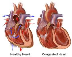
“The heart’s pumping action moves oxygen-rich blood as it travels from the lungs to the left atrium, then on to the left ventricle, which pumps it to the rest of the body. The left ventricle supplies most of the heart’s pumping power, so it’s larger than the other chambers and essential for normal function. In left-sided or left ventricular (LV) heart failure, the left side of the heart must work harder to pump the same amount of blood.

Right-sided or right ventricular (RV) heart failure usually occurs as a result of left-sided failure. When the left ventricle fails, increased fluid pressure is, in effect, transferred back through the lungs, ultimately damaging the heart’s right side. When the right side loses pumping power, blood backs up in the body’s veins. This usually causes swelling or congestion in the legs, ankles and swelling within the abdomen such as the GI tract and liver (causing ascites).”

Americasn Heart Association (www.heart.org)

Left sided and Right sided Congestive Heart Failures!

chf-part-i  chf1

chf-part-1a

The definition of heart failure, it occurs when the heart loses its ability to pump enough blood through the body. Usually, the loss in pumping action is a symptom of an underlying heart problem, such as hypertension and CAD = coronary artery disease. The term heart failure suggests a sudden and complete stop of heart activity but actually the heart does not suddenly or abruptly stop. Instead the way it works is heart failure usually develops over time, years. The heart first compensates with the disease or illness the individual has but, just like a car, after wear and tear the heart goes into decompensating to heart failure due to the heart decline. How serious is this condition? It varies from person to person depending on factors like an individual with obesity & unhealthy versus a person in healthier condition. All people diagnosed or not diagnosed with heart failure lose a pumping capacity of the heart happens as they age but diagnosed with heart failure makes the engine of the body a challenge in doing its function properly. The pump loss is more significant in the person with heart failure and often results from a heart attack (actual scaring to the tissue=death to that tissue area) or from other diseases that can damage the heart. The severity of the condition determines the impact it has on a person’s life. At the other end, extremes, treatment often helps people lead full lives if the person follows the meds ordered by the doctor including the diet and activity/exercise the doctor orders to the patient with heart failure (compliance so important). There are different levels of heart failure but even the mildest form is a serious health problem, which must be treated. If not the pump (the heart) will just get worse in doing its function properly. To improve the chance of living longer in an individual with heart failure, patients must take care of themselves, see their physician (cardiologist) on a regular basis, and closely follow treatments (as ordered) with knowing what heart failure actually to understanding how the disease works (is the failure on the right side or left side? Which in time will effect the other side in time). In knowing what side the failure is on will make you understand what signs and symptoms to expect.


Types of Heart Failure


The term congestive heart failure (CHF) is often used to describe all patients with heart failure. In reality, congestion=the buildup of fluids in the heart for not pumping correctly, just like pipes in a home not working properly=back up of water in the pipes, happens with CHF also to the fluids (blood) backing up in the lungs.   This is just one feature of the condition and does not occur in all patients. There are two main categories of heart failure although within each category, symptoms and effects may differ from patient to patient. The two categories are:    1-Systolic heart failure (systolic is the top number of your blood pressure=the heart at work). This occurs when the heart’s (muscle-myocardium) ability to contract (pump=being active) decreases, particularly starting on the L side of the heart where the muscle of the heart is thickest and most powerful (myocardium=heart muscle). The left side of the heart sends the highly oxygenated blood that just came from the lungs to be pumped out throughout the body to send oxygen (nutrients) to all our tissues.   When the heart is in left sided failure it cannot pump blood with enough force to push a sufficient amount out of the heart into the circulation through the aorta. This is not sending nutrients=oxygen like it does when not in failure.  The aorta is a artery (vessel) that leaves the L side of the heart (left side of the heart=highly oxygenated rich blood). Due to the heart not using enough force pushing the blood forward in the aorta this causes the blood to back up and cause it to go back up from the aorta into the L lower to the L upper chamber of the heart that goes further back up into the pulmonary vein into the lungs=congestion in the lungs due to the heart failure.


2-Diastolic heart failure (diastolic is the bottom number of your blood pressure which is the pressure when the heart is at rest). This failure occurs when the heart has a problem relaxing. The heart cannot properly fill with blood because the muscle of the heart due to trying so hard to compensate over a long period of time with disease (ex. High B/P, Obesity, etc…) strains the heart in doing its function that failure finally starts that the muscle of the heart (myocardium) becomes stiff. This causes the heart to lose its ability to relax to allow proper filling of the heart in upper and lower chambers=back up of the blood.   This failure starts on the right side of the heart causing the blood to back up away from the heart and may lead this blood that is highly concentrated with carbon dioxide to accumulation especially in the feet, ankles and legs. Some patients may have lung congestion.


Causes of Heart Failure:


As stated, the heart loses some of its blood pumping ability as a natural consequence of aging. How- ever, a number of other factors can lead to a potentially life-threatening loss of pumping activity.


As a symptom of underlying heart disease, heart failure is closely associated with the major risk factors for coronary heart disease: smoking, high cholesterol levels, hypertension (persistent high blood pressure), diabetes= abnormal blood sugar levels, and obesity. A person can change or eliminate those risk factors and thus lower their risk of developing or aggravating their heart disease and heart failure through healthy habits performed routinely, proper dieting, and balancing rest with exercise.


Among prominent risk factors, hypertension-HTN (high blood pressure) and diabetes are PARTICULARLY IMPORTANT. Uncontrolled HTN increases the risk of heart failure by 200 %, compared to those who do not have hypertension.   Moreover, the degree of risk appears directly related to the severity of the high blood pressure.


Persons with diabetes have about a two to eight fold greater risk of heart failure than those without diabetes. Women with diabetes have a greater risk of heart failure than men with diabetes. Part of the risk comes from the diabetes association with other risk factors for heart disease such as high cholesterol or obesity or other risk factors. However, the disease process of diabetes also damages the heart muscle.


The presence of coronary disease is among the greatest risks for heart failure. Muscle damage and scarring caused by a heart attack greatly increase the risk of heart failure. Cardiac arrhythmias, or irregular heartbeats, also raise heart failure risk. Any disorder that causes abnormal swelling or thickening of the heart sets the stage for heart failure.


In some people, heart failure arises from problems with heart valves, the flap-like structures that help regulate blood flow through the heart. Infections in the heart are another source of increased risk for heart failure.


A single risk factor may be sufficient to cause heart failure, but a combination of factors dramatically increases the risk. Advanced age adds to the potential impact of any heart failure risk.


Finally, genetic abnormalities contribute to the risk for certain types of heart disease, which in turn may lead to heart failure. However, in most instances, a specific genetic link to heart failure has not been identified.


SO LIVE AS HEALTHY AS POSSIBLE IN YOUR ROUTINE HABITS, YOUR DIETING OF THE 4 FOOD GROUPS, MAINTAINING YOUR WEIGHT IN A THEREPEUTIC RANGE (look as calculating BMI online for free to find out what your weight range for your height is), and BALANCING REST WITH EXERCISE TO HELP DECREASE THE CHANCE OF GETTING HEART FAILURE=PREVENTION!


QUOTE FOR THURSDAY:

“Your risk for some eye diseases and conditions increases as you grow older, and some eye changes are more serious. Keep your eyes as healthy as possible by getting regular eye exams so any problems can be spotted early.”

NIH – Natonal Institute on Aging  (https://www.nia.nih.gov)

Eye health for those at age 60 and over!

Eyes over 601Eyesover602

Women-Higher Risk for Some Eye Diseases

Women are more likely than men to have glaucoma and women are also more likely to be visually impaired or blind due to glaucoma. Also, women are 24 percent less likely to be treated for glaucoma. Cataract is somewhat more common in women, as well. Women should be sure to follow the Academy’s screening guidelines and adhere to their Eye M.D.’s follow-up appointment recommendations and treatment plans.

Low Vision

The term low vision describes vision loss that makes daily tasks difficult. Normal aging of the eye does not lead to low vision; it is a result of eye diseases, injuries or both. Low vision symptoms nclude loss of central and/or peripheral (side) vision, blurred or hazy vision or night blindness. A person may have trouble recognizing faces, reading, driving and shopping. If you experience any of these problems, it is important to see your Eye M.D., who will check for and treat any underlying conditions and advise on low vision resources and low vision aids and devices to help with reading and other daily tasks. Most people with low vision need brighter lighting in their living areas.

Avoid Falls and Related Eye Injuries

About half of all eye injuries occur in or around the home, most often during improvement projects (44 percent). The good news is that nearly all eye injuries can be prevented by using protective eyewear, so every household needs to have at least one pair of certified safety glasses on hand.

It’s also important to reduce the risk of falls, which become more likely as we age, due to changes in vision and balance. Consider taking these safety steps around the home to diminish the risks of injuring your eyes:

  • Make sure that rugs and shower/bath/tub mats are slip-proof.
  • Secure railings so that they are not loose.
  • Cushion sharp corners and edges of furnishings and home fixtures.

Systemic health problems

Systemic health problems like high blood pressure and diabetes that may be diagnosed or become more problematic in midlife can also affect eye health. One warning sign of both high blood pressure and diabetes is when the ability to see clearly changes frequently. Be sure to keep your Eye M.D. informed about your health conditions and use of medications and nutritional supplements, as well as your exercise, eating, sleeping and other lifestyle choices.

Exercise

Our eyes need good blood circulation and oxygen intake, and both are stimulated by regular exercise. Regular exercise also helps keep our weight in the normal range, which reduces the risk of diabetes and of diabetic retinopathy. Gentler exercise, including walking, yoga, tai chi, or stretching and breathing, can also be effective ways to keep healthy. Remember to use sun safety and protective eyewear when enjoying sports and recreation.

Sleep

As we sleep, our eyes enjoy continuous lubrication. Also during sleep the eyes clear out irritants such as dust, allergens, or smoke that may have accumulated during the day.

Some research suggests that light-sensitive cells in the eye are important to our ability to regulate our wake-sleep cycles. This becomes more crucial as we age, when more people have problems with insomnia. While it’s important that we protect our eyes from over-exposure to UV light, our eyes also need exposure to some natural light every day to help maintain normal sleep-wake cycles.

QUOTE FOR WEDNESDAY:

“As you age, it is normal to notice changes in your vision. A few common changes for older adults include:

  • Losing the ability to see up close
  • Having trouble distinguishing colors, such as blue from black
  • Needing more time to adjust to changing levels of light”

NIH – National Institute of Hearing  (https://www.nia.nih.gov)