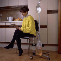
What is cystic fibrosis?


Cystic fibrosis is a hereditary disease that affects the lungs and digestive system. The body produces thick and sticky mucus that can clog the lungs and obstruct the pancreas. Cystic fibrosis (CF) can be life-threatening, and people with the condition tend to have a shorter-than-normal life span.

What is this disease?

Cystic fibrosis (CF) is a progressive, genetic disease that causes persistent lung infections and limits the ability to breathe over time.

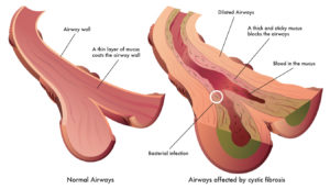
In people with CF, mutations in the cystic fibrosis transmembrane conductance regulator (CFTR) gene cause the CFTR protein to become dysfunctional. When the protein is not working correctly, it’s unable to help move chloride — a component of salt — to the cell surface. Without the chloride to attract water to the cell surface, the mucus in various organs becomes thick and sticky.

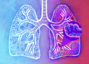
In the lungs, the mucus clogs the airways and traps germs, like bacteria, leading to infections, inflammation, respiratory failure, and other complications. For this reason, minimizing contact with germs is a top concern for people with CF.

In the pancreas, the buildup of mucus prevents the release of digestive enzymes that help the body absorb food and key nutrients, resulting in malnutrition and poor growth. In the liver, the thick mucus can block the bile duct, causing liver disease. In men, CF can affect their ability to have children.

Symptoms of CF

People with CF can have a variety of symptoms, including:

  • Very salty-tasting skin
  • Persistent coughing, at times with phlegm
  • Frequent lung infections including pneumonia or bronchitis
  • Wheezing or shortness of breath
  • Poor growth or weight gain in spite of a good appetite
  • Frequent greasy, bulky stools or difficulty with bowel movements
  • Male infertility

Cystic fibrosis is a genetic disease. People with CF have inherited two copies of the defective CF gene — one copy from each parent. Both parents must have at least one copy of the defective gene.

People with only one copy of the defective CF gene are called carriers, but they do not have the disease. Each time two CF carriers have a child, the chances are:

  • 25 percent (1 in 4) the child will have CF
  • 50 percent (1 in 2) the child will be a carrier but will not have CF
  • 25 percent (1 in 4) the child will not be a carrier and will not have CF

The defective CF gene contains a slight abnormality called a mutation. There are more than 1,700 known mutations of the disease. Most genetic tests only screen for the most common CF mutations. Therefore, the test results may indicate a person who is a carrier of the CF gene is not a carrier.

Diagnosing cystic fibrosis is a multistep process, and should include a newborn screening, a sweat test, a genetic or carrier test, and a clinical evaluation at a CF Foundation-accredited care center. Although most people are diagnosed with CF by the age of 2, some are diagnosed as adults. A CF specialist can order a sweat test and recommend additional testing to confirm a CF diagnosis.

Read the CF Foundation’s clinical care guidelines for diagnosing CF.

According to the Cystic Fibrosis Foundation Patient Registry, in the United States:

  • More than 30,000 people are living with cystic fibrosis (more than 70,000 worldwide).
  • Approximately 1,000 new cases of CF are diagnosed each year.
  • More than 75 percent of people with CF are diagnosed by age 2.
  • More than half of the CF population is age 18 or older.


QUOTE FOR WEDNESDAY:

“It’s inevitable that when kids mix — returning from camp or heading back to school — germs spread. And in a pandemic year fueled by the delta variant, some of those germs may cause COVID-19. The Centers for Disease Control and Prevention has advice for keeping your child protected from this highly contagious version of the coronavirus now and this fall: Mask up in schools and other crowded venues, and make sure everyone age 12 and older in the family gets a COVID-19 shot.”

NPR.org (WAMC Northeast public radio)

How to make the end of the summer better and safer the kids!

We’ve all had to adjust to and accept pandemic living.  When it comes to addressing canceled trips, playdates and a drastically different summer overall, the key is to take a direct approach and not sugarcoat things.  Kids are smart. So, give them a little credit during those tough parent-child conversations.  The most important thing to remember when you’re talking to your child is that it’s not necessarily what you say, but how you say it. You need to be direct and to the point. Let them know the reason behind why they can’t do the things that they use to.  It is also the same for adults but the majority have the key factors already major enough in knowing how to deal with it.  It is harder on the child.

To help your child understand why being around a lot of people isn’t a good idea right now, she suggests an approach like this.

“Nearly all children can remember being sick at some point in their lives, and germs are something that most children can understand the concept of. Start a discussion with your child about how germs are very tiny and when they enter our bodies, they make us sick.  Explain what covid is, how you get it but most important is how to prevent it.  They have been saying it for months to social distance and get the VACCINE!

Physical distancing can be a difficult concept for some children to understand.  You can also use items to illustrate how far six feet away really is. Use their toys, the length of a pet or even their favorite foods to show the distance (for example, six medium pizzas equal six feet).  Kids notice everything. So, they’re bound to notice when people are too close and know when someone is wearing a MASK!

It’s hard for little kids to resist touching things (Ex. public things like a public toilet, public counter, etc…). They can’t help it.  Kids explore their world through touch. If you think about babies, they’re putting everything in their mouths.  Parents urge your little ones including the little ones they play with to not touch their face.

Should your child struggle with the concept of not touching things, their face or others, try not to get upset. If you respond in a negative way like angry or anxious, it will only make your child more anxious and forgetful due to there reaction of your negative reaction to them.  You want them to remember this in not touching their face.

Washing the hands and using purex or something like it to help keep the hands clean!

QUOTE FOR TUESDAY:

“Know the extent of the disease;  The identified incidence of Legionnaires’ disease varies widely according to the level of surveillance and reporting. Since many countries lack appropriate methods of diagnosing the infection or sufficient surveillance systems, the rate of occurrence is unknown. In Europe, Australia and the USA there are about 10–15 cases detected per million population per year.

Of the reported cases 75–80% are over 50 years and 60–70% are male. Other risk factors for community-acquired and travel-associated legionellosis include: smoking, a history of heavy drinking, pulmonary-related illness, immuno-suppression, and chronic respiratory or renal illnesses.”

World Health Organization – WHO

QUOTE FOR MONDAY:

“The bacterium L. pneumophila was first identified in 1977, as the cause of an outbreak of severe pneumonia in a convention centre in the USA in 1976.  Legionnaires’ disease has an incubation period of 2 to 10 days (but up to 16 days has been recorded in some outbreaks).  Untreated Legionnaires’ disease usually worsens during the first week.Of the reported cases 75–80% are over 50 years and 60–70% are male.”

World Health Organization – WHO

QUOTE FOR THE WEEKEND:

“Although Legionnaires’ disease primarily affects the lungs, it occasionally can cause infections in wounds and in other parts of the body, including the heart.  Most people catch Legionnaires’ disease by inhaling the bacteria from water or soil. Older adults, smokers and people with weakened immune systems are particularly susceptible to Legionnaires’ disease.  Although prompt treatment with antibiotics usually cures Legionnaires’ disease, some people continue to have problems after treatment.”

MAYO CLINIC

QUOTE FOR FRIDAY:

You can get Legionnaire’s disease at any time of the year, but more cases are usually found in the summer and early fall. While Legionnaires‘ disease can be very serious, most cases can be treated successfully. The Legionella pneumophila bacteria can also cause a less severe, flu-like condition known as Pontiac fever. Legionnaires’ disease typically affects people older than 45, especially if they smoke or have a long-term lung disease such as asthma.

Michigan Medicine University of Michigan


QUOTE FOR THURSDAY:

“Lung congestion is emerging as a pervasive, insidious problem in end-stage renal disease (ESRD) patients on dialysis. Pulmonary congestion and congestive heart failure are pervasive in ESRD, in ESRD the kineys don’t work partly to 100% in filtering out waste products + water dumping them into the bladder to urinating them out of the body.  Due to this water & waste products build up in the circulatory system & in time water builds up in the lungs.”

Karger (blood purification-karger.com)

How Lung Congestion is a common probem with Peritoneal Dialysis Patients.

Peritoneal dialysis is a commonly used form of renal replacement therapy worldwide, although less frequently utilized in the United States (around 10% of prevalent dialysis patients). Two major variations of peritoneal dialysis are commonly used:

  • Continuous ambulatory peritoneal dialysis (CAPD): The patient performs exchanges manually three to four times per day.
  • Automated peritoneal dialysis (APD): An automated cycler performs multiple nighttime exchanges. At the end of this time the patient either instills fluid in the abdomen for a daytime dwell- continuous cycling peritoneal dialysis (CCPD) or leaves no dialysate in the abdomen for the daytime- nocturnal intermittent peritoneal dialysis (NIPD). NIPD is probably not suitable for patients with minimal residual kidney function (RKF).Important goals for a PD patient to achieve include: normalization of acid-base abnormalities, bone and mineral metabolism abnormalities, blood pressure, nutritional status, and functional status.Ultra-filtration failure is clinically recognized as the inability to maintain normal fluid homeostasis. While peripheral and pulmonary edema are specific findings of volume overload, more sensitive signs include hypertension and weight gain. In the evaluation of volume overload, the provider must assess contributing factors such as dietary sodium excess, non-compliance with medications or with the dialysis regimen, episodes of hyperglycemia (which decrease the osmotic stimulus for water removal), and decreased residual kidney function.

Difficulty with ultra-filtration may be associated with dialysate leaks. Leaks can occur into the pleural space (hydrothorax), abdominal wall (causing localized edema), or into a hernia. Due to sequestration of fluid and increased lymphatic absorption, ultrafiltration is decreased.

A rare condition, encapsulating peritoneal sclerosis (EPS) may present with ultrafiltration failure. The patient may also have symptoms of uremia (due to inadequate solute removal), nausea/vomiting, decreased appetite, and weight loss.

Soon after an episode of peritonitis, a decrease in drain volume can be detected in many patients. This may explain the correlation between peritonitis and a high rate of cardiovascular events.

Tests Performed:

Peritoneal equilibration test (PET)

After an overnight dwell, 2 liters of 2.5% dextrose solution is instilled and dwells for 4 hours. At time 0, 2 hrs, and 4 hours, samples of dialysate urea, glucose, sodium, and creatinine are measured along with serum values at 2 hours. One can then calculate the ratio of dialysate/plasma (D/P) creatinine and the ratio of dialysate glucose at 4 hrs to time 0 (D/Do glucose).

Patients who have rapid absorption of glucose and/or rapid removal of creatinine are classified as rapid (or high) transporters while patients who have slow equilibration of urea, creatinine, and dextrose are slow transporters. Using published nomograms, patients can be classified in one of four categories: High, High-Average, Low-Average, and Low transporters.

PET allows the provider to tailor a dialysis prescription to the patient’s peritoneal characteristics. In general, fast transporters will have better ultrafiltration with shorter exchanges and usually are treated with CCPD or NIPD. One should perform the first PET test 4 – 6 weeks after initiating dialysis as it may be inaccurate immediately after starting PD. Similarly, a PET should not be performed within one month of an episode of peritonitis. A PET only needs to be repeated for a clinical change. There is no role for routine monitoring of transport status.

Dialysate and urine collection for urea

  • Based on a 24 hour collection of urine and sample of drained dialysate over 24 hours.
  • Dialysis dose is typically quantified by the removal of urea (Kt/Vurea). The daily peritoneal Kt urea is calculated by multiplying D/P urea by 24 hour drain volume. To normalize for urea distribution volume (V), which is assumed to be total body water, Kt urea is divided by V which can be calculated through many methods (such as Watson method). Finally, to arrive at the weekly Kt/V (std Kt/V) the daily Kt/V is multiplied by 7. To calculate renal (residual) Kt/V, U/P urea is multiplied by 24 hour urine volume and divided by V. The renal Kt/V is multiplied by 7 as well. The renal Kt/V and peritoneal Kt/V can then be added together to give the total Kt/V result.
  • In patients who rely on residual kidney function to achieve the minimum acceptable weekly Kt/V urea of 1.7, urine collection should be done every two months; dialysate collection and urine collection is otherwise typically done every four months.
  • Observational studies suggested that higher Kt/V urea values are correlated with decreased mortality. However, the early studies did not separate renal urea removal from dialytic urea removal. Subsequent analysis of large cohorts, such as CANUSA, have demonstrated that the presence of residual kidney function is far more important to survival than peritoneal urea removal.
  • Three randomized studies have compared different targets of solute removal in PD patients
    • ADEMEX (Mexico): Achieved peritoneal Kt/V 2.2 vs 1.8. No difference in mortality or hospitalizations were seen although more patients withdrew from the low dose group due to uremia.
    • Lo WK et al (Hong Kong): Patients randomized to three total (renal + peritoneal) Kt/V targets-1.5 to 1.7, 1.7 to 2.0, and > 2.0. This study demonstrated no difference in survival or hospitalizations but patients in the lowest dose group had worse anemia and higher erythropoietin requirements. Patients in that group were more likely to be removed from the study by their physician due to uremic symptoms.
    • Mak et al (Hong Kong): Patients on CAPD randomized to extra exchange or not. Achieved peritoneal Kt/V was 1.56 vs 1.92. There was no difference in serum albumin but higher dose group had fewer hospitalizations.
  • Most national and international guidelines recommend achieving total Kt/V urea of >=1.7

Abdominal X- ray/Peritoneography

  • Plain abdominal X-rays are performed to evaluate for malposition of the peritoneal catheter.
  • The normal position of catheter is in the pelvic gutter.
  • In patients with normal inflow of dialysate but problems with outflow, the most common underlying cause is constipation. However, if symptoms do not improve after resumption of normal bowel movements, abdominal X-ray should be done to assess catheter position.
  • For peritoneography, the initial X ray is taken, then 100-200 ml non-ionic contrast is mixed into a 2L dialysate bag and instilled in the patient. The patient changes positions to mix dialysate and a repeat X ray is taken.
  • Can be used to diagnose an entrapped catheter or a peritoneal leak.

Abdominal computed tomography scan

  • Can be used to evaluate the presence of dialysate leaks. Contrast injection as per peritoneography can help to delineate the leak.
  • Abdominal computed tomography (CT) scan can also be used when EPS is suspected. Typical imaging findings include peritoneal calcifications, thick-walled “cocoon” encasing the intestines, and bowel dilatation.
 

QUOTE FOR WEDNESDAY:

“Did you know you have more than 600 muscles in your body? These muscles help you move, lift things, pump blood through your body, and even help you breathe.  Keeping your muscles healthy will help you to be able to walk, run, jump, lift things, play sports, and do all the other things you love to do. Exercising, getting enough rest, and eating a balanced diet will help to keep your muscles healthy for life.  Keeping your muscles healthy will help you to be able to walk, run, jump, lift things, play sports, and do all the other things you love to do.  Strong muscles also help to keep your joints in good shape. If the muscles around your knee, for example, get weak, you may be more likely to injure that knee. Strong muscles also help you keep your balance, so you are less likely to slip or fall.”

National Institute of Arthritis and Musculoskeletal and Skin Diseases