QUOTE FOR TUESDAY:

“At every stage of life, men are encouraged to consider three equally important aspects of their health—physical health, mental health and social connection. Men’s Health Month in the United States is observed every June. This month aims to raise awareness of the same health concerns of Men’s Health Week but lasts the whole month. It is different from November, which is held in November and focuses on men’s mental health as well as prostate cancer. During the month of June, men are encouraged to set goals for their own health and wellness and begin to create a roadmap for achieving those goals.  This is for men to address medical or health problems that may be starting or already there or hopefully cleared as healthy and to continue yearly to make sure its cleared healthy or if not address before the issue gets severe.”

Lifespan / Miriam Hospital (https://www.lifespan.org/lifespan-living/importance-mens-health-month)

Men’s Health Month – Looking at when and at what age in a man’s life to get screening done by a doctor with why.

Men in this age range are encouraged to discuss the health concerns below with their doctors. These discussions can be part of a yearly annual wellness visit. While you may think you don’t need some of these tests, establishing a base line can be useful for continued health monitoring as you age, or as more acute health concerns arise.

  • Physical exam: check blood pressure, screen for obesity and assess body composition (waist circumference). Testicular exam and testicular self-exam are important at this age.
  • Metabolic screening: fasting blood sugar and fasting lipid profile based on risk and family history.
  • Vaccines: influenza, COVID-19, Hepatitis A/B, HPV, Tdap and MMR should be considered.
  • STI screening: HIV, Hepatitis B/C, syphilis, gonorrhea and chlamydia screening should be considered, and pre-exposure prophylaxis for HIV (PreP) should be discussed.
  • Assessment of risky behaviors: discuss any use of tobacco, alcohol, recreational drugs, anabolic steroids, as well as use of seatbelts and helmets and gun safety.
  • Family planning: “pre-conception” counseling to educate men that adopting a healthy lifestyle—exercising, eating healthy foods, and avoiding substances—at an early age improves the chances of conceiving and having a healthy pregnancy and a healthy child.

Recommended screenings for adult men over 40

These screenings are similar to those recommended for younger men but start to look at health concerns that most often appear in middle age.

  • Physical exam: check blood pressure, screen for obesity, measure body composition and consider prostate exam (in some cases).
  • Metabolic screening: fasting blood sugar and fasting lipid profile and estimation of cardiovascular risk.
  • Vaccines: influenza, covid-19, Hepatitis A/B, HPV (through age 45), Tdap and MMR. Shingles vaccine is recommended for adults over 50.
  • STI screening: HIV, Hepatitis B/C, syphilis, gonorrhea and chlamydia screening should be considered, and pre-exposure prophylaxis for HIV (PreP) should be discussed.
  • Cardiovascular screening: based on risk and symptoms (may include stress testing or coronary artery calcium score).
  • Cancer screening: based on family history and personal risks. May include prostate, colon and lung cancer screening as well as skin exam.
  • Eye exam.

Recommended screenings for adult men over 65

Older men should continue to evaluate their health and make lifestyle changes based on conversations with their doctors to ensure they are able to live life to the fullest.

  • Physical exam: blood pressure, height and weight, waist circumference and prostate exam.
  • Metabolic screening: fasting blood sugar, fasting lipid profile, thyroid function (in some cases).
  • Vaccines: influenza, covid-19, Hepatitis A/B, Tdap, Pneumovax/Prevnar and Shingles.
  • STI screening: based on risk.
  • Cardiovascular screening: abdominal ultrasound, coronary artery calcium score and stress testing based on risk and symptoms.
  • Cancer screening: prostate, colon and lung as well as skin exam.
  • Osteoporosis: screening should be considered in men over 70, men who lose height over time or have a low impact fracture. Fall risk assessment should be completed.
  • Eye exam.

Mental health is an important determinant of overall health and quality of life at every age. Although men are more likely to suffer “deaths of despair” including alcoholism, overdose and suicide, they are far less likely than women to seek out mental health services. Undiagnosed and untreated mood disorders in young men are associated with impaired learning, risk-taking behaviors, use of substances and violence. Adult men with chronic diseases like diabetes and cardiovascular disease have worse outcomes when they also suffer from depression, and depression is associated with decreased longevity in older men.  Men are less likely to get treated than a women in screening or when signs or symptoms arise until they get in the way or regarding mental health do men make a move and help yourself since no one may do it for you.  Help yourself its June make a change for the best in your health!

QUOTE FOR MONDAY:

“Cataracts affect more than 20.5 million Americans age 40 and older, and 6.1 million Americans have had cataract surgery.  Prevalence of Age Related Macular Degeneration-AMD increases with age. For people age 80 and older, approximately 3 in 10 Americans have early AMD and 1 in 10 have late AMD.”

American Academy of Ophthalmology (https://www.aao.org/newsroom/eye-health-statistics)

Part II Cataract Awareness Month – meds that can cause them, s/s, how to prevent them, and treatments.

Medications that can cause Cataracts:

Certain medications are well-known causes of cataracts, and some drugs can also accelerate their development. Steroid medications – whether pills, injections, or eye drops – are most frequently associated with cataract formation. If you are taking steroid medications to manage a long-term condition, it is important to note any visual changes and to have your ocular health managed by a qualified ophthalmologist. The medications that can cause cataracts include:  Corticosteroids, Eye drops containing steroids, Glaucoma medications, Certain antipsychotics and antidepressants, Certain medications for autoimmune conditions, and Medication to control heart arrhythmia

Signs and Symptoms of Cataracts:

You may not have any symptoms at first, when cataracts are mild. But as cataracts grow, they can cause changes in your vision. For example, you may notice that:

  • Your vision is cloudy or blurry
  • Colors look faded
  • You can’t see well at night
  • Lamps, sunlight, or headlights seem too bright
  • You see a halo around lights
  • You see double (this sometimes goes away as the cataract gets bigger)
  • You have to change the prescription for your glasses often

These symptoms can be a sign of other eye problems, too. Be sure to talk to your eye doctor if you have any of these problems.

HOW YOU CAN PREVENT CATARACTS:

Maintaining healthy eating habits can help to prevent cataracts in two ways. First, a good diet will control your weight, thus eliminating one of the significant risk factors. Second, increasing your antioxidant intake can also inhibit the oxidation process. In a 2013 Swedish, researchers observed 30,000 women over age 49, and found that those who consumed the highest amounts of antioxidants had a 13 percent lower chance of developing cataracts than those who consumed the least amounts.

To reduce your risk for cataracts, be sure to eat plenty of foods high in antioxidants.

TREATMENTS FOR CATARACTS:

Surgery is the only way to get rid of a cataract, but you may not need to get surgery right away. 

Home treatment. Early on, you may be able to make small changes to manage your cataracts. You can do things like:

  • Use brighter lights at home or work
  • Wear anti-glare sunglasses
  • Use magnifying lenses for reading and other activities

New glasses or contacts. A new prescription for eyeglasses or contact lenses can help you see better with cataracts early on.

Surgery. Your doctor might suggest surgery if your cataracts start getting in the way of everyday activities like reading, driving, or watching TV. During cataract surgery, the doctor removes the clouded lens and replaces it with a new, artificial lens (also called an intraocular lens, or IOL). This surgery is very safe, and 9 out of 10 people who get it can see better afterwards.

Talk about your options with your doctor. Most people don’t need to rush into surgery. Waiting to have surgery usually won’t harm your eyes or make surgery more difficult later. Remember these tips:

  • Tell your doctor if cataracts are getting in the way of your everyday activities
  • See your doctor for regular check-ups
  • Ask your doctor about the benefits and risks of cataract surgery
  • Encourage family members to get checked for cataracts, since they can run in families

Latest research on cataracts:

Scientists are studying what causes cataracts and how we can find them earlier and treat them better.


QUOTE FOR THE WEEKEND:

“June is Cataracts Awareness Month. As the year nears its midpoint, aging adults are advised to pause and learn more about a vision-worsening condition that occurs around age 40 when proteins in the eye lens begin to break down, clump and form a cloudy area over time. Cataracts affect 70 percent of adults by age 75, and recent CDC data* indicate more than 17% of adults above age 40 have a cataract in one or both eyes.

Risk factors linked to early development and quicker progression of cataracts include excessive sunlight exposure, smoking, and long-term use of steroid medication. Medical conditions linked to higher risk include diabetes, obesity, high blood pressure, and prior eye surgery or eye injuries. Aging adults should discuss their health behaviors and history with their eye specialist to evaluate risk for developing cataracts and other age-related eye diseases to prevent blindness.”

UNC school of medicine / school of medicine ophthalmology (https://www.med.unc.edu/ophth/2023/06/june-is-cataracts-awareness-month/)


Part I Cataract Awareness Month – Learn the 3 types and risk factors!


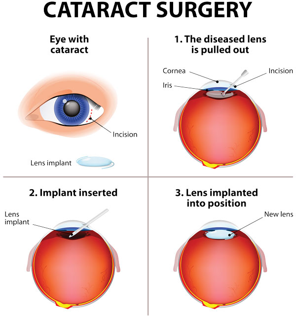
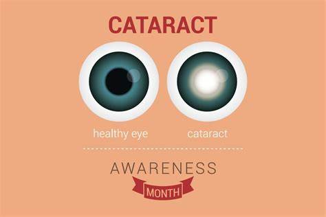
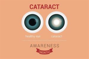
A cataract is a cloudy area in the lens of your eye. Cataracts are very common as you get older. In fact, more than half of all Americans age 80 or older either have cataracts or have had surgery to get rid of cataracts.

At first, you may not notice that you have a cataract. But over time, cataracts can make your vision blurry, hazy, or less colorful. You may have trouble reading or doing other everyday activities.

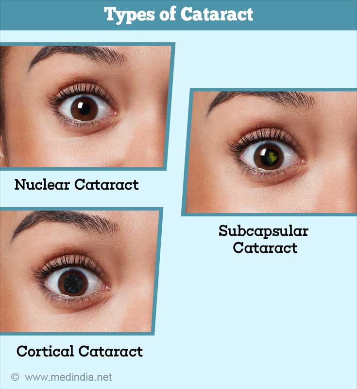
There are 3 types of Cataracts:

1-Nuclear cataracts, which form in the lens’ nucleus, are the most common type of cataracts. Because opacity develops in the center of the lens, known as the central nucleus, nuclear cataracts interfere with a person’s ability to see objects in the distance. Usually the result of advancing age, nuclear cataracts can take years to develop and often give the nucleus a yellow tint.

2=Cortical cataracts begin at the outer rim of the lens and gradually work toward the central core. Thus, this type of cataract resembles spokes of a wheel. Patients with cortical cataracts often notice problems with glare, or a “halo” effect around lights. They may also experience a disruption of both near and distance vision.

3-Subcapsular cataracts progress the most rapidly. While nuclear cataracts take years to develop, subcapsular cataracts reach an advanced stage within a matter of months. Posterior subcapsular cataracts affect the back of the lens, causing glare and blurriness. This type of cataract is usually seen in patients who suffer from diabetes, extreme nearsightedness or retinitus pigmentosa, as well as among those who take steroid medication.

Obesity is most commonly linked to the development of posterior subcapsular cataracts. According to researchers at Harvard University, individuals with a BMI of 33 had at least a 30 percent greater likelihood of developing cataracts, compared to subjects with a BMI of 23 or below.

**Congenital cataracts exist and refer to cataracts that are present from birth, as well as to those that develop in early childhood. These cataracts can be nuclear, cortical, or subcapsular. Congenital cataracts may be linked to an infection contracted by the mother during pregnancy or to a genetic condition such as Fabry disease, Alport syndrome, or galactosemia. Because clear vision is essential to the development of the child’s eyes and brain, it is important to diagnose congenital cataracts as early as possible.

Most cataracts form as a result of advancing age. Other possible causes of cataracts include environmental factors and certain medications, such as antidepressants. If your medical history or lifestyle increases your risk of developing cataracts, it is important to have your eye health monitored regularly by a qualified ophthalmologist.

Over 50 percent of Americans over the age of 80 have cataracts

RISK FACTORS:

1 Age

Age is the main reason cataracts form. According to the American Academy of Ophthalmology, the eye disease appears in over 22 million Americans over the age of 40. Over 50 percent of Americans over the age of 80 have cataracts. In fact, if we live long enough, nearly all of us will eventually develop this condition. Because the lens of the eye cannot shed old cells naturally, protein that accumulates on the lens gradually builds up over time, progressively obscuring vision by preventing light from reaching the retina.

2 History of Cataracts in the family

3 Ultraviolet Radiation (UVA or UVB)

According to the U.S. Environmental Protection Agency, long-term exposure to ultraviolet radiation, especially UVB rays, can cause changes in pigment that lead to the formation of cataracts. This is especially common in tropical climates, where high concentrations of UV exposure occur year-round. To protect the eyes from sun damage, it is important to wear sunglasses with a high level of UVA/UVB protection.

4 Trauma to the eye

Injury or trauma to the eye increases a patient’s risk of developing cataracts. Individuals who have experienced inflammation in the eye, either post-operatively or as the result of another eye disease, are also more likely to eventually develop cataracts. For instance, iritis is an ocular condition that causes chronic inflammation inside the eye, and is commonly linked to early and rapid cataract formation.

5 Have certain health problems, like diabetes

6 Smoking and Alcohol

Lifestyle habits such as smoking or consuming alcohol are often considered causes of cataracts.

7 Medications

Certain medications are well-known causes of cataracts, and some drugs can also accelerate their development. Steroid medications – whether pills, injections, or eye drops – are most frequently associated with cataract formation. If you are taking steroid medications to manage a long-term condition, it is important to note any visual changes and to have your ocular health managed by a qualified ophthalmologist.

Most cataracts are caused by normal changes in your eyes as you get older.

When you’re young, the lens in your eye is clear. Around age 40, the proteins in t he lens of your eye start to break down and clump together. This clump makes a cloudy area on your lens — or a cataract. Over time, the cataract gets more severe and clouds more of the lens.


QUOTE FOR FRIDAY:

“Actinic Keratosis a rough, scaly patches of skin that are considered precancerous and are due to sun exposure. Prevention is to cut sun exposure and wear sunscreen. Treatments include performing cryosurgery (freezing with liquid nitrogen), cutting the keratoses away, burning them, putting 5-fluorouracil on them, and using photodynamic therapy (injecting into the bloodstream a chemical that collects in actinic keratoses and makes them more sensitive to destruction by a specialized form of light).

Keratoacanthoma this is a harmless, hard nodule that appears on the skin, most commonly on the face or arm of elderly people. The nodule may grow to up to 2 centimeters in diameter over about 8 weeks before gradually disappearing. However, the unsightly nodule is often surgically removed. The cause of keratoacanthoma is unknown, although exposure to sunlight appears to be a factor.”

Medicine Net (https://www.medicinenet.com/image-collection/actinic_keratosis_solar_keratosis_picture/picture.htm)

Part III Skin Cancer Awareness – Akinetic Keratosis & Keratoacanthoma including risk factors and treatments.

Continuation of Types of skin cancer:


5-Akinetic Keratosis:

Actinic keratosis (AK) is a skin disorder that causes rough, scaly patches of skin. Another name for AK is solar keratosis. AK is a type of precancer, which means that if you don’t treat the condition, it could turn into cancer. Without treatment, AK can lead to a type of skin cancer called squamous cell carcinoma.

A condition which causes scaly patches on the skin from exposure to the sun over the years. It is commonly found on face, lips, ears, neck, back of the hand and forearms.  Very common (More than 3 million cases per year in US)
Rarely requires lab test or imaging.  Treatable by a medical professional.  Can last several months or years.
Knowing the causes, risk factors and warning signs can help you detect them early, when they are easiest to treat and cure.

The risk factors of Akinetic Keratosis are:

UV exposure from the sun or indoor tanning.

-History of skin cancer in particular history of actinic keratosis.

– Age over 40.

-Fair skin: People with fair skin including lighter color hair or eyes have an increased risk.

Warning Signs can help with early detection and treatment this can be successfully removed without complications. Look out for any new, changing or unusual skin growths, so you can spot skin cancers like BCC when they are easiest to treat and cure.

Treatments Akinetic Keratosis:

An actinic keratosis sometimes disappears on its own but might return after more sun exposure. It’s hard to tell which actinic keratoses will develop into skin cancer, so they’re usually removed as a precaution.

Medicines-

If you have several actinic keratoses, your health care provider might prescribe a medicated cream or gel to remove them, such as fluorouracil (Carac, Efudex others), imiquimod (Aldara, Zyclara) or diclofenac. These products might cause inflamed skin, scaling or a burning sensation for a few weeks.

Surgical and other procedures-

Many methods are used to remove actinic keratosis, including:

  • Freezing (cryotherapy). Actinic keratoses can be removed by freezing them with liquid nitrogen. Your health care provider applies the substance to the affected skin, which causes blistering or peeling. As your skin heals, the damaged cells slough off, allowing new skin to appear. Cryotherapy is the most common treatment. It takes only a few minutes and can be done in your health care provider’s office. Side effects may include blisters, scarring, changes to skin texture, infection and changes in skin color of the affected area.
  • Scraping (curettage). In this procedure, your health care provider uses a device called a curet to scrape off damaged cells. Scraping may be followed by electrosurgery, in which a pencil-shaped instrument is used to cut and destroy the affected tissue with an electric current. This procedure requires local anesthesia. Side effects may include infection, scarring and changes in skin color of the affected area.
  • Laser therapy. This technique is increasingly used to treat actinic keratosis. Your health care provider uses an ablative laser device to destroy the patch, allowing new skin to appear. Side effects may include scarring and discoloration of the affected skin.
  • Photodynamic therapy. Your health care provider might apply a light-sensitive chemical solution to the affected skin and then expose it to a special light that will destroy the actinic keratosis. Side effects may include inflamed skin, swelling and a burning sensation during therapy.


6-Keratocanthoma (KA)

The term “Keratoacanthoma” (KA) was coined by Freudenthal in the year 1936. It was first described way back in 1889 by Hutchinson and was called molluscum sebaceum and self-limiting epithelioma. KA is benign, self-limiting squamo-proliferative lesion.

It shows male preponderance and most commonly arises on the sun-exposed parts predominantly face, neck forearms, hands and legs. Cutaneous lesions arise from hair follicles whereas mucosal lesions originate from ectopic sebaceous glands. This is a slow growing cancer of the skin that looks like a dome or crater.  This is common; more than 200,000 cases per year in US.  Regarding treatment from medical professional is advised.  This condition often requires lab test or imaging.  Keratoacanthoma last several months.  It is common for ages 60 and older and is more common in males.

KA is benign despite its similarities to squamous cell carcinoma (SCC), or the abnormal growth of cancerous cells on the skin’s most outer layer.

The risk factors of Keratocanthoma (KA):

UV exposure from the sun or indoor tanning.

-contact with chemical carcinogens, or cancer-causing chemicals

 -trauma 

-Infection with some strains of a wart virus, such as papillomavirus

-History of skin cancer in particular history of Keratoacanthoma.

Age over 60.

-People with fair skin.

Warning Signs can help with early detection and treatment, this can be successfully removed without complications if caught early. Look out for any new, changing or unusual skin growths, so you can spot skin cancers like BCC when they are easiest to treat and cure.

Treatments:

If your medical professional suspects a keratoacanthoma, they will first want to establish the correct diagnosis by performing a biopsy.  Than treatments could include the following:

  • Removal (excision), in which a scalpel is used to cut away the keratoacanthoma and then place stitches to bring the wound edges together.
  • Mohs micrographic surgery, in which tiny slivers of skin are removed until there are no more cancer cells. This technique is particularly useful for keratoacanthomas located on the nose, ears, lips, and hands.
  • Electrodesiccation and curettage, also known as “scrape and burn.” After numbing the lesion, the medical professional uses a sharp instrument called a curette to scrape away the skin cancer cells, followed by an electric needle to burn (cauterize) the tissue. The electrodesiccation helps kill the cancer cells and stop bleeding at the site.
  • Radiation treatment, where x-ray therapy is often useful for patients who may have difficulty with a surgical procedure due to other health issues.

Very rarely, keratoacanthomas are treated with medicine injected directly into the skin lesion (intralesional chemotherapy). In patients with more than one keratoacanthoma, the medical professional may suggest taking oral medication (ie, isotretinoin) to reduce their size and number.

Once the skin cancer has been removed, frequent follow-up appointments with a dermatologist or medical professional trained to examine the skin are essential to ensure that the keratoacanthoma has not returned and that no new skin cancer has developed elsewhere on your body. In addition, good sun protection habits (as noted in the Self-Care section) are vital to preventing further damage from UV light.


QUOTE FOR THURSDAY:

“Most of the cells on the skin’s surface are flat, scale-like squamous cells. Squamous cell carcinomas are the second most common type of skin cancer, accounting for about 20 percent of all diagnoses. These cancers also are slowing growing, but squamous cell carcinomas can spread to lymph nodes and even internal organs. Research indicates that the metastasis rate of squamous cell carcinoma of the skin is less than 10 percent within two years of diagnosis.

More than 150 years ago, a German scientist named Friedrich Merkel documented what he identified as tastzellen, German for “touch cells.” They would later be named Merkel cells. These cells are found below the epidermis, where they interact with nerve cells and help the skin feel light touch, textures and fine details.

Merkel cell carcinomas may appear as bumps or nodules—often red in color—on sun-exposed skin. This is a rare cancer, but it is often aggressive and may metastasize.  About 2,000 to 3,000 cases of Merkel cell carcinoma are diagnosed in the United States every year, according to the American Cancer Society, and the numbers are on the rise. The disease is rarely diagnosed in patients under 50 years old and is mostly found in white men older than 70.”

Cancer Center/ City of Hope (https://www.cancercenter.com/community/blog/2023/10/skin-cells-merkel-basal-squamous#Q2)


Part II Skin Cancer Awareness – Squamous Cell Carcinoma & Merkel Cell Carcinoma including risk factors and treatment.

Continuation of Types of Skin Cancer:


3-Squamous cell carcinoma – SCC

Squamous cell carcinoma is the second most common form of skin cancer in the United States. It accounts for about 15 percent of all skin cancers.  It is caused due to over production of skin cells. Squamous cell carcinoma of the skin is caused by DNA damage that leads to abnormal changes (mutations) in the squamous cells in the outermost layer of skin. This cancer is common (More than 200,000 cases per year in US).  The majority of squamous cell skin cancers are easily and successfully treated with current therapies.

Knowing the causes, risk factors and warning signs can help you detect them early, when they are easiest to treat and cure.

The risk factors of SCC:

UV exposure from the sun or indoor tanning.

-History of skin cancer, including squamous cell carcinoma (SCC) or melanoma

– Age over 50: Most BCCs appear in people over age 50.

-Fair skin: People with fair skin have an increased risk.

Warning Signs can help with early detection and treatment this can be successfully removed without complications. Look out for any new, changing or unusual skin growths, so you can spot skin cancers like BCC when they are easiest to treat and cure.

IT’S A FACT, Squamous cell carcinoma is considered more aggressive than basal cell carcinoma.  If squamous cell carcinoma does spread to internal organs it can be life threatening. The quicker the treatment when SCC is in the one layer of skin only the better the results.  The primary symptom to look out for with SCC is a growing bump or lesion on the skin which has a rough scaly surface or flat red patches.

Treatments for SCC:

If the skin cancer is small, not deep into the skin, called superficial, and has a low risk of spreading, less-invasive treatment choices include:

  • Curettage and electrodessication. This treatment involves removing the top of the skin cancer with a scraping tool called a curet. Then an electric needle is used to sear the base of the cancer.
  • Laser therapy. This treatment uses an intense beam of light to destroy growths. There’s usually little damage to nearby tissue. And there’s a reduced risk of bleeding, swelling and scarring.
  • Freezing. This treatment, called cryosurgery, involves freezing cancer cells with liquid nitrogen. Freezing might be done after using a scraping tool, called a curet, to remove the surface of the skin cancer.
  • Photodynamic therapy. During photodynamic therapy, a liquid medicine that makes the cancer cells sensitive to light is applied to the skin. Later, a light that destroys the skin cancer cells is shined on the area. This treatment might be used with surgery or other treatments.

Treatments for larger skin cancers:

More-invasive treatments might be recommended for larger squamous cell carcinomas and those that go deeper into the skin. Options might include:

  • Simple excision. This involves cutting out the cancer and a margin of healthy skin around it. Sometimes more skin around the tumor is removed, called a wide excision.
  • Mohs surgery. Mohs surgery involves removing the cancer layer by layer and looking at each layer under the microscope until no cancer cells are left. This allows the surgeon to remove the whole growth without taking too much of the healthy skin around it.
  • Radiation therapy. Radiation therapy uses powerful energy beams to kill cancer cells. Radiation therapy is sometimes used after surgery when there is an increased risk that the cancer might return. It also might be an option for people who can’t have or don’t want surgery.

Treatments for skin cancer that spreads past the skin:

When squamous cell carcinoma spreads to other parts of the body, medicines might be recommended, including:

  • Chemotherapy. Chemotherapy uses strong medicines to kill cancer cells. If squamous cell carcinoma spreads to the lymph nodes or other parts of the body, chemotherapy can be used alone or with other treatments, such as targeted therapy and radiation therapy.
  • Targeted therapy. Targeted therapy uses medicines that attack specific chemicals in the cancer cells. By blocking these chemicals, targeted treatments can cause cancer cells to die. Targeted therapy is usually used with chemotherapy.
  • Immunotherapy. Immunotherapy is a treatment with medicine that helps the body’s immune system kill cancer cells. The immune system fights off diseases by attacking germs and other cells that shouldn’t be in the body. Cancer cells survive by hiding from the immune system. Immunotherapy helps the immune system cells find and kill the cancer cells.For squamous cell carcinoma of the skin, immunotherapy might be considered when the cancer is advanced and other treatments aren’t an option.


4-Merkel Cell Carcinoma

This is a type of skin cancer characterized by flesh-colored nodule that occurs on the face, head or neck. It begins in the cells at the base of the uppermost layer of the skin (epidermis).  A normal Merkel cell is a cross between a nerve cell and an endocrine (or hormone-producing) cell located on or just below the skin in the underlying tissue, and functions predominantly as a touch receptor. Merkel cell carcinoma occurs when these cells begin to grow uncontrollably.

Merkel cell tumors typically arise on, but are not limited to, sun-exposed parts of the body such as the face and neck. Their shape and color are less distinctive than other skin cancers, and they can often appear as an innocent pink pearly nodule. As a result, it is usually only the speed with which they grow that attracts the attention of patients and their doctors.

With early detection and treatment, Merkel cell carcinoma can be well contained and even cured. Treatment becomes more difficult as the tumor grows and spreads, but aggressive therapy can still lead to high rates of survival.

Again, Warning Signs can help with early detection and treatment this can be successfully removed without complications. Look out for any new, changing or unusual skin growths, get yourself to the doctor immediately so you can spot skin cancers like BCC when they are easiest to treat and cure.

Risk Factors of Merkel Cell Carcinoma:

UV exposure from the sun or indoor tanning.

-History of skin cancer, including squamous cell carcinoma (SCC) or melanoma

– Age over 50: Most BCCs appear in people over age 50.

-Fair skin: People with fair skin have an increased risk.

-Male gender: Men are more likely to develop Basal Cell Carcinoma.

-Chronic infections and skin inflammation from burns, scars and other conditions-weakens the immune system.

-Merkel Cell Virus.  Recently, researchers have linked a virus to many cases of Merkel cell carcinoma. However, it remains to be determined if the Merkel cell polyomarvirus causes the disease, and if it might help guide future treatment. If so, the virus could offer promising new targets for immunotherapy.

IT’S A FACT, Memorial Sloan Kettering Hospital in NYC states  “Merkel cell carcinoma, also called neuroendocrine cancer of the skin, is an aggressive type of skin cancer that affects only about 400 people in the United States each year. But like other skin cancers, that number is growing.”.

Treatments:

Treatment of stage I and stage II Merkel cell carcinoma:

  • Surgery to remove the tumor, such as wide local excision with or without lymph node dissection.
  • Radiation therapy after surgery.

Treatment of stage III:

  • Wide local excision with or without lymph node dissection.
  • Radiation therapy.
  • Immunotherapy (immune checkpoint inhibitor therapy using pembrolizumab), for tumors that cannot be removed by surgery.
  • A clinical trial of chemotherapy.
  • A clinical trial of immunotherapy (nivolumab).

Treatment of Stage IV Merkel Cell Carcinoma

  • Immunotherapy (immune checkpoint inhibitor therapy using avelumab or pembrolizumab).
  • Chemotherapy, surgery or radiation therapy as palliative treatment to relieve symptoms and improve quality of life.
  • A clinical trial of immunotherapy (nivolumab and ipilimumab).