Part II Bladder Cancer – the prevention, how its diagnosed, the staging. treatments and after treatments!


Prevention of bladder cancer:

Although there’s no guaranteed way to prevent bladder cancer, you can take steps to help reduce your risk. For instance:

  • Don’t smoke. If you don’t smoke, don’t start. If you smoke, talk to your doctor about a plan to help you stop. Support groups, medications and other methods may help you quit.
  • Take caution around chemicals. If you work with chemicals, follow all safety instructions to avoid exposure.
  • Choose a variety of fruits and vegetables. Choose a diet rich in a variety of colorful fruits and vegetables. The antioxidants in fruits and vegetables may help reduce your risk of cancer.

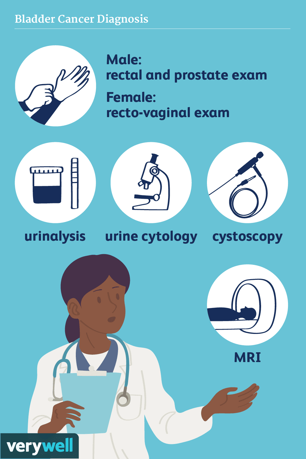
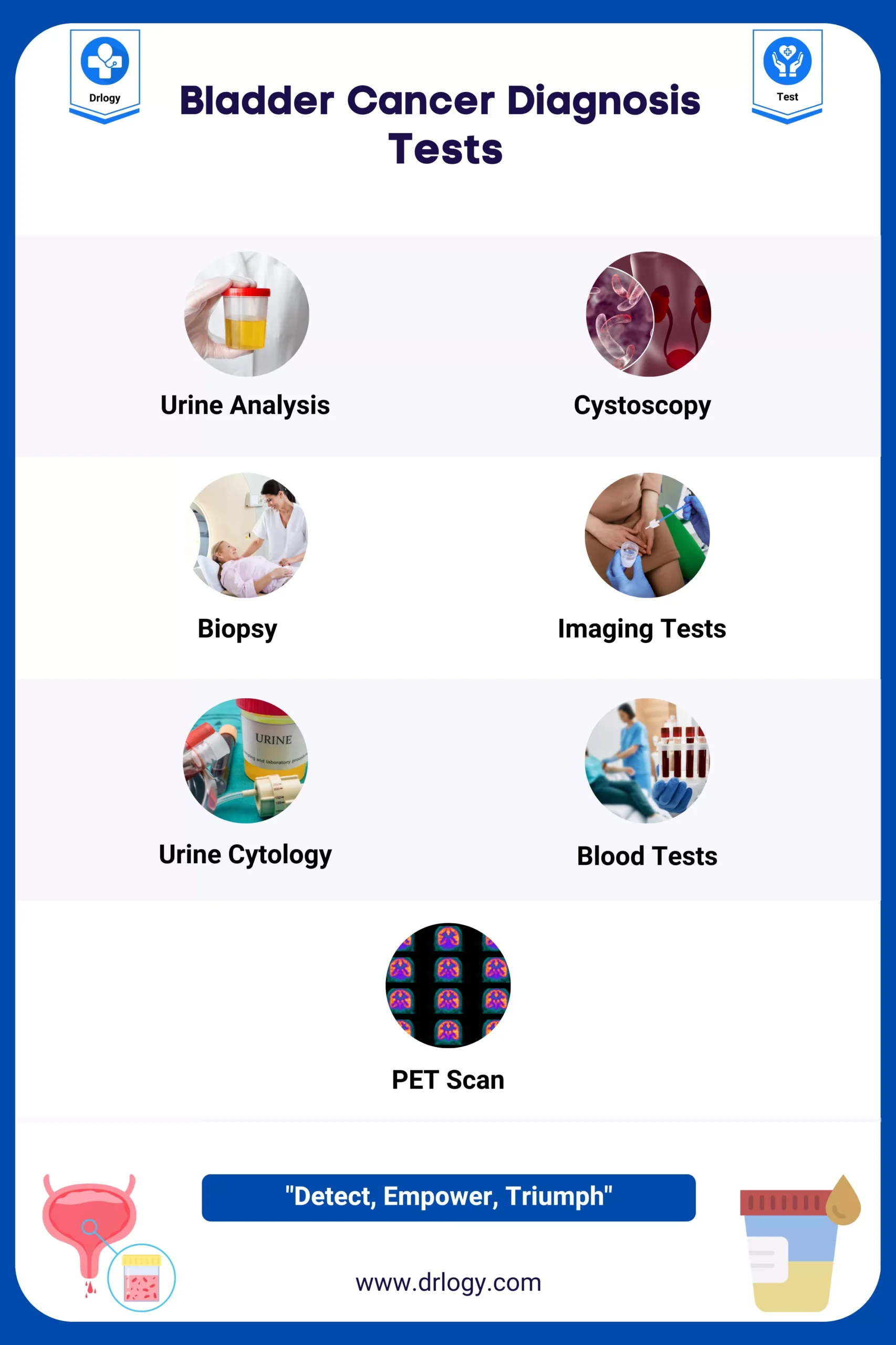
How bladder cancer is diagnosed could include the following:

  • Using a scope to examine the inside of your bladder (cystoscopy). To perform cystoscopy, your doctor inserts a small, narrow tube (cystoscope) through your urethra. The cystoscope has a lens that allows your doctor to see the inside of your urethra and bladder, to examine these structures for signs of disease. Cystoscopy can be done in a doctor’s office or in the hospital.
  • Removing a sample of tissue for testing (biopsy). During cystoscopy, your doctor may pass a special tool through the scope and into your bladder to collect a cell sample (biopsy) for testing. This procedure is sometimes called transurethral resection of bladder tumor (TURBT). TURBT can also be used to treat bladder cancer.
  • Examining a urine sample (urine cytology). A sample of your urine is analyzed under a microscope to check for cancer cells in a procedure called urine cytology.
  • Imaging tests. Imaging tests, such as computerized tomography (CT) urogram or retrograde pyelogram, allow your doctor to examine the structures of your urinary tract.During a CT urogram, a contrast dye injected into a vein in your hand eventually flows into your kidneys, ureters and bladder. X-ray images taken during the test provide a detailed view of your urinary tract and help your doctor identify any areas that might be cancer.

    Retrograde pyelogram is an X-ray exam used to get a detailed look at the upper urinary tract. During this test, your doctor threads a thin tube (catheter) through your urethra and into your bladder to inject contrast dye into your ureters. The dye then flows into your kidneys while X-ray images are captured.

Determining the extent of the cancer

After confirming that you have bladder cancer, your doctor may recommend additional tests to determine whether your cancer has spread to your lymph nodes or to other areas of your body.

Tests may include:

  • CT scan
  • Magnetic resonance imaging (MRI)
  • Positron emission tomography (PET)
  • Bone scan
  • Chest X-ray

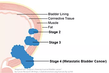
Staging of Bladder Cancer:

Your doctor uses these diagnostic tests listed above for information from these procedures to assign your cancer a stage.

The stages of bladder cancer are indicated by Roman numerals ranging from 0 to IV. The lowest stages indicate a cancer that’s confined to the inner layers of the bladder and that hasn’t grown to affect the muscular bladder wall. The highest stage — stage IV — indicates cancer that has spread to lymph nodes or organs in distant areas of the body, like a lot of other cancers are staged I through IV.

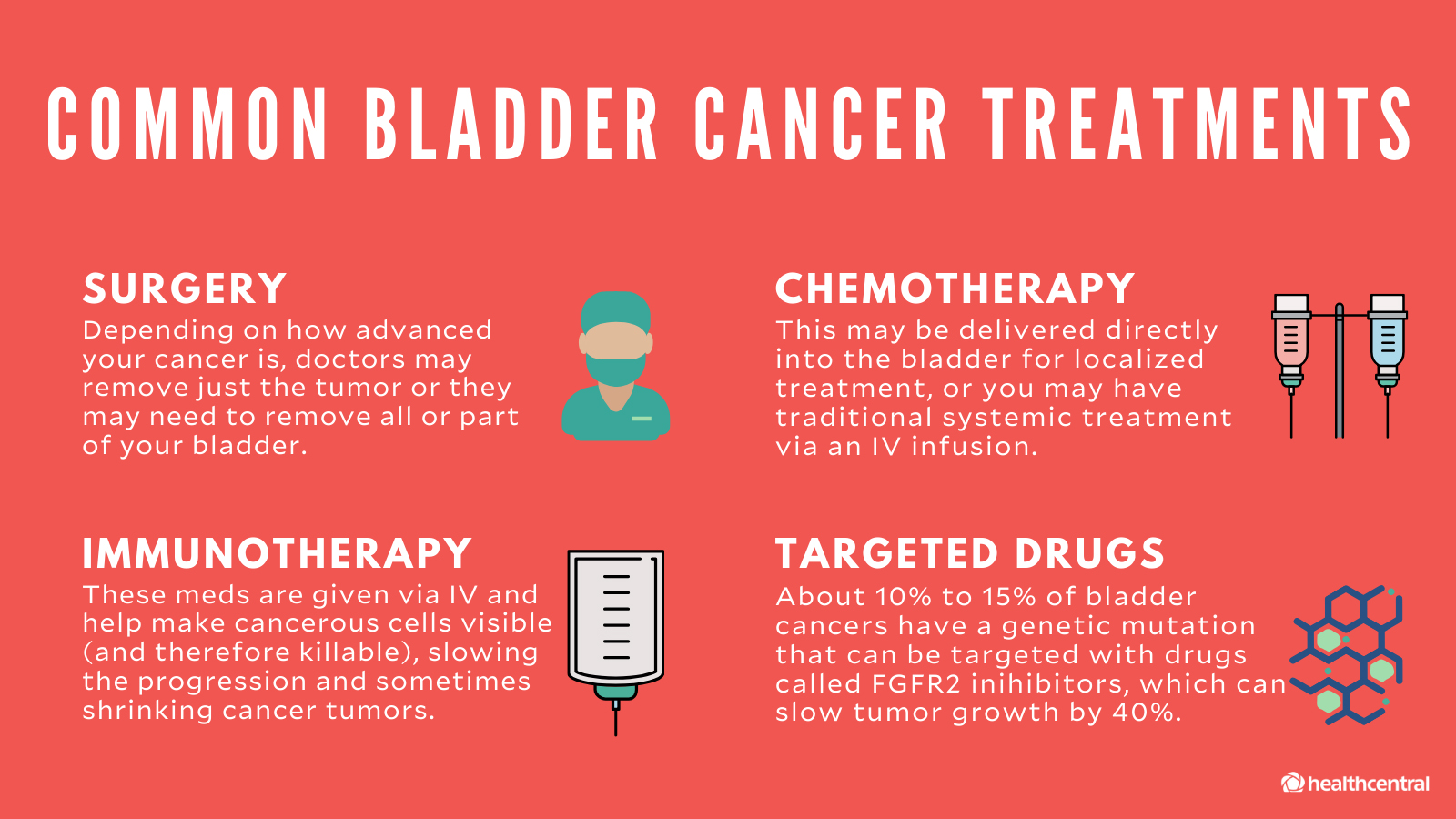
Treatments of bladder cancer:

If cancer invades the muscles of the bladder, doctors will usually treat it with chemotherapy to shrink the tumor, followed by surgery to remove the bladder. However, a recent clinical trial found that adding immunotherapy to chemotherapy may allow certain patients to avoid surgery.

Bladder cancer treatment may include: Surgery, to remove the cancer cells. Chemotherapy in the bladder (intravesical chemotherapy), to treat cancers that are confined to the lining of the bladder but have a high risk of recurrence or progression to a higher stage.

Approaches to bladder cancer surgery might be used could include:

  • Transurethral resection of bladder tumor (TURBT). TURBT is a procedure to diagnose bladder cancer and to remove cancers confined to the inner layers of the bladder — those that aren’t yet muscle-invasive cancers. During the procedure, a surgeon passes an electric wire loop through a cystoscope and into the bladder. The electric current in the wire is used to cut away or burn away the cancer. Alternatively, a high-energy laser may be used.Because doctors perform the procedure through the urethra, you won’t have any cuts (incisions) in your abdomen.

    As part of the TURBT procedure, your doctor may recommend a one-time injection of cancer-killing medication (chemotherapy) into your bladder to destroy any remaining cancer cells and to prevent cancer from coming back. The medication remains in your bladder for a period of time and then is drained.

  • Cystectomy. Cystectomy is surgery to remove all or part of the bladder. During a partial cystectomy, your surgeon removes only the portion of the bladder that contains a single cancerous tumor.A radical cystectomy is an operation to remove the entire bladder and the surrounding lymph nodes. In men, radical cystectomy typically includes removal of the prostate and seminal vesicles. In women, radical cystectomy may involve removal of the uterus, ovaries and part of the vagina.

    Radical cystectomy can be performed through an incision on the lower portion of the belly or with multiple small incisions using robotic surgery. During robotic surgery, the surgeon sits at a nearby console and uses hand controls to precisely move robotic surgical instruments.

  • Neobladder reconstruction. After a radical cystectomy, your surgeon must create a new way for urine to leave your body (urinary diversion). One option for urinary diversion is neobladder reconstruction. Your surgeon creates a sphere-shaped reservoir out of a piece of your intestine. This reservoir, often called a neobladder, sits inside your body and is attached to your urethra. The neobladder allows most people to urinate normally. A small number of people difficulty emptying the neobladder and may need to use a catheter periodically to drain all the urine from the neobladder.
  • Ileal conduit. For this type of urinary diversion, your surgeon creates a tube (ileal conduit) using a piece of your intestine. The tube runs from your ureters, which drain your kidneys, to the outside of your body, where urine empties into a pouch (urostomy bag) you wear on your abdomen.
  • Continent urinary reservoir. During this type of urinary diversion procedure, your surgeon uses a section of intestine to create a small pouch (reservoir) to hold urine, located inside your body. You drain urine from the reservoir through an opening in your abdomen using a catheter a few times each day.

Chemotherapy drugs can be given:

  • 1-Through a vein (intravenously). Intravenous chemotherapy is frequently used before bladder removal surgery to increase the chances of curing the cancer. Chemotherapy is going in your system generally through the blood stream and chemo may also be used to kill cancer cells that might remain after surgery. In certain situations, chemotherapy may be combined with radiation therapy.
  • 2-Directly into the bladder (intravesical therapy). During intravesical chemotherapy, a tube is passed through your urethra directly to your bladder. The chemotherapy is placed in the bladder for a set period of time before being drained. It can be used as the primary treatment for superficial bladder cancer, where the cancer cells affect only the lining of the bladder and not the deeper muscle tissue.

Radiation therapy:

Radiation therapy uses beams of powerful energy, such as X-rays and protons, to destroy the cancer cells. Radiation therapy for bladder cancer usually is delivered from a machine that moves around your body, directing the energy beams to precise points.

Radiation therapy is sometimes combined with chemotherapy to treat bladder cancer in certain situations, such as when surgery isn’t an option or isn’t desired at that time or ever depending on your stage of cancer.

Immunotherapy:

Immunotherapy is a drug treatment that helps your immune system to fight cancer.

Immunotherapy can be given:

  • Directly into the bladder (intravesical therapy). Intravesical immunotherapy might be recommended after TURBT for small bladder cancers that haven’t grown into the deeper muscle layers of the bladder. This treatment uses bacillus Calmette-Guerin (BCG), which was developed as a vaccine used to protect against tuberculosis. BCG causes an immune system reaction that directs germ-fighting cells to the bladder.
  • Through a vein (intravenously). Immunotherapy can be given intravenously for bladder cancer that’s advanced or that comes back after initial treatment. Several immunotherapy drugs are available. These drugs help your immune system identify and fight the cancer cells.

Targeted therapy:

Targeted therapy drugs focus on specific weaknesses present within cancer cells. By targeting these weaknesses, targeted drug treatments can cause cancer cells to die. Your cancer cells may be tested to see if targeted therapy is likely to be effective.

Targeted therapy may be an option for treating advanced bladder cancer when other treatments haven’t helped.

Bladder preservation:

In certain situations, people with muscle-invasive bladder cancer who don’t want to undergo surgery to remove the bladder may consider trying a combination of treatments instead. Known as trimodality therapy, this approach combines TURBT, chemotherapy and radiation therapy.

First, your surgeon performs a TURBT procedure to remove as much of the cancer as possible from your bladder while preserving bladder function. After TURBT, you undergo a regimen of chemotherapy along with radiation therapy.

If, after trying trimodality therapy, not all of the cancer is gone or you have a recurrence of muscle-invasive cancer, your doctor may recommend a radical cystectomy.

After bladder cancer treatment:

Bladder cancer may recur, even after successful treatment. Because of this, people with bladder cancer need follow-up testing for years after successful treatment. What tests you’ll have and how often depends on your type of bladder cancer and how it was treated, among other factors.

In general, doctors recommend a test to examine the inside of your urethra and bladder (cystoscopy) every three to six months for the first few years after bladder cancer treatment. After a few years of surveillance without detecting cancer recurrence, you may need a cystoscopy exam only once a year. Your doctor may recommend other tests at regular intervals as well.

People with aggressive cancers may undergo more-frequent testing. Those with less aggressive cancers may undergo testing less often.


QUOTE FOR THURSDAY:

“Bladder cancer makes up about 4% of cancers in the US. It is the fourth most common cancer in men, but it’s less common in women.

The American Cancer Society’s estimates for bladder cancer in the United States for 2024 are:

  • About 83,190 new cases of bladder cancer (about 63,070 in men and 20,120 in women)
  • About 16,840 deaths from bladder cancer (about 12,290 in men and 4,550 in women)

The rates of both new bladder cancers and deaths from bladder cancer have been dropping in recent years.

Bladder cancer occurs mainly in older people. About 9 out of 10 people with this cancer are over the age of 55. The average age of people when they are diagnosed with bladder cancer is 73.

Overall, the chance men will develop this cancer during their lifetime is about 1 in 28. For women, the chance is about 1 in 89. However, each person’s chances of getting bladder cancer might be higher or lower than this, depending on their risk factors.”

American Cancer Society (https://www.cancer.org/cancer/types/bladder-cancer/about/key-statistics.html)


Part I Bladder Cancer-including signs/symptoms, how common is this cancer, types of bladder cancer, the survival rate, and risk factors!

Bladder cancer is a common type of cancer that begins in the cells of the bladder. The bladder is a hollow muscular organ in your lower abdomen that stores urine.

Bladder cancer most often begins in the cells (urothelial cells) that line the inside of your bladder. Urothelial cells are also found in your kidneys and the tubes (ureters) that connect the kidneys to the bladder. Urothelial cancer can happen in the kidneys and ureters, too, but it’s much more common in the bladder.

Most bladder cancers are diagnosed at an early stage, when the cancer is highly treatable. But even early-stage bladder cancers can come back after successful treatment like many other cancers. For this reason, people with bladder cancer typically need follow-up tests for years after treatment to look for bladder cancer that recurs.

Signs and symptoms of Bladder Cancer may include the following:

  • Blood in urine (hematuria), which may cause urine to appear bright red or cola colored, though sometimes the urine appears normal and blood is detected on a lab test
  • Frequent urination
  • Painful urination
  • Back pain

Bladder cancer begins when cells in the bladder develop changes (mutations) in their DNA. A cell’s DNA contains instructions that tell the cell what to do. The changes tell the cell to multiply rapidly and to go on living when healthy cells would die. The abnormal cells form a tumor that can invade and destroy normal body tissue. In time, the abnormal cells can break away and spread (metastasize) through the body.

How common is Bladder cancer:

Urinary Bladder Cancer is the sixth most common type of cancer in the United States. In 2024, approximately 83,190 people will receive a diagnosis of bladder cancer, and some 16,840 people are expected to die from it, according to estimates by the Surveillance, Epidemiology, and End Results Program (SEER) of the National Cancer Institute (NCI).

Bladder cancer is most likely to affect white men. In fact, the incidence rate of bladder cancer is four times higher among men than among women. It is also twice as high in white males as among Black, Hispanic or Asian/Pacific Islander men.

The NCI explains that there are three major types of bladder cancer. The name of each type of cancer indicates the kind of cells that become malignant.

  • Transitional cell carcinoma begins in cells in the innermost tissue layer of the bladder.
  • Squamous cell carcinoma begins in the squamous cells. This type may form after long-term infection or irritation.
  • Adenocarcinoma begins in glandular (secretory) cells in the lining of the bladder.

Cancer in the lining of the bladder is called superficial bladder cancer. Cancer that has spread through the lining of the bladder and has invaded the muscle wall of the organ, or has spread to nearby organs and lymph nodes, is called invasive bladder cancer.

Survival Rate:

Approximately 78 percent of people diagnosed with bladder cancer survived five years or more after diagnosis between 2012 and 2019, according to federal estimates.

Types of bladder cancer:

Different types of cells in your bladder can become cancerous. The type of bladder cell where cancer begins determines the type of bladder cancer. Doctors use this information to determine which treatments may work best for you.

Types of bladder cancer include:

  • Urothelial carcinoma. Urothelial carcinoma, previously called transitional cell carcinoma, occurs in the cells that line the inside of the bladder. Urothelial cells expand when your bladder is full and contract when your bladder is empty. These same cells line the inside of the ureters and the urethra, and cancers can form in those places as well. Urothelial carcinoma is the most common type of bladder cancer in the United States.
  • Squamous cell carcinoma. Squamous cell carcinoma is associated with chronic irritation of the bladder — for instance, from an infection or from long-term use of a urinary catheter. Squamous cell bladder cancer is rare in the United States. It’s more common in parts of the world where a certain parasitic infection (schistosomiasis) is a common cause of bladder infections.
  • Adenocarcinoma. Adenocarcinoma begins in cells that make up mucus-secreting glands in the bladder. Adenocarcinoma of the bladder is very rare.

Some bladder cancers include more than one type of cell.

Risk factors:

Factors that may increase bladder cancer risk include:

  • Smoking. Smoking cigarettes, cigars or pipes may increase the risk of bladder cancer by causing harmful chemicals to accumulate in the urine. When you smoke, your body processes the chemicals in the smoke and excretes some of them in your urine. These harmful chemicals may damage the lining of your bladder, which can increase your risk of cancer.
  • Increasing age. Bladder cancer risk increases as you age. Though it can occur at any age, most people diagnosed with bladder cancer are older than 55.
  • Being male. Men are more likely to develop bladder cancer than women are.
  • Exposure to certain chemicals. Your kidneys play a key role in filtering harmful chemicals from your bloodstream and moving them into your bladder. Because of this, it’s thought that being around certain chemicals may increase the risk of bladder cancer. Chemicals linked to bladder cancer risk include arsenic and chemicals used in the manufacture of dyes, rubber, leather, textiles and paint products.
  • Previous cancer treatment. Treatment with the anti-cancer drug cyclophosphamide increases the risk of bladder cancer. People who received radiation treatments aimed at the pelvis for a previous cancer have a higher risk of developing bladder cancer.
  • Chronic bladder inflammation. Chronic or repeated urinary infections or inflammations (cystitis), such as might happen with long-term use of a urinary catheter, may increase the risk of a squamous cell bladder cancer. In some areas of the world, squamous cell carcinoma is linked to chronic bladder inflammation caused by the parasitic infection known as schistosomiasis.
  • Personal or family history of cancer. If you’ve had bladder cancer, you’re more likely to get it again. If one of your blood relatives — a parent, sibling or child — has a history of bladder cancer, you may have an increased risk of the disease, although it’s rare for bladder cancer to run in families. A family history of Lynch syndrome, also known as hereditary nonpolyposis colorectal cancer (HNPCC), can increase the risk of cancer in the urinary system, as well as in the colon, uterus, ovaries and other organs.

QUOTE FOR WEDNESDAY:

“Arthritis includes approximately 100 conditions that affect the joints and surrounding tissues. It is a leading cause of activity limitations, disability, and chronic pain, and is associated with dispensed opioid prescriptions, substantially contributing to health care costs.

Combined 2019–2021 National Health Interview Survey data were analyzed to update national prevalence estimates of self-reported diagnosed arthritis. An estimated 21.2% (18.7% age-standardized) of U.S. adults aged ≥18 years (53.2 million) had diagnosed arthritis during this time frame. Age-standardized arthritis prevalences were higher among women (20.9%) than men (16.3%), among veterans (24.2%) than nonveterans (18.5%), and among non-Hispanic White (20.1%) than among Hispanic or Latino (14.7%) or non-Hispanic Asian adults (10.3%). Adults aged ≥45 years represent 88.3% of all U.S. adults with arthritis. Unadjusted arthritis prevalence was high among adults with chronic obstructive pulmonary disease (COPD) (57.6%), dementia (55.9%), a disability (54.8%), stroke (52.6%), heart disease (51.5%), diabetes (43.1%), or cancer (43.1%).

Approximately one half of adults aged ≥65 years with COPD, dementia, stroke, heart disease, diabetes, or cancer also had a diagnosis of arthritis. These prevalence estimates can be used to guide public health policies and activities to increase equitable access to physical activity opportunities within the built environment and other arthritis-appropriate, evidence-based interventions.”

National Library of Medicine (https://www.ncbi.nlm.nih.gov/pmc/articles/PMC10578950/)

Arthritis – what it is, the types of arthritis, signs/symptoms, how its diagnosed and the treatments!


Arthritis is a joint disease ((Medically Arthro means joint / itis means inflammation). The main symptoms of arthritis are joint pain and stiffness, which typically worsen with age. A joint exists where the ends of two or more bones meet. The knee joint, for example, is formed between the bones of the lower leg (tibia and fibula) and the thighbone (femur). The hip joint is located where the top of the thighbone (femoral head) meets the cup portion  of the pelvis (acetabulum).

Cartilage. A smooth layer of cartilage covers the ends of bones in a joint. Cartilage cushions the bone and allows the joint to move easily without the friction that would occur with bone-on-bone contact.

Synovium. A joint is enclosed by a fibrous capsule that is lined with a tissue called the synovium, which produces a fluid that also helps to reduce friction and wear in a joint.

Muscles, tendons, and ligaments. Ligaments connect the bones and keep the joint stable. Muscles and tendons power the joint and enable it to move.

Osteoarthritis causes cartilage — the hard, slippery tissue that covers the ends of bones where they form a joint — to break down. Rheumatoid arthritis is a disease in which the immune system attacks the joints, beginning with the lining of joints.

Uric acid crystals, which form when there’s too much uric acid in your blood, can cause gout. Infections or underlying disease, such as psoriasis or lupus, can cause other types of arthritis.

Treatments vary depending on many factors but first the type of arthritis. The main goals of arthritis treatments are to reduce symptoms and improve quality of life.

Types of Arthritis:

1-Rheumatoid Arthritis (RA) symptoms are joint inflammation that comes from pain, warmth, and swelling. The inflammation is typically symmetrical that is occurring on both sides of the body at the same time (such as hands, wrists, or knees). Other signs of Rheumatoid Arthritis include joint stiffness that is particularly in the AM upon awakening or after periods of inactivity; ongoing fatigue, and low-grade fever. Signs and symptoms come about gradually over years but can come on rapidly for some other people. RA is while is an autoimmune disorder.

2-Osteoarthritis is usually caused by normal wear and tear, while rheumatoid arthritis is an autoimmune disorder. Other types of arthritis can be caused by uric acid crystals, infections or even an underlying disease, such as psoriasis or lupus.

3-Autoimmune Inflammatory Arthritis

A healthy immune system is protective. It generates inflammation to clear infections and heal injuries. But in inflammatory arthritis, the immune system is overactive, attacking healthy tissue, including joints in the spine, hands and feet. In some people, inflammation becomes systemic, damaging the eyes, skin, heart and other organs. Many, but not all types of inflammatory arthritis are considered autoimmune diseases because the immune system loses the ability to distinguish self from not-self and attacks the body it’s supposed to protect.

Rheumatoid arthritis (RA) is the most common form of autoimmune inflammatory arthritis. Psoriatic arthritis (PsA) axial spondyloarthritis (axSpA), gout and juvenile arthritis are less common and can be more challenging to diagnose.

4-Infectious Arthritis

A bacterial, viral or fungal infection triggers infectious arthritis. It usually starts when an infection from another part of the body travels to a joint, usually the knee. Symptoms like swelling, pain and fever can be sudden and intense, but treatment with antibiotics or antifungals usually clears the infection pretty quickly. Most viral infections last a week or two and go away on their own. Some people with infectious arthritis may need to have their joint fluid drained to remove infected synovial fluid, reduce pain and inflammation and prevent joint damage.

5-Gout (Metabolic Arthritis)

Metabolic or gouty arthritis — commonly known as gout — results from a buildup in joints of painful uric acid crystals. These are a byproduct of the breakdown of purines — substances normally found in human cells and many foods, especially red meat, organ meats, some seafoods and alcohol. Normally the body gets rid of excess uric acid, but when it doesn’t, it can accumulate in joints, causing sudden and intense bouts of pain, especially the big toe.

However, most people with high uric acid levels never develop gout and many gout patients have normal uric acid. Some research suggests that certain factors in addition to uric acid might trigger gout. Possible culprits include damage from OA, disruptions in the microbiome and even white blood cells in the fluid inside joints.

Some people experience only one gout attack, or flare, and never have other symptoms. They don’t typically require medication. People who have more than one gout flare or severe symptoms are typically prescribed uric acid-lowering drugs. Those drugs can have serious side effects (and may not address the real problem), so in addition to taking medication, patients are advised to adopt a mostly plant-based, low-purine diet, rich in fruit, vegetables, whole grains, olive oil and low-purine fish.

6-Ankylosing spondylitis is a type of arthritis that causes inflammation in the joints and ligaments of the spine.

Regardless of whether arthritis is caused by injury, normal wear and tear, or systemic disease, the affected joint becomes inflamed, causing swelling, pain, and stiffness. Inflammation is one of the body’s normal reactions to injury or disease. In arthritic joints, however, inflammation may cause long-lasting or permanent disability when it destroys the joint’s cartilge.

The most common types of arthritis are osteoarthritis and rheumatoid arthritis.

The most common signs and symptoms of arthritis involve the joints:

  • Pain
  • Stiffness
  • Swelling
  • Redness
  • Decreased range of motion

How arthritis is diagnosed:

Laboratory tests

The analysis of different types of body fluids can help pinpoint the type of arthritis you may have. Fluids commonly analyzed include blood, urine and joint fluid. To obtain a sample of joint fluid, doctors cleanse and numb the area before inserting a needle in the joint space to withdraw some fluid.

Imaging

These types of tests can detect problems within the joint that may be causing your symptoms. Examples include:

  • X-rays. Using low levels of radiation to visualize bone, X-rays can show cartilage loss, bone damage and bone spurs. X-rays may not reveal early arthritic damage, but they are often used to track progression of the disease.
  • Computerized tomography (CT). CT scanners take X-rays from many different angles and combine the information to create cross-sectional views of internal structures. CTs can visualize both bone and the surrounding soft tissues.
  • Magnetic resonance imaging (MRI). Combining radio waves with a strong magnetic field, MRIs can produce more-detailed cross-sectional images of soft tissues such as cartilage, tendons and ligaments.
  • Ultrasound. This technology uses high-frequency sound waves to image soft tissues, cartilage and fluid-containing structures near the joints (bursae). Ultrasound is also used to guide needle placement for removing joint fluid or injecting medications into the joint.

Treatments for arthritis:

Arthritis treatment focuses on relieving symptoms and improving joint function. You may need to try several different treatments, or combinations of treatments, before you determine what works best for you.

Medications

The medications used to treat arthritis vary depending on the type of arthritis. Commonly used arthritis medications include:

  • NSAIDs. Nonsteroidal anti-inflammatory drugs (NSAIDs) can relieve pain and reduce inflammation. Examples include ibuprofen (Advil, Motrin IB, others) and naproxen sodium (Aleve). Stronger NSAIDs can cause stomach irritation and may increase your risk of heart attack or stroke. NSAIDs are also available as creams or gels, which can be rubbed on joints.
  • Counterirritants. Some varieties of creams and ointments contain menthol or capsaicin, the ingredient that makes hot peppers spicy. Rubbing these preparations on the skin over your aching joint may interfere with the transmission of pain signals from the joint itself.
  • Steroids. Corticosteroid medications, such as prednisone, reduce inflammation and pain and slow joint damage. Corticosteroids may be given as a pill or as an injection into the painful joint. Side effects may include thinning of bones, weight gain and diabetes.
  • Disease-modifying antirheumatic drugs (DMARDs). These drugs can slow the progression of rheumatoid arthritis and save the joints and other tissues from permanent damage. In addition to conventional DMARDs, there are also biologic agents and targeted synthetic DMARDs. Side effects vary but most DMARDs increase your risk of infections.

Therapy

Physical therapy can be helpful for some types of arthritis. Exercises can improve range of motion and strengthen the muscles surrounding joints. In some cases, splints or braces may be warranted.

Surgery

If conservative measures don’t help, doctors may suggest surgery, such as:

  • Joint repair. In some instances, joint surfaces can be smoothed or realigned to reduce pain and improve function. These types of procedures can often be performed arthroscopically — through small incisions over the joint.
  • Joint replacement. This procedure removes the damaged joint and replaces it with an artificial one. Joints most commonly replaced are hips and knees.
  • Joint fusion. This procedure is more often used for smaller joints, such as those in the wrist, ankle and fingers. It removes the ends of the two bones in the joint and then locks those ends together until they heal into one rigid unit.


QUOTE FOR TUESDAY

”More than 100 million people in the United States have asthma and/or allergies. Some people may have more than one of these conditions.

  • More than 27 million people in the U.S. have asthma (more than 22 million adults and 4.5 million children).1,2
  • About 20 million people in the U.S. have food allergies (16 million adults and 4 million children).3,4
  • About 81 million people in the U.S. have rhinitis due to nasal allergies, also called “hay fever” (67 million adults and 114 million children).3,4
  • There is no cure for asthma or allergies.”

Asthma and Allergy Foundation of America (https://aafa.org/get-involved/asthma-and-allergy-awareness-month/)

May is asthma and allergy month!

More than 65 million Americans overall have asthma and allergies. Some people may have one or both of these conditions.  About 25 million Americans have asthma (20 million adults and 5 million children)  About 32 million Americans have food allergies (26 million adults and 6 million children).  About 24 million Americans have rhinitis (hay fever), or nasal allergies (19.2 million adults and 5.2 million children)  There is no cure for asthma or allergies.

Rates of asthma are highest among African Americans and Puerto Ricans, young boys, and people living below the poverty line. Each year, asthma leads to more than 1.6 million emergency department visits and 170,000 hospital stays. For many, the change in seasons—with more allergens in the environment—can increase wheezing, chest tightness, shortness of breath, coughing, or severe asthma attacks.

Asthma is a chronic (long-term) condition that affects the airways in the lungs. The airways are tubes that carry air in and out of your lungs. If you have asthma, the airways can become inflamed and narrowed at times. This makes it harder for air to flow out of your airways when you breathe out.  Many of those affected with Covid 19 with asthma have a harder time with dealing with it compared to those with no asthma due to the condition affecting breathing to begin with.

Normally, the body’s immune system helps fight infections. But it may also respond to other things you breathe in, such as pollen or mold.  These are things that trigger symptoms of asthma to come on.   In some people, the immune system reacts strongly by creating inflation.  Ending result is the airways narrow causing it more difficult to breath. Over time, the airway walls can become thicker again making oxygen and carbon dioxide exchange not be as effective as someone without asthma.

Symptoms of asthma may include:

  • Chest tightness
  • Coughing, especially at night or early morning
  • Shortness of breath
  • Wheezing, which is a whistling sound when you breathe out

Other conditions can cause these symptoms. but in asthma, the symptoms often follow a pattern:

  • They come and go over time or within the same day.
  • They start or get worse with viral infections, such as a cold.
  • They are triggered by exercise, allergies, cold air, or breathing too fast from laughing or crying.
  • They are worse at night or in the morning.

About 1 in 13 people in the United States has asthma, according to the Centers for Disease Control and Prevention external link . It affects people of all ages and often starts during childhood. Certain things can set off or worsen asthma symptoms, such as pollen, exercise, viral infections, or cold air. These are called asthma triggers. When symptoms get worse, it is called an asthma attack.

There is no cure for asthma, but treatment and an asthma action plan can help you manage it.

ALLERGY MONTH TOO:

Seasonal allergies often get lumped into one category. However, each season has its own unique allergens. Follow the guide below to see which months you can expect to see a flare up of which allergens.

For spring allergy sufferers, the joys of warmer weather, birds chirping and flowers blooming come at a price. Bothersome nose and eye symptoms, breathing difficulties and skin allergies can set in as trees begin to pollinate. Tree pollen season occurs between February and May.

Season length and timing varies each year depending on weather.  Look at this winter this year how long it has lasted and finally spring has started this month.  Due to a long, harsh winter, trees did not begin pollinating until March. Because pollen is microscopic, we cannot see it in the air and often do not know when the season has started until symptoms begin.

A common myth regarding spring allergies is that because symptoms often start in correlation with blooming flowers, the flower pollens contribute to the problem. Our allergies are due to plants that spread pollen by wind (anemophilous plants), which is how the pollen enters our eyes, noses, mouths or skin. These plants are not showy or eye-catching because they do not need to be. The plants we typically notice are usually flowering plants that are pretty for the purpose of catching the attention of pollinators like bees and other insects. These plant pollens are spread from plant to plant by the insects that visit them (entomophilous plants). For this reason, most of our pollen exposure is due to pollen in the air outdoors, and thus our allergies are to wind-pollinated plants.

Many trees are primarily pollinated by wind, and tree pollens are the main springtime allergen. Mold spores also contribute to spring allergies but are most bothersome in the fall. Common trees in the northwest Ohio region that contribute to allergy symptoms include oak, cottonwood, birch, maple, sycamore, ash, elm, hickory, walnut, beech and mulberry. There is limited cross-reactivity between tree pollens. This means that while some trees are related and pollens are somewhat similar, many tree pollens have unique features that prevent the ability to create a single treatment for tree pollen allergy. Allergists are specially trained physicians who can test patients to multiple different tree pollens and treat each patient uniquely for their specific tree pollen allergies.

For seasonal allergy sufferers, it is important to meet with a board-certified allergist to identify which allergens are most bothersome and to allow for more focused attention on avoidance measures and treatment options.

QUOTE FOR MONDAY:

“There is no treatment to reverse damage to motor neurons or cure ALS at this time. However, some treatments may slow progression of the disease, improve quality of life, and extend survival. New treatments have become available in the past several years, and researchers continue to explore diverse avenues to slow or stop progression of ALS.

Supportive health care is best provided by integrated, multi-disciplinary teams of professionals.  Doctors may use the medications approved by the U.S. Food and Drug Administration (FDA) to support a treatment plan for ALS.”

National Institute of Neurological Disorders and Stroke – NIH (https://www.ninds.nih.gov/health-information/disorders/amyotrophic-lateral-sclerosis-als)


Part III ALS awareness month-What is the Rx?


Over the past decade, understanding of the multiple destructive pathways that lead to neuronal death in amyotrophic lateral sclerosis (ALS) has greatly improved.  There is still no cure for ALS unfortunately with all the technology medicine has but there are therapies.

Home Care at some point in middle to beginning of late stage.

This would include a Home Health Assistant followed under a Visiting Nurse/RN with an Attending Doctor for follow up on the pt care=A whole team of medical care including as soon as needed:

PT and OT:

Physical therapy (PT) is the use of exercises and treatments to improve physical movement and overall mobility. Occupational therapy (OT) is similar but is focused on developing or maintaining the physical skills needed to perform everyday tasks. For example, a physical therapist might prescribe stretching exercises to limit discomfort and preserve range of motion or use pool therapy to help you walk and improve joint function. An occupational therapist may help you find new ways to brush your teeth or recommend equipment that can make your activities of daily living easier to perform.

Speech Therapy:

For people living with ALS with bulbar symptoms, ALS leads to speech problems when it attacks bulbar neurons. These are the nerve cells responsible for bringing messages from the lower parts of the brain (bulbar region) to the muscles that move the lips, tongue, soft palate (back of roof of mouth), jaw, and vocal folds (voice box). As nerves are lost to the disease, the muscles they control become weak and tight. This causes dysarthria-difficulty speaking).  A speech-language pathologist can help with both speaking and swallowing difficulties. This may include finding devices to help you communicate as your speech becomes harder for others to understand.

Respiratory Therapy:

A respiratory therapist can teach you new techniques for breathing and coughing, helping you keep your airway and lungs clear and healthy. When mechanical ventilators are needed, they can help you evaluate the options and choose the best ventilator for your needs.  An ALS diagnosis is shocking and frightening, but as with any disease, knowledge is power. Being aware of symptoms and how you can prepare for – and manage – them is key to quality of life and often, for peace of mind. The impact of ALS on breathing is one of the most daunting aspects of the disease and one for which you and your family can and should prepare for early in the ALS journey.

Psychotherapy:

Feeling sad or scared after being diagnosed with ALS is completely natural.

In time the patient with ALS has there life completed turned upside down when they need assistance with there activitities of daily living from independent in their ADLs.  It’s not unusual to feel depressed or anxious after getting diagnosed with ALS. If you have difficulty coping with the mental and emotional side of ALS, a counselor or psychiatrist can help.

Potential symptoms of depression include:  Prolonged feelings of sadness, hopelessness, worthlessness, anxiety, or guilt.   Irritability or angry outbursts over small matters. Changes in sleeping patterns, including insomnia or sleeping too much.  Changes in appetite.

Medications:

There are a number of medications that can help treat the various symptoms of ALS and new drugs are being developed all the time. Talk with your doctor or therapist to find out what is currently available and whether any such medications might be right for you.

There are currently four drugs approved by the U.S. Food and Drug Administration to treat ALS (Radicava, Rilutek, Tiglutik, and Nuedexta). Studies all over the world, many funded by The ALS Association, are ongoing to develop more treatments and a cure for ALS.

Radicava™ (edaravone)

The FDA approved Radicava™ in 2017, less than a year after Mitsubishi Tanabe Pharma America submitted a new drug application, making it the first new treatment specifically for ALS in 22 years.

Rilutek (riluzole, now generic)

This was the first FDA-approved drug available to treat ALS — in 1995. It inhibits glutamate release and prolongs life approximately three months. Riluzole is the generic name of Rilutek.

Tiglutik (thickened riluzole)

The first and only thickened liquid form of riluzole, Tiglutik was approved by the FDA in September 2018. This formulation contrasts with the oral pill form of riluzole that has been on the market for ALS for more than 20 years. It is designed to avoid potential problems of crushing tablets.

Nuedexta®

Indicated for the treatment of pseudobulbar affect (PBA), which is characterized by frequent, involuntary, and often sudden episodes of crying and/or laughing that are exaggerated and/or don’t match how you feel. PBA occurs secondary to a variety of otherwise unrelated neurologic conditions. Nuedexta® (dextromethorphan HBr and quinidine sulfate) was FDA-approved in 2011.


QUOTE FOR THE WEEKEND:

“ALS is a disease that typically involves a gradual onset. The initial symptoms of ALS can be quite varied. One person may have trouble grasping a pen or lifting a coffee cup, while another may experience a change in vocal pitch when speaking.

The progression rate of ALS can be quite variable, as well. Although the mean survival time with ALS is two to five years after diagnosed with ALS, some people go longer for live five years, 10 years or even longer.  It varies from individual to individual after diagnosed with ALS.”

ALS Association (https://www.als.org/understanding-als/symptoms-diagnosis)