Part I National Craniofacial and Cleft Palate Awareness and Prevention Month!

What is cleft lip and palate?

National Cleft and Craniofacial Awareness and Prevention Month is observed in July. This is an opportunity to raise awareness and improve understanding of orofacial clefts, clefts of the lip and palate, and other conditions of the head and face. In the United States, approximately 2,600 babies are born with a cleft palate and 4,400 babies are born with a cleft lip, with or without a cleft palate each year. There are other craniofacial birth defects including craniosynostosis (skull sutures fusing prematurely), anotia/microtia (ear is missing or underdeveloped), and anophthalmia/microphthalmia (missing or abnormally small eye).

There are several forms of cleft lip and palate, and each one requires a slightly different treatment. The three types of cleft lip and palate are

  • Unilateral incomplete
  • Unilateral complete
  • Bilateral complete

What causes cleft lip and cleft palate?

We’re not sure what causes cleft lip and cleft palate. They may be caused by a combination of factors, like genes and things in your everyday life, like certain medicines you take. Risk factors include:

  • Having a family history of cleft lip and cleft palate
  • Smoking or drinking alcohol during pregnancy
  • Having diabetes before pregnancy
  • Taking certain anti-seizure medicines during the first trimester of pregnancy, like topiramate or valproic acid
  • Being obese during pregnancy.
  • Having certain infections during pregnancy, like rubella (also called German measles)

How are Cleft lip and palate treated?

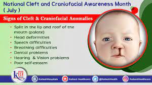
Cleft lip and palate can cause problems with speech, eating, hearing, and social development. Most of the time, cleft lip and palate can be easily treated with corrective surgery, allowing children born with the condition to live normal lives. However, in some cases – particularly in developing countries where access to medical care is not as widely available – children do not receive treatment. This can result in further problems as the child grows.

July is National Cleft and Craniofacial Awareness and Prevention Month.

Cleft lip and cleft palate happen when a baby’s lip or mouth doesn’t form completely during pregnancy. Cleft lip is an opening in a baby’s upper lip. Cleft palate is an opening in the roof of a baby’s mouth. Cleft lip and cleft palate are birth defects. These conditions affect thousands of babies, children, teens and adults in the United States each year.

Cleft lip and palate happen very early in pregnancy. Your baby’s lips form between 4 and 7 weeks of pregnancy, and the palate forms between 6 and 9 weeks of pregnancy. Cleft lip and palate don’t have to happen together — a baby can have one without the other.

Prevention:

To help reduce the chances of having a baby with an orofacial cleft or other craniofacial condition health care providers must encourage patients who are thinking about becoming pregnant to commit to a healthy lifestyle. Among certain healthy habits, we have: control diabetes, quit smoking, exercise regularly, practice yoga, etc. before becoming pregnant. Moreover, health care providers should also work with prospective parents to make informed decisions about medical treatment during pregnancy. This condition can occur in the first three months of pregnancy. Therefore, steps before becoming pregnant are essential for the health of the baby.

 

QUOTE FOR THURSDAY:

“Group B streptococcus (also called Group B strep or GBS) is a common type of bacteria (tiny organisms that live in and around your body) that can cause infection. Usually GBS is not serious for adults, but it can hurt newborns.

Many people carry Group B strep bacteria and don’t know it. It may never make you sick. GBS in adults usually doesn’t have any symptoms, but it can cause some minor infections, like a bladder or urinary tract infection (UTI).

While GBS may not be harmful to you, it can be very harmful to your baby. If you’re pregnant, you can pass it to your baby during labor and childbirth.

About 10 to 30 percent of pregnant women carry GBS bacteria. The best way to know if you have GBS is to get tested.”

March of Dimes (https://www.marchofdimes.org/find-support/topics/planning-baby/group-b-strep-infection)


International Group B Strep Awareness


Group B Streptococcus (GBS), or Streptococcus agalactiae, is a type of bacteria that is naturally found in the digestive and lower reproductive tracts of both men and women.  About 1 in 4 pregnant women “carry” or are “colonized” with GBS.

Carrying GBS does not mean that you are unclean.  Anyone can carry GBS. GBS is not considered to be a sexually transmitted disease or infection as it can occur on its own even in someone with no prior sexual experience. However, bacteria can be passed between sexual partners, including through oral contact.

The awareness month for group B strep is July. This is a common bacterium that affects adults and it does not present with any symptoms or signs. The pregnant women can pass these bacteria to their unborn children during the first few months after delivery or during child birth and this can be fatal. Most people who suffer from this bacterial infection are not aware of it and pregnant women get to know about it because of the screening that is done during prenatal visits. The screening tests is usually done during the third trimester and this test was started in the mid 1990s. Research indicates that since that time, the number of children who are affected by this infection has dropped from 1.7 to 0.28 in every 1000 births.

This month helps in raising awareness to women who are pregnant and those who are in the child rearing period to prevent Group B Strep infection. This helps women to keep their unborn babies healthy and safe as the infection can be fatal.

The National Charity that is associated with group B strep advices all pregnant women to be aware of this infection caused by streptococcus group B. This is the most common type of infection in new born babies as it causes meningitis, pneumonia and sepsis which can be fatal in the first few weeks of life. The bacterium is carried by women who do not present with any symptoms and therefore it can be easily passed to the baby during child birth.

The main message that is shared on this month awareness is that Group B Strep is fatal and that the infection can be prevented. People are taught on the need to go for testing as the bacterial colonization can be carried by people in the vagina or the lower intestines. Most people are carriers of this infection but they are not aware because they have never been tested for the same. This is because the infection does not present with any signs or symptoms. However, in some cases, the infection can get access into the blood stream and this can trigger fatal infections. When a pregnant woman gets this infection it can be spread to the unborn baby in the following ways:

While the baby is still in the womb, during a normal or a cesarean section birth, through the urinary tract or amniotic fluid, or through direct contact with the bacteria that is present in the birth canal.

The infants who are infected by this bacterium during birth are prone to infections and this includes meningitis, pneumonia and sepsis. The children who survive this infection are at risk of getting long term complications especially in cases where they suffered from meningitis. The common complications that can occur include cerebral palsy, hearing loss, mental and physical disabilities.

Pregnant women should be aware of the need for testing for each pregnancy and it does not matter whether they were tested for the previous pregnancies. This is because all pregnancies are different and it is vital to ensure that the baby is safe in all pregnancies.


QUOTE FOR WEDNESDAY:

“Health care providers can treat hemochromatosis safely and effectively by removing blood from the body on a regular basis. This is similar to donating blood. The process is known as phlebotomy.

The goal of phlebotomy is to lower your iron levels. The amount of blood removed and how often it’s removed depend on your age, your overall health and the severity of iron overload.”

MAYO CLINIC (https://www.mayoclinic.org/diseases-conditions/hemochromatosis/diagnosis-treatment/drc-20351448)

Part II Hemochromatosis – How its Diagnosed & Rx.

Diagnosis

It can be tricky for your doctor to diagnose hemochromatosis, because other conditions have the same symptoms. He might want you to get tested if:

  • You’re having symptoms.
  • You have one of the problems listed above.
  • A family member has the disorder.

There are some other ways your doctor can figure out if you have it:

Checking your history. He’ll ask about your family and if anyone has hemochromatosis or signs of it. He might also ask about things like arthritis and liver disease, which might mean you or someone in your family has hemochromatosis but doesn’t know it.

Physical exam. Your doctor will examine your body. This involves using a stethoscope to listen to what’s going on inside. He might also tap on different parts of your body.

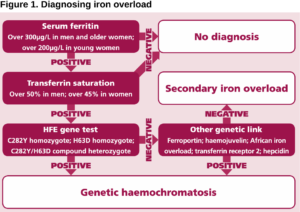
Blood tests. Two tests can give your doctor a clue about hemochromatosis:

  • Transferrin saturation. This shows how much iron is stuck to transferrin, a protein that carries iron in your blood.
  • Serum ferritin. This test measures the amount of ferritin, a protein that stores iron, in your blood. – If either of these show you have more iron than you should, your doctor might order a third test to see if you have a gene that causes hemochromatosis.
  • Liver biopsy. Your doctor will take a small piece of your liver. He’ll look at it under a microscope to see if there’s any liver damage.
  • MRI. This is a scan that uses magnets and radio waves to take a picture of your organs.

    Treatment

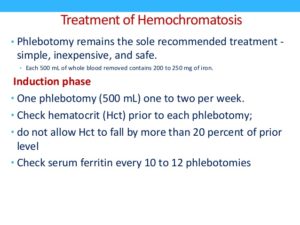
    If you have primary hemochromatosis, doctors treat it by removing blood from your body on a regular basis. It’s alot like donating blood. Your doctor will insert a needle into a vein in your arm or leg. The blood flows through the needle and into a tube that’s attached to a bag.

    • Initial treatment schedule. In the beginning, you may have a pint (about 470 milliliters) of blood taken once or twice a week — usually in a hospital or your provider’s office. While you lean back in a chair, a needle is inserted into a vein in your arm. The blood flows from the needle into a tube that’s attached to a blood bag. The process of removing blood is referred to as therapeutic blood removal.  In the Initial treatment. You’ll visit your doctor’s office or a hospital once or twice a week to have your blood drawn. You may have up to a pint taken at a time.
    • Maintenance treatment schedule. Once your iron levels go down, blood can be removed less often, typically every 2 to 3 months. Some people may maintain typical iron levels without having any blood taken. Some may need to have blood removed monthly. The schedule depends on how quickly iron builds up in your body.  So Maintenance treatment starts when once your blood iron levels have gone back to normal, you’ll still have to have to have blood taken, but not as often. It’ll be based on how fast iron builds back up in your body.

    The goal is to remove some of your blood so that your iron levels return to normal. This could take up to a year or more. Blood removal is divided into two parts: initial treatment and maintenance treatment.


QUOTE FOR TUESDAY:

“Hemochromatosis is a metabolic disorder in which your organs accumulate excess iron, leading to organ damage. Hereditary hemochromatosis affects one in 300 people in the United States.”

John Hopkins Medicine (https://www.hopkinsmedicine.org/health/conditions-and-diseases/hemochromatosis)

What is Hemochromatosis and the symptoms?


This is a problem with the Liver and in a nutshell it is IRON OVERLOAD!

Hemochromatosis (Hemo meaning blood, Chroma meaning change in color and Osis meaning swelling)  is a condition where there is abnormal accumulation of iron in the organs resulting in organ toxicity. It is the most common autosomal recessive genetic condition along with being the most common cause of severe iron overload. 75 percent of patients who have hereditary hemochromatosis are asymptomatic. The diagnosis can be made based on the clinical symptoms of the disease. Since most patients are asymptomatic, they are diagnosed when their serum iron levels are noted to be elevated during a routine chemistry screening panel. Some of the tests performed on an individual suspected with hemochromatosis include genetic testing to test for HFE mutations, transferrin saturation levels, hepatic iron concentration, and serum ferritin studies. Imaging studies such as a chest radiography and echocardiography might be beneficial to help evaluate cardiac disease in patients with hemochromatosis.

Early diagnosis is important in hemochromatosis, like in most every other diagnosis.

The aim of therapy is to remove iron before it causes irreversible parenchymal damage. Once the diagnosis is confirmed, it is treated by phlebotomy to maintain normal iron stores and rid the body of excess iron. Chelation therapy uses agents such as deferoxamine, deferiprone, or deferasirox. Surgery may be required if there is end stage liver disease or severe arthropathy. In the United States, hereditary hemochromatosis is found in 1 of 200 to 500 individuals. Most of these cases are seen in individuals of northern European origin. It has the same prevalence in Australia, Europe, and other western countries. The highest prevalence is among individuals of Celtic origin and less among those of African descent.

Symptoms would be:

1.) Fatigue and 2 types of fatigue, that would be:

Physical fatigue occurs when there is transient inability to achieve peak physical performance.

Mental fatigue is the temporary inability to maintain optimal cognitive performance. It is a common and non-specific symptom that can be seen in stress, overwork, depression, boredom, anemia, inadequate sleep, malnutrition, pregnancy, infections, and more. It can manifest as somnolence in patients.

2.) Impotence in patients with hemochromatosis is due to a result of hypogonadism. Hypogonadism can occur in hemochromatosis patients due to pituitary or testicular iron deposition.

3.) Arthropathy that’s a disease of a joint. In arthropathy, there can be joint pain, stiffness, swelling, and decreased range of motion. Patients can see this occuring because of the accumulation of iron in the joint tissues. The most commonly affected joints are the knees, feet, wrists, neck, back, metacarpophalangeal joints, and proximal interphalangeal joints.

4.) It has been observed that 62 percent of patients with hemochromatosis experience partial loss of body hair. The most commonly affected area is the pubic area. Approximately 12 percent of patients experience total body hair loss. In some patients, the thinning and loss of hair may be reversed through therapy.

5.) Koilonychia refers to spoon nails where the nails are abnormally thin and have lost their convexity resulting in flat or concave nails. Causes of koilonychia include iron deficiency anemia and it can be seen in 25 to 50 percent of these patients.

6.) Skin Hyperpigmentation or Bronzing of the skin refers to darkening of the skin. It is due to the combination of melanin and iron deposition which is characteristic. It is one of the classic triad symptoms with the other two being cirrhosis and diabetes in the late stages of the disease. This usually occurs only when the total iron content in the body is more than 5 times the normal levels.

7.) Amenorrhea refers to the absence of menstruation in females who are in their reproductive age.

8.) In the heart it can cause the following: Atrial flutter is a problem with the way your heart beats. Such problems, whether in the rhythm or speed of the heartbeat, are known as arrhythmias. Heart Failure is the other.

In addition to these symptoms it can also cause problems with the spleen, adrenal glands, pituitary gland, gallbladder or thyroid.

For helping in possible PREVENTION of this diagnosis and most others stay as healthy as possible and get yearly check ups to pick up this or any other diagnosis EARLY!! In so many cases of diseases the early picked up or diagnosed the better the results turn out in most cases.  When a disease just start in your body is being treated compared to late in the disease higher the odds will resolve or you will last longer with a better quality of life, just like with so many other diseases!

QUOTE FOR MONDAY:

“Preeclampsia is a serious condition that can happen after the 20th week of pregnancy or after giving birth (called postpartum preeclampsia). Most people who have preeclampsia have dangerously high blood pressure and may have problems with their kidneys or liver. Blood pressure is the force of blood that pushes against the artery walls. An artery is a blood vessel that carries blood away from the heart to other parts of the body. High blood pressure (also called hypertension) can stress the heart and cause problems during pregnancy.”

March of Dimes (https://www.marchofdimes.org/find-support/topics/pregnancy/preeclampsia)

Pre-eclampsia : What it is, whose at risk for it, causes,symptoms, when to go to MD, complications and prevention!

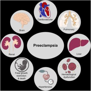
The Box below shows the organs Pre-eclampsia can effect:

What is Preeclampsia?

Preeclampsia is a condition that occurs only during pregnancy. Some symptoms may include high blood pressure and protein in the urine, occurring after week 20 of pregnancy. Preeclampsia is often precluded by gestational hypertension. While high blood pressure during pregnancy does not necessarily indicate preeclampsia, it may be a sign of another problem. The condition affects at least 5-8% of pregnancies.

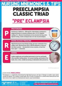
Preeclampsia, formerly called toxemia, is when a pregnant woman has high blood pressure, protein in her urine, and swelling in her legs, feet, and hands. It can range from mild to severe. It usually happens late in pregnancy, though it can come earlier or just after delivery.

Preeclampsia can lead to eclampsia, a serious condition that can have health risks for mom and baby and, in rare cases, cause death. Women with preeclampsia who have seizures have eclampsia.

If you have a mild case and your baby has not reached full development, your doctor will probably recommend you do the following:

  • Rest, lying on your left side to take the weight of the baby off your major blood vessels.
  • Increase prenatal checkups.
  • Consume less salt
  • Drink at least 8 glasses of water a day
  • Change your diet to include more protein

If you have a severe case, your doctor may try to treat you with blood pressure medication until you are far enough along to deliver safely, along with possibly bed rest, dietary changes, and supplements.

The only cure for preeclampsia is to give birth. Even after delivery, symptoms of preeclampsia can last 1 to 6 weeks or more.

You can help protect yourself by learning the symptoms of preeclampsia and by seeing your doctor for regular prenatal care. Catching preeclampsia early may lower the chances of long-term problems for both mom and baby.

For many years preeclampsia has been considered to be a two-stage disease. The first stage comprises poor placentation. The second stage is the clinical expression of the disease namely new hypertension and new proteinuria in the patient.

Who is at risk for preeclampsia?

  • A first-time mom
  • Previous experience with gestational hypertension or preeclampsia
  • Women whose sisters and mothers who had preeclampsia (family history)
  • Women carrying multiple babies
  • Women younger than 20 years and older than age 35
  • Women who had high blood pressure or kidney disease prior to pregnancy
  • Women who are obese or have a BMI of 30 or greater
  • Complications in previous pregnancy.

Preeclampsia Causes

Many experts think preeclampsia and eclampsia happen when a woman’s placenta doesn’t work the way it should, but they don’t know exactly why. Some think poor nutrition or high body fat might contribute. A lack of blood flow to the uterus could play a role. Genes are also a factor.

How do I know if I have preeclampsia?

At each prenatal checkup, your healthcare provider will check your blood pressure, urine levels, and may order blood tests which may show if you have preeclampsia. Your physician may also perform other tests that include: checking kidney and blood-clotting functions; ultrasound scan to check your baby’s growth; and Doppler scan to measure the efficiency of blood flow to the placenta.

Symptoms:

Preeclampsia sometimes develops without any symptoms. High blood pressure may develop slowly, or it may have a sudden onset. Monitoring your blood pressure is an important part of prenatal care because the first sign of preeclampsia is commonly a rise in blood pressure. Blood pressure that exceeds 140/90 millimeters of mercury (mm Hg) or greater — documented on two occasions, at least four hours apart — is abnormal.

Other signs and symptoms of preeclampsia may include:

  • Excess protein in your urine (proteinuria) or additional signs of kidney problems
  • Severe headaches
  • Changes in vision, including temporary loss of vision, blurred vision or light sensitivity
  • Upper abdominal pain, usually under your ribs on the right side
  • Nausea or vomiting
  • Decreased urine output
  • Decreased levels of platelets in your blood (thrombocytopenia)
  • Impaired liver function
  • Shortness of breath, caused by fluid in your lungs

When to go to the doctor:

Make sure you attend your prenatal visits so that your health care provider can monitor your blood pressure. Contact your provider immediately or go to an emergency room if you have severe headaches, blurred vision or other visual disturbances, severe belly pain, or severe shortness of breath.

Because headaches, nausea, and aches and pains are common pregnancy complaints, it’s difficult to know when new symptoms are simply part of being pregnant and when they may indicate a serious problem — especially if it’s your first pregnancy. If you’re concerned about your symptoms, contact your doctor.

Complications may include:

  • Fetal growth restriction. Preeclampsia affects the arteries carrying blood to the placenta. If the placenta doesn’t get enough blood, the baby may receive inadequate blood and oxygen and fewer nutrients. This can lead to slow growth known as fetal growth restriction.
  • Preterm birth. Preeclampsia may lead to an unplanned preterm birth — delivery before 37 weeks. Also, planned preterm birth is a primary treatment for preeclampsia. A baby born prematurely has increased risk of breathing and feeding difficulties, vision or hearing problems, developmental delays, and cerebral palsy. Treatments before preterm delivery may decrease some risks.
  • Placental abruption. Preeclampsia increases your risk of placental abruption. With this condition, the placenta separates from the inner wall of the uterus before delivery. Severe abruption can cause heavy bleeding, which can be life-threatening for both the mother and baby.
  • hemolysis elevated liver enzymes and low platelet count (HELLP) syndrome. HELLP stands for hemolysis (the destruction of red blood cells), elevated liver enzymes and low platelet count. This severe form of preeclampsia affects several organ systems. HELLP syndrome is life-threatening to the mother and baby, and it may cause lifelong health problems for the mother.Signs and symptoms include nausea and vomiting, headache, upper right belly pain, and a general feeling of illness or being unwell. Sometimes, it develops suddenly, even before high blood pressure is detected. It also may develop without any symptoms.
  • Eclampsia. Eclampsia is the onset of seizures or coma with signs or symptoms of preeclampsia. It is very difficult to predict whether a patient with preeclampsia will develop eclampsia. Eclampsia can happen without any previously observed signs or symptoms of preeclampsia.Signs and symptoms that may appear before seizures include severe headaches, vision problems, mental confusion or altered behaviors. But, there are often no symptoms or warning signs. Eclampsia may occur before, during or after delivery.
  • Other organ damage. Preeclampsia may result in damage to the kidneys, liver, lung, heart, or eyes, and may cause a stroke or other brain injury. The amount of injury to other organs depends on how severe the preeclampsia is.
  • Cardiovascular disease. Having preeclampsia may increase your risk of future heart and blood vessel (cardiovascular) disease. The risk is even greater if you’ve had preeclampsia more than once or you’ve had a preterm delivery.

Prevention of preeclampsia:

Medication

The best clinical evidence for prevention of preeclampsia is the use of low-dose aspirin. Your primary care provider may recommend taking an 81-milligram aspirin tablet daily after 12 weeks of pregnancy if you have one high-risk factor for preeclampsia or more than one moderate-risk factor.

It’s important that you talk with your provider before taking any medications, vitamins or supplements to make sure it’s safe for you.

Lifestyle and healthy choices

Before you become pregnant, especially if you’ve had preeclampsia before, it’s a good idea to be as healthy as you can be. Talk to your provider about managing any conditions that increase the risk of preeclampsia.


QUOTE FOR THE WEEKEND:

July is recognized as Cord Blood Awareness Month. This month aims to educate people about cord blood donation and storage benefits. Cord blood is the blood that remains in the umbilical cord and placenta after a baby is born.

Public cord blood banking is free and will provide life-saving benefits to a family in need. Once you donate your cord blood, however, you no longer have rights to those stem cells. If your child or family member is in need of cord blood stem cells, there is no guarantee that you or children can use their own cells. With private cord blood banking, there are fees but you own the cells. Your full rights to use it are preserved, and it is always immediately available to you.”

New England Cord Blood Bank – NECBB (https://www.cordbloodbank.com/july-cord-blood-awareness-month/)