QUOTE FOR FRIDAY:

“Your risk of getting colorectal cancer increases as you get older. Other risk factors include having:

  • Inflammatory bowel disease such as Crohn’s disease or ulcerative colitis.
  • A personal or family history of colorectal cancer or colorectal polyps.
  • A genetic syndrome such as familial adenomatous polyposis (FAP) or hereditary non-polyposis colorectal cancer (Lynch syndrome).”

Center for Disease Control and Prevention – CDC (https://www.cdc.gov/colorectal-cancer/risk-factors/index.html)

The risk factors of Colon Cancer!

How does colon-recto cancer even start:

Most colorectal cancers start as a growth on the inner lining of the colon or rectum. These growths are called polyps.

Some types of polyps can change into cancer over time (usually many years), but not all polyps become cancer. The chance of a polyp turning into cancer depends on the type of polyp it is. There are different types of polyps.

  • Adenomatous polyps (adenomas): These polyps sometimes change into cancer. Because of this, adenomas are called a pre-cancerous condition. The 3 types of adenomas are tubular, villous, and tubulovillous.
  • Hyperplastic polyps and inflammatory polyps: These polyps are more common, but in general they are not pre-cancerous. Some people with large (more than 1cm) hyperplastic polyps might need colorectal cancer screening with colonoscopy more often.
  • Sessile serrated polyps (SSP) and traditional serrated adenomas (TSA): These polyps are often treated like adenomas because they have a higher risk of colorectal cancer.

Other factors that can make a polyp more likely to contain cancer or increase someone’s risk of developing colorectal cancer include:

  • If a polyp larger than 1 cm is found
  • If more than 3 polyps are found
  • If dysplasia is seen in the polyp after it’s removed. Dysplasia is another pre-cancerous condition. It means there’s an area in a polyp or in the lining of the colon or rectum where the cells look abnormal, but they haven’t become cancer.

The body is made up of trillions of living cells. Normal body cells grow, divide into new cells, and die in an orderly way. During the early years of a person’s life, while they are still growing, their normal cells divide faster. Once the person becomes an adult, most cells divide only to replace worn-out or dying cells or to repair injuries.

Cancer begins when cells in a part of the body start to grow out of control. There are many kinds of cancer, but they all start because of out-of-control growth of abnormal cells.

Cancer cell growth is different from normal cell growth. Instead of dying, cancer cells continue to grow and form new, abnormal cells. In most cases the cancer cells form a tumor. Cancer cells can also invade (grow into) other tissues, something that normal cells cannot do. Growing out of control and invading other tissues are what makes a cell a cancer cell.

Cells become cancer cells because of damage to DNA. DNA is in every cell and directs all its actions. In a normal cell, when DNA is damaged the cell either repairs the damage or the cell dies. In cancer cells, the damaged DNA is not repaired, but the cell doesn’t die like it should. Instead, this cell goes on making new cells that the body does not need. These new cells will all have the same damaged DNA as the first abnormal cell does.

People can inherit damaged DNA, but most often the DNA damage is caused by mistakes that happen while the normal cell is reproducing or by something in our environment. Sometimes the cause of the DNA damage is something obvious, like cigarette smoking. But often no clear cause is found.

Cancer cells often travel to other parts of the body, where they begin to grow and form new tumors that replace normal tissue. This process is called metastasis. It happens when the cancer cells get into the bloodstream or lymph vessels of our body.

No matter where a cancer may spread, it is always named for the place where it started. For example, breast cancer that has spread to the liver is still called breast cancer, not liver cancer

Different types of cancer can behave very differently. For example, lung cancer and breast cancer are very different diseases. They grow at different rates and respond to different treatments. This is why people with cancer need treatment that is aimed at their particular kind of cancer.

Not all tumors are cancerous. Tumors that aren’t cancer are called benign. Benign tumors can cause problems – they can grow very large and press on healthy organs and tissues. But they cannot grow into (invade) other tissues. Because they can’t invade, they also can’t spread to other parts of the body (metastasize). These tumors are rarely life threatening.

Their are risk factors in getting colon cancer as well as other cancer and noncancerous diseases BUT remember their are modifiable risk factors leading to disease which are factors we can’t control being 4 areas 1.)Race 2.)Age 3.) Sex 4.) Heredity in the family (particularly nuclear and grandparents meaning higher risk than a first cousin or second cousin and down the family tree).

Risk Factors to colon cancer can be:

Most colorectal cancers occur in people without a family history of colorectal cancer. Still, as many as 1 in 5 people who develop colorectal cancer have other family members who have been affected by this disease.  People with a history of colorectal cancer in one or more first-degree relatives (parents, siblings, or children) are at increased risk. The risk is about doubled in those with only one affected first-degree relative=Nuclear Family. It is even higher if that relative was diagnosed with cancer when they were younger than 45, or if more than one first-degree relative is affected.

The reasons for the increased risk are not clear in all cases. Cancers can “run in the family” because of inherited genes, shared environmental factors, or some combination of these.

Having family members who have had adenomatous polyps is also linked to a higher risk of colon cancer. (Adenomatous polyps are the kind of polyps that can become cancerous.)

1-Inherited syndromes

About 5% to 10% of people who develop colorectal cancer have inherited gene defects (mutations) that can cause family cancer syndromes and lead to them getting the disease. These syndromes often lead to cancer that occurs at a younger age than is usual.

The most common inherited syndromes linked with colorectal cancers are familial adenomatous polyposis (FAP) and hereditary non-polyposis colorectal cancer (HNPCC), but other rarer syndromes can also increase colorectal cancer risk.

Familial adenomatous polyposis (FAP): FAP is caused by changes (mutations) in the APC gene that a person inherits from his or her parents. About 1% of all colorectal cancers are due to FAP.

The most common type of FAP causes people to develop hundreds or thousands of polyps in their colon and rectum, usually in their teens or early adulthood.

Hereditary non-polyposis colon cancer (HNPCC): HNPCC, also known as Lynch syndrome, accounts for about 2% to 4% of all colorectal cancers.

The cancers in this syndrome also develop when people are relatively young, although not as young as in FAP. People with HNPCC may also have polyps, but they only have a few, not hundreds as in FAP. The lifetime risk of colorectal cancer in people with this condition may be as high as 80%.

Turcot syndrome: This is a rare inherited condition in which people are at increased risk of adenomatous polyps and colorectal cancer, as well as brain tumors. There are actually 2 types of Turcot syndrome:

  • One can be caused by gene changes similar to those seen in FAP, in which cases the brain tumors are medulloblastomas.
  • The other can also be caused by gene changes similar to those seen in HNPCC, in which cases the brain tumors are glioblastomas.
  • MUTYH-associated polyposis:Racial and ethnic background  1. Jews of Eastern European descent (Ashkenazi Jews) have one of the highest colorectal cancer risks of any ethnic group in the world. Several gene mutations leading to an increased risk of colorectal cancer have been found in this group. People with type 2 (usually non-insulin dependent) diabetes have an increased risk of developing colorectal cancer. Both type 2 diabetes and colorectal cancer share some of the same risk factors (such as excess weight). But even after taking these factors into account, people with type 2 diabetes still have an increased risk. They also tend to have a less favorable prognosis (outlook) after diagnosis.  

2-Night shift workers are prone to cancers (colon/rectal and breast cancer).

3-Previous treatment for certain cancers –

Several studies have suggested that men who had radiation therapy to treat prostate cancer might have a higher risk of rectal cancer because the rectum receives some radiation during treatment. Men should consider the many possible side effects of prostate cancer treatment when making treatment decisions. Some doctors recommend that the risk of rectal cancer should be considered as one of those possible side effects but the patient is always responsible in doing research on their own before making the final decisions of treatment.  They choose the treatment they feel is the best choice; not the doctor.

  • Some studies have found that men who survive testicular cancer seem to have a higher rate of colorectal cancer and some other cancers. This might be because of the treatments they have received.

4-Factors less clear but can effect risk for colon cancer:

        1-Type 2 diabetes

       2-Racial Group African Americans have the highest colorectal cancer incidence and mortality rates of all       racial groups in the United States. The reasons for this are not yet understood.

        3-Peutz-Jeghers syndrome: People with this rare inherited condition tend to have freckles around the mout (and sometimes on the hands and feet) and a special type of polyp in their digestive tracts (called hamartoma). They are at greatly increased risk for colorectal cancer, as well as several other cancers, which usually appear at a younger than normal age. This syndrome is caused by mutations in the gene STK1.  People with this syndrome develop colon polyps which will become cancerous if the colon is not removed.

Patients undergo lifelong surveillance of organs to monitor for cancer and prevent secondary problems from the polyps.

REVISED 3/05/2023

QUOTE FOR THURSDAY:

“A significant challenge for diagnosing women with heart disease is the lack of recognition of symptoms that might be related to heart disease, or that don’t fit into classic definitions. Women can develop symptoms that are subtler and harder to detect as a heart attack, especially if the physician is only looking for the “usual” heart attack symptoms.

“Women are much more likely to have atypical heart attack symptoms,” says Dr. Lili Barouch, director of the Johns Hopkins Columbia Heart Failure Clinic. “So while the classical symptoms, such as chest pains, apply to both men and women, women are much more likely to get less common symptoms such as indigestion, shortness of breath, and back pain, sometimes even in the absence of obvious chest discomfort.”

John Hopkins Medicine (https://www.hopkinsmedicine.org/health/conditions-and-diseases/heart-disease-differences-in-men-and-women)

Women with Heart Disease and how it differs compared to men diagnosed with it.

                             Women and Heart DIsease

Many many women and their doctors don’t know that heart disease is the number one killer of women. Furthermore, the heart disease that is seen in women is often not quite the same as heart disease in men.

Let’s remember that Heart disease is an umbrella term that includes heart failure, coronary artery disease (CAD), arrhythmias, angina, and other heart-related infections, irregularities, and birth defects

These facts lead to two common (and sometimes tragic) misapprehensions held by many women and their doctors: That women don’t really get much heart disease, and when they do, it behaves pretty much like the heart disease that men get.

The truth is that not only is heart disease very common in women, but also, when women get heart disease it often acts quite differently than it does in men. Failing to understand these two fundamental truths leads to a lot of preventable deaths and disability in women with heart disease.

If you are a woman, you need to know the basics about heart disease – especially heart disease as it behaves in women.

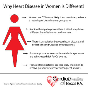
When women have angina, they are more likely than men to experience “atypical” symptoms. Instead of chest pain, they are more likely to experience a hot or burning sensation, or even tenderness to touch, which may be located in the back, shoulders, arms or jaw – and often women have no chest discomfort at all. An alert doctor will think of angina whenever a patient describes any sort of fleeting, exertion-related discomfort located anywhere above the waist, and they really shouldn’t be thrown off by such “atypical” descriptions of symptoms. However, unless doctors are thinking specifically of the possibility of CAD, they are all too likely to write such symptoms off to mere musculoskeletal pain or gastrointestinal disturbances.

Women are more likely than men to have heart attack symptoms unrelated to chest pain, such as:

      • Neck, jaw, shoulder, upper back or abdominal discomfort.
      • Shortness of breath.
      • Right arm pain.
      • Nausea or vomiting.
      • Sweating.
      • Lightheadedness or dizziness.
      • Unusual fatigue.

Heart attacks (or myocardial infarctions)  also tend to behave differently in women.

Frequently, instead of the crushing chest pain that is considered typical for a heart attack, women may experience nausea, vomiting, indigestion, shortness of breath or extreme fatigue – but no chest pain. Unfortunately, these symptoms are also easy to attribute to something other than the heart. Furthermore, women (especially women with diabetes) are more likely than men to have “silent” heart attacks – that is, heart attacks without any acute symptoms at all, and which are diagnosed only at a later time, when subsequent cardiac symptoms occur.

The Diagnosis Of CAD in Women Can Be More Difficult.

Diagnostic tests that work quite well in men can be misleading in women. The most common problem is seen with stress testing – in women, the electrocardiogram (ECG) during exercise can often show changes suggesting CAD, whether CAD is present or not, making the study difficult to interpret. Many cardiologists routinely add an echocardiogram or a thallium study when doing a stress test in a woman, which greatly improves diagnostic accuracy.

In women with typical CAD, coronary angiography is every bit as useful as in men; it identifies the exact location of any plaques (i.e., blockages) within the coronary arteries, and guides therapeutic decisions. However, in women with atypical coronary artery disorders (to be discussed in the next section), coronary angiograms often appear misleadingly normal. Thus, in women angiography is often not the gold standard for diagnosis, as it is for most men.

CAD In Women Can Take Atypical Forms.

At least four atypical coronary artery disorders can occur in women, usually in younger (i.e., pre-menopausal) women. Each of these conditions can produce symptoms of angina with apparently “normal” coronary arteries (that is, coronary arteries that often appear normal on angiogram). The problem, obviously, is that if the physician trusts the results of the angiogram, he/she is likely to miss the real diagnosis.

DALLAS, February 19, 2013 — A new study show women’s heart disease awareness is increasing.  A study with the number of women aware that heart disease is the leading cause of death nearly is doubling in the last 15 years, but that this knowledge still lags in minorities and younger women, according to the American Heart Association (AHA).

Among the study’s major findings, researchers comparing women’s views about heart disease in 1997 and today found:

  • In 2012, 56 percent of women identified heart disease as the leading cause of death compared with 30 percent in 1997.
  • In 1997, women were more likely to cite cancer than heart disease as the leading killer (35 percent versus 30 percent); but in 2012, only 24 percent cited cancer.
  • In 2012, 36 percent of black women and 34 percent of Hispanic women identified heart disease as the top killer — awareness levels that white women had in 1997 (33 percent).
  • Women 25-34 years old had the lowest awareness rate of any age group at 44 percent.

Among the women surveyed in 2012, researchers found:

  • Racial and ethnic minorities reported higher levels of trust in their healthcare providers compared with whites, and were also more likely to act on the information provided—dispelling the myth that mistrust of providers contributes to disparities.
  • Compared with older women, younger women were more likely to report not discussing heart disease risk with their doctors (6 percent among those 25-34 versus 33 percent for those 65 and older).

Risk Factors for Heart Disease in Women – Those we can’t change = Nonmodifiable Factors:

Age and Family History, Gender, Ethnicity.

The risk of having heart disease increases with age and this is due to stiffening of heart muscles which makes the heart less efficient in pumping blood around the body. You can determine your heart age by using this tool, developed by the British Heart Foundation: https://www.bhf.org.uk/heart-health/risk-factors/check-your-heart-age.

Another risk factor you cannot change is if you have a history of heart disease among family members. This can double your risk, so if your mother, father, sister or brother has suffered from heart disease before the age of 60 you are at a greater risk of developing heart disease.

Modifiable Risk Factors – Those we can change are:

1-Smoking is the single largest preventable cause of death in Australia, and approximately 40% of women who smoke die due to heart disease, stroke or blood vessel disease. Smokers are 2-4 times more at risk of developing heart disease compared to non-smokers. In 2011/2012, over 1.3 million women in Australia smoked, and 89% of them did this on a daily basis. While these numbers are for women aged 15 and over, the largest group were in the 25-34 age group.

Passive smoking (exposure to the cigarette smoke of others) also causes an increase in the risk of developing heart disease, which increases further in people having high blood pressure or high cholesterol. Women who smoke and also take the contraceptive pill have a 10 times higher risk of having a heart attack.

2-Alcohol. Do you know that drinking too much alcohol increases the risk of heart disease? Excessive drinking causes more weight gain (due to increased calories!), increase in blood pressure and blood lipids. Over a long period of time it can weaken the heart muscle and cause abnormal heart rhythms. Try and not drink alcohol every day, limit it to two standard drinks at a time and aim for at least two alcohol free days a week and make sure you don’t increase the amount you drink on the other days. Periodically take a break from any alcohol for a week or more and you will notice many benefits including a better nights sleep.

3.High Blood Pressure or Hypertension. Your blood pressure is a measurement of how ‘hard’ your heart is working to push blood around your body, through the blood vessels. It can be a ‘silent’ killer and if you do not know your blood pressure then it is worth having it checked by your GP. Changing your lifestyle will reduce your blood pressure. A recent study suggests that keeping your blood pressure under 140/90 can increase your life expectancy by 5 years at the age of 50 years. You can assess your high blood pressure through your MD monthly or less expensive buy a b/p machine and check your b/p everyday especially if your on antihypertensive meds to make sure your b/p isn’t under 100/60 to prevent hypotension.

4.Diabetes. Do you have diabetes and if so, is it under control?

Diabetes doubles your risk of having heart disease. People who have uncontrolled diabetes are at risk of having heart disease at an earlier age. For pre-menopausal women, having diabetes cancels the protective effects of hormone present in women and significantly increases the risk of heart disease. Taking steps to find out what your blood sugar is and keeping it well-controlled is essential.

5.Obesity- Do you know your body fat content?  If you think that you are overweight then you put yourself at risk of having heart disease. Being overweight will increase your blood pressure and contribute to developing diabetes. In addition to that, women who carry weight around their middle (belly fat) as opposed to their hips are twice as likely to develop heart disease.

By taking the steps to reduce your weight, you can reduce your risk of heart disease. A great tool developed by National Heart Foundation of Australia calculates if you might be at risk: http://www.heartfoundation.org.au/healthy-eating/Pages/bmi-calculator.aspx

6- INACTIVE-Are you physically active every day? Recent research indicates that “sitting is the new smoking” and being physically inactive can double your risk of having heart disease. It is important to get some exercise every day, such as a 30 minute walk where you raise your heart rate. It also raises your serotonin levels (feel-good hormone) and can reduce depression

7- STRESS-We could almost ask – do you know anyone who is not stressed?! However, while everyday life is stressful, those people who are almost constantly stressed are at risk of adopting unhealthy behaviours in order to reduce their stress levels. Examples include increasing their alcohol intake or smoking in order to relax; or tending to eat more junk food because they are often short of time. All of these factors increase their risk of heart disease.

Women, stress and the risk of heart disease

Along with poor diet, lack of exercise and smoking, unmanaged stress may increase the risk for heart disease. Now medical experts are discovering that mental stress affects women in different, and in some cases, more devastating ways, especially if they already have coronary conditions. One study that

Heart disease is the leading cause of death for men and women in the United States. Every year, 1 in 4 deaths are caused by heart disease. The good news? Heart disease can often be prevented when people make healthy choices and manage their health conditions. Communities, health professionals, and families can work together to create opportunities for people to make healthier choices. Make a difference in your community: Spread the word about strategies for preventing heart disease and encourage people to live heart healthy lives


QUOTE FOR WEDNESDAY:

“The Foundation for Women’s Cancer (FWC) understands the importance of bringing awareness to all gynecologic cancers — cervical, ovarian, uterine/ endometrial, vaginal and vulvar cancer. The month of September has been established as Gynecologic Cancer Awareness Month (GCAM), with a goal of reaching more and more people each year.

These resources give you the tools to share knowledge about gynecologic cancer symptoms, risk factors, prevention and early detection — because every five minutes, someone will be diagnosed with one of these cancers.”

Foundation for Women’s Cancer (https://foundationforwomenscancer.org/gcam/)

Gynecological Cancer Awareness Month

According to the CDC, uterine cancer (cancer which develops in the uterus) is the most commonly diagnosed gynecological cancer in the U.S. and the fourth most common cancer in U.S. women overall.

Types of Gynecological Cancer

  • Cervical cancer.
  • Ovarian cancer.
  • Uterine cancer.
  • Vaginal cancer.
  • Vulvar cancer.

September is Gynecologic Cancer Awareness


It is estimated that within the past 2 years 98,000 women would be diagnosed with a gynecologic cancer and some 30,000 would die from the disease.

Gynecological Cancer Awareness Month provides an important opportunity to draw attention to this important women’s health issue and offer vital information on risk cancers, warning signs, and prevention strategies.

Uterine Cancer

Uterine cancer forms in the tissues of the uterus, the organ in which a fetus develops. The two types of uterine cancer are endometrial cancer and uterine sarcoma.

Endometrial cancer forms in the tissues of the endometrium – the lining of the uterus. Obesity, high blood pressure, and diabetes may increase the risk of endometrial cancer.

Uterine Sarcoma is a rare type of cancer that forms in the uterine muscles or in tissues that support the uterus. Exposure to X-rays during radiation therapy can increase the risk of uterine sarcoma.

Treatment with the breast cancer drug tamoxifen is a risk factor for both types of uterine cancer.

According to the CDC, uterine cancer (cancer which develops in the uterus) is the most commonly diagnosed gynecological cancer in the U.S. and the fourth most common cancer in U.S. women overall. While any woman can develop uterine cancer, it is most commonly diagnosed in women who have gone through menopause. Risk factors for uterine cancer include age (being age 50 or older), obesity, taking estrogen alone as hormone replacement, and having a family history of uterine, ovarian or colon cancer.

Routine testing is not recommended for uterine cancer, so it is important for women to be aware of symptoms—such as abnormal vaginal discharge or bleeding and/or pain and pressure in the pelvic area—and talk to a healthcare provider if they experience these.

Ovarian Cancer

While ovarian cancer accounts for about 3% of cancers among women, it causes more deaths than any other gynecological cancer, according to the American cancer Society. While the survival rates for ovarian cancer are excellent when the disease is diagnosed early, only about 20% of ovarian cancers are found at this early stage. One reason for this is that there is no recommended routine screening for ovarian cancer on women without symptoms. And the symptoms of ovarian cancer—including abnormal abdominal bloating, abdominal pain or pressure, and feeling full quickly when eating—can also be easily be ignored or mistaken for other problems.

For women who experience these symptoms, or who at at higher risk (including women who have had breast cancer or have a family history of ovarian, breast or colorectal cancer), a healthcare provider may recommending further screening. Testing may include rectovaginal pelvic exam, a transvaginal ultrasound,or a CA-125 blood test.

There are three types of ovarian cancer in adults, including ovarian epithelial cancer, which begins in the tissue covering the ovary, lining of the fallopian tube, or the peritoneum; ovarian germ cell tumors, which start in the egg or germ cells; and ovarian low malignant potential tumors, which begin in the tissue covering the ovary.

Cervical Cancer

According to the American Sexual Health Organization, the vast majority of cases of cervical cancer—cancer that develops on the cervix, the opening to the uterus—are linked to human papillomavirus (HPV) infection. The majority of women with an HPV infection will not develop cervical cancer, but regular screening is essential. In most cases cervical cancer can be prevented through early detection and treatment of abnormal cell changes that occur in the cervix years before cervical cancer develops. These changes are typically detected through a Pap test or an HPV test. HPV vaccines can also prevent cervical cancer.

In its early stages, cervical cancer typically doesn’t have any symptoms, which is why regular screening is so important. At later stages, symptoms may include abnormal vaginal discharge or bleeding or pain during sex. While these can also be signs of other health issues, if a woman experiences these symptoms, she should report them to her healthcare provider.

Cervical Cancer. Carcinoma of Cervix. Malignant neoplasm arising from cells in the cervix uteri. Vaginal bleeding. Vector diagram

Vaginal and Vulvar Cancer

There are two main types of vaginal cancer: squamous cell carcinoma and adenocarcinoma. Adenocarcinoma is more likely than squamous cell cancer to spread to the lungs and lymph nodes. A rare type of adenocarcinoma is linked to being exposed to diethylstilbestrol (DES) before birth.

Adenocarcinomas not linked with being exposed to DES are most common in women after menopause.

According to the American Sexual Health Organization, vaginal and vulvar cancers are rare—an estimated 1,000 women are diagnosed with vaginal cancer and 3,500 women with vulvar cancer each year. Like cervical cancer, vaginal and vulvar cancers are also associated with HPV infection, with up to 90% of vaginal cancers and pre-cancers and more than 50% of vulvar cancers linked to infection with the high-risk HPV types.

Vulvar cancer forms in a woman’s external genitalia. Vulvar cancer most often affects the outer vaginal lips.

Abnormal cells can grow on the surface of the vulvar skin for a long time. This condition is called vulvar intraepithelial neoplasia (VIN). Because it is possible for VIN to become vulvar cancer, it is important to get treatment.

Risk factors for vulvar cancer include having VIN, HPV infection, and having a history of genital warts.

HPV vaccines, which prevent some of the high-risk types of HPV, can also help prevent vaginal and vulvar cancers.


QUOTE FOR TUESDAY:

“Leukemia is the most common cancer in children and teens, accounting for almost 1 out of 3 cancers. Most childhood leukemias are acute lymphocytic leukemia (ALL). Most of the remaining cases are acute myeloid leukemia (AML). Chronic leukemias are rare in children.”

American Cancer Society (https://www.cancer.org/cancer/types/leukemia-in-children.html)

Leukemia

leukemia1leukemia2

Leukemia is the most common cancer in children and teens, accounting for almost 1 out of 3 cancers. Most childhood leukemias are acute lymphocytic leukemia (ALL). Most of the remaining cases are acute myeloid leukemia (AML). Chronic leukemias are rare in children.

A risk factor is anything that affects a person’s chance of getting a disease such as cancer. Different cancers have different risk factors.

Lifestyle-related risk factors such as tobacco use, diet, body weight, and physical activity play a major role in many adult cancers. But these factors usually take many years to influence cancer risk, and they are not thought to play much of a role in childhood cancers, including leukemias.

There are a few known RISK FACTORS for childhood leukemia.

Inherited syndromes

-Some inherited disorders increase a child’s risk of developing leukemia:

  • Down syndrome (trisomy 21): Children with Down syndrome have an extra (third) copy of chromosome 21. They are many times more likely to develop either acute lymphocytic leukemia (ALL) or acute myeloid leukemia (AML) than are other children, with an overall risk of about 2% to 3%. Down syndrome has also been linked with transient leukemia (also known as transient myeloproliferative disorder) – a leukemia-like condition within the first month of life, which often resolves on its own without treatment.
  • Li-Fraumeni syndrome: This is a rare condition caused by a change in the TP53 tumor suppressor gene. People with this change have a higher risk of developing several kinds of cancer, including leukemia, bone or soft tissue sarcomas, breast cancer, adrenal gland cancer, and brain tumors.Other genetic disorders (such as neurofibromatosis and Fanconi anemia) also carry an increased risk of leukemia, as well as some other types of cancers.Having a parent who develops leukemia as an adult does not seem to raise a child’s risk of leukemia.The possible risks from fetal or childhood exposure to lower levels of radiation, such as from x-ray tests or CT scans, are not known for sure. Some studies have found a slight increase in risk, while others have found no increased risk. Any risk increase is likely to be small, but to be safe, most doctors recommend that pregnant women and children not get these tests unless they are absolutely needed.1-Acute lymphocytic leukemia (ALL) is a type of cancer of the blood and bone marrow — the spongy tissue inside bones where blood cells are made.Acute lymphocytic leukemia is the most common type of cancer in children, and treatments result in a good chance for a cure. Acute lymphocytic leukemia can also occur in adults, though the chance of a cure is greatly reduced.
  • The word “acute” in acute lymphocytic leukemia comes from the fact that the disease progresses rapidly and creates immature blood cells, rather than mature ones. The “lymphocytic” in acute lymphocytic leukemia refers to the white blood cells called lymphocytes, which ALL affects. Acute lymphocytic leukemia is also known as acute lymphoblastic leukemia.
  • What is Leukemia?  First their are types of leukemia, which are cancers of the bone marrow and blood and this is the most common childhood cancers unfortunately.  They account for about 30% of all cancers in children.  The most common types that are found in children they are 1.) acute lymphocytic leukemia (ALL) 2.) acute myelogenous leukemia (AML).
  • Exposure to high levels of radiation is a risk factor for childhood leukemia. Japanese atomic bomb survivors had a greatly increased risk of developing AML, usually within 6 to 8 years after exposure. If a fetus is exposed to radiation within the first months of development, there may also be an increased risk of childhood leukemia, but the extent of the risk is not clear.
  • Siblings (brothers or sisters) with leukemia have a slightly increased chance (2 to 4 times normal) of developing leukemia, but the overall risk is still low. The risk is much higher among identical twins. If one twin develops childhood leukemia, the other twin has about a 1 in 5 chance of getting leukemia as well. This risk is much higher if the leukemia develops in the first year of life.

2-Acute myelogenous leukemia (AML) is a cancer of the blood and bone marrow — the spongy tissue inside bones where blood cells are made.

The word “acute” in acute myelogenous leukemia denotes the disease’s rapid progression. It’s called myelogenous (my-uh-LOHJ-uh-nus) leukemia because it affects a group of white blood cells called the myeloid cells, which normally develop into the various types of mature blood cells, such as red blood cells, white blood cells and platelets.

Acute myelogenous leukemia is also known as acute myeloid leukemia, acute myeloblastic leukemia, acute granulocytic leukemia and acute nonlymphocytic leukemia.

QUOTE FOR MONDAY:

“Leukemia is a broad term for cancers of the blood cells. The type of leukemia depends on the type of blood cell that becomes cancer and whether it grows quickly or slowly. Leukemia occurs most often in adults older than 55, but it is also the most common cancer in children younger than 15.  There is Acute Lymphoblastic Leukemia, Acute Myeloid Leukemia, Chronic Lymphocytic Leukemia , Chronic Myelogenous Leukemia, Hairy Cell Leukemia, Childhood Acute Lymphoblastic Leukemia, and Childhood Acute Myeloid Leukemia.”

National Cancer Institute (https://www.cancer.gov/types/leukemia#: ~:text=Leukemia%20is%20a%20broad%20term,in%20children%20younger%20than%2015.)

Childhood Cancer Awareness Month – Leukemia


Leukemia is the most common cancer in children and teens, accounting for almost 1 out of 3 cancers. Most childhood leukemias are acute lymphocytic leukemia (ALL). Most of the remaining cases are acute myeloid leukemia (AML). Chronic leukemias are rare in children.

A risk factor is anything that affects a person’s chance of getting a disease such as cancer. Different cancers have different risk factors.

Lifestyle-related risk factors such as tobacco use, diet, body weight, and physical activity play a major role in many adult cancers. But these factors usually take many years to influence cancer risk, and they are not thought to play much of a role in childhood cancers, including leukemias.

There are a few known RISK FACTORS for childhood leukemia.

Inherited syndromes

-Some inherited disorders increase a child’s risk of developing leukemia:

  • Down syndrome (trisomy 21): Children with Down syndrome have an extra (third) copy of chromosome 21. They are many times more likely to develop either acute lymphocytic leukemia (ALL) or acute myeloid leukemia (AML) than are other children, with an overall risk of about 2% to 3%. Down syndrome has also been linked with transient leukemia (also known as transient myeloproliferative disorder) – a leukemia-like condition within the first month of life, which often resolves on its own without treatment.
  • Li-Fraumeni syndrome: This is a rare condition caused by a change in the TP53 tumor suppressor gene. People with this change have a higher risk of developing several kinds of cancer, including leukemia, bone or soft tissue sarcomas, breast cancer, adrenal gland cancer, and brain tumors.

Other genetic disorders (such as neurofibromatosis and Fanconi anemia) also carry an increased risk of leukemia, as well as some other types of cancers.

Siblings (brothers or sisters) with leukemia have a slightly increased chance (2 to 4 times normal) of developing leukemia, but the overall risk is still low. The risk is much higher among identical twins. If one twin develops childhood leukemia, the other twin has about a 1 in 5 chance of getting leukemia as well. This risk is much higher if the leukemia develops in the first year of life.

Having a parent who develops leukemia as an adult does not seem to raise a child’s risk of leukemia.

Exposure to high levels of radiation is a risk factor for childhood leukemia. Japanese atomic bomb survivors had a greatly increased risk of developing AML, usually within 6 to 8 years after exposure. If a fetus is exposed to radiation within the first months of development, there may also be an increased risk of childhood leukemia, but the extent of the risk is not clear.

The possible risks from fetal or childhood exposure to lower levels of radiation, such as from x-ray tests or CT scans, are not known for sure. Some studies have found a slight increase in risk, while others have found no increased risk. Any risk increase is likely to be small, but to be safe, most doctors recommend that pregnant women and children not get these tests unless they are absolutely needed.

What is Leukemia?  First their are types of leukemia, which are cancers of the bone marrow and blood and this is the most common childhood cancers unfortunately.  They account for about 30% of all cancers in children.  The most common types that are found in children they are 1.) acute lymphocytic leukemia (ALL) 2.) acute myelogenous leukemia (AML).

1.)-Acute lymphocytic leukemia (ALL) is a type of cancer of the blood and bone marrow — the spongy tissue inside bones where blood cells are made.

The word “acute” in acute lymphocytic leukemia comes from the fact that the disease progresses rapidly and creates immature blood cells, rather than mature ones. The “lymphocytic” in acute lymphocytic leukemia refers to the white blood cells called lymphocytes, which ALL affects. Acute lymphocytic leukemia is also known as acute lymphoblastic leukemia.

Acute lymphocytic leukemia is the most common type of cancer in children, and treatments result in a good chance for a cure. Acute lymphocytic leukemia can also occur in adults, though the chance of a cure is greatly reduced.

2.)-Acute myelogenous leukemia (AML) is a cancer of the blood and bone marrow — the spongy tissue inside bones where blood cells are made.

The word “acute” in acute myelogenous leukemia denotes the disease’s rapid progression. It’s called myelogenous (my-uh-LOHJ-uh-nus) leukemia because it affects a group of white blood cells called the myeloid cells, which normally develop into the various types of mature blood cells, such as red blood cells, white blood cells and platelets.

Acute myelogenous leukemia is also known as acute myeloid leukemia, acute myeloblastic leukemia, acute granulocytic leukemia and acute nonlymphocytic leukemia.

2.) Acute myelogenous leukemia (AML) is a cancer of the blood and bone marrow — the spongy tissue inside bones where blood cells are made.

The word “acute” in acute myelogenous leukemia denotes the disease’s rapid progression. It’s called myelogenous (my-uh-LOHJ-uh-nus) leukemia because it affects a group of white blood cells called the myeloid cells, which normally develop into the various types of mature blood cells, such as red blood cells, white blood cells and platelets.

Acute myelogenous leukemia is also known as acute myeloid leukemia, acute myeloblastic leukemia, acute granulocytic leukemia and acute nonlymphocytic leukemia.