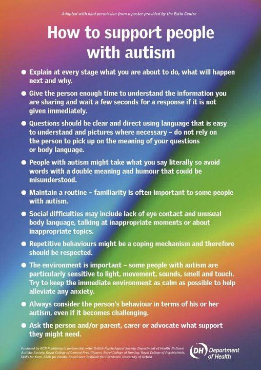
Part III March is kidney month – Chronic Kidney Failure


Chronic Renal or Kidney Disease (CRF):

In giving a short and easily understandable definition Chronic kidney disease happens when your kidneys no longer filter your blood the way they should, so wastes (toxins, usually end products of an acid) build up in your blood. This has probably been going on for years, and it may keep getting worse over time. Just like a car engine damaged but still using the car without getting the engine repaired sooner or later in time the engine no longer functions the same with any organ of the body getting damaged by some long term condition. If your disease gets worse and worse over time, you could have kidney failure or some multi organ failure, depending on the condition causing this.

Regarding Chronic Kidney Failure the causes can be:

** Diabetes (uncontrolled diabetes (Type 1 or 2) for many years.

** High blood pressure for many years.

These are the top 2 causes of most chronic kidney disease. Controlling these diseases can help slow or stop the damage to the individual’s kidneys who has one of these, if not both.

Other common causes of chronic renal failure (CRF) include:

-recurring pyelonephritis (kidney infection)

-polycystic kidney disease (multiple cysts in the kidneys

-autoimmune disorders such as systemic lupus erythematosus.

-hardening of the arteries, which can damage blood vessels in the kidney.

-A narrowed or blocked renal artery. A renal artery carries blood to the kidneys. Know this for starters, each of your kidneys has about a million tiny filters, called nephrons. The nephron is the tiny filtering structure in your kidneys. Each of your kidneys contain more than a million tiny filtering nephrons that help clean your blood removing toxins dumping them into your urinary bladder so you can evacuate them though urine (urea, urine; get it). Your nephrons play a vital role to our essential daily living. If over a long time you have a renal artery blocked the nephrons stop their function and die.

Remember the nephrons help all humans do the following if there kidneys or one kidney is functioning properly:

-Remove excess water, wastes (like urea, ammonia, etc.) & other substances from your blood.

-Return substances like sodium, potassium or phosphorus to the body whenever any of these substances run llow in your body or do the opposite if they run high to evacuate them through voiding dumping the sodium or phosphorus or potassium in the urinary bladder through the tube from the kidneys to the urinary bladder called ureters.

**If nephrons are damaged by the high sugar content or high blood pressure in the kidneys, they stop working. For a while, healthy nephrons can take on the extra work or overload. But if the damage continues, more and more nephrons shut down. After a certain point, the nephrons that are left cannot filter your blood well enough to keep you’re blood filtered properly to keep you healthy. Just like running from a bear in the street chancing you. We can run only so long but sooner or later we will run out of energy and not be able to run anymore, same concept for the kidney nephrons when they run out of enough energy due to the kidneys not properly working.**

The symptoms can be:

Urinate less than normal.

Have swelling and weight gain from fluid buildup in your tissues. This is called edema.

Feel very tired or sleepy.

Not feel hungry, or you may lose weight without trying.

Often feel sick to your stomach (nauseated) or vomit.

Have trouble sleeping.

Have headaches or trouble thinking clearly.

So what will you do GO TO A DOCTOR OR CALL 911 AND GO TO THE ER:

Your doctor will do blood and urine tests to help find out how well your kidneys are working. These tests can show signs of kidney disease and anemia. (You can get anemia from having damaged kidneys.) You may have other tests to help rule out other problems that could cause your symptoms.

To diagnose chronic renal failure is pretty much the same tests that are listed above on acute renal failure plus:

Chronic kidney disease is also called chronic renal failure or chronic renal insufficiency.There are five stages of kidney disease, from kidney damage with normal GFR to kidney failure.  So GFR will help the MD rule out acute versus chronic to give the MD direction on Rx.Your doctor will ask questions about any past kidney problems. He or she will also ask whether you have a family history of kidney disease and what medicines you take, both prescription and over-the-counter drugs.

Diagnosis & Treatment (Rx) for Chronic Kidney (Renal) Failure (CRF):

There are things you can do to slow or stop the damage to your kidneys. Taking medicines and making some lifestyle changes can help you manage your disease, prevent further damage to the kidneys, if their functioning at all and make you possibly feel better.

Kidney disease is a complex problem. You will probably need to take a number of medicines and have many tests. To stay as healthy as possible, take your medicines just the way your doctor says to and work closely with your doctor.

Go to all your appointments for the MD to see a increase in function or decrease in function of your kidney or kidneys you have still functioning to a level. To do that you can’t just go every 6 months especially when first diagnosed with it or with a collapse of an exacerbation of kidney failure in a worse level that brought on new symptoms that brought you to the ER.

Lifestyle changes are an important part of your treatment. Taking these steps can help slow down kidney disease and reduce your symptoms. These steps may also help with high blood pressure, diabetes, and other problems that make kidney disease worse or made the kidney disease happen with the secondary diagnosis you had originally for years (ex. Hypertension or Diabetes if not both especially is uncontrolled)

Very hard, never a complete 100 % resolution. It is like emphysema done by smokers the damage is done or like a heart attack the area of the infarction=damage is already done to the heart muscle.

Scared now, understandable but unfortunately the damage is done, so its get the organ to its optimal level of functioning or replace the damaged kidney (s) through a transplant of one.  You need to know this, do it if you want to live LONGER.  Remember fear is fear itself, the fear run it over and deal with what you have and make your life longer & better!  It’s all up to you.  HANDSOME I know you can do it , I am there for you as a good friend and professional RN and I would love you to last longer John!! XO

You may have a test done that lets your doctor look at a picture of your kidneys, such as an ultrasound or CT (Cat Scan of the kidneys). These tests can help your doctor measure the size of your kidneys, estimate blood flow to the kidneys, and see if urine flow is blocked. In some cases, your doctor may take a tiny sample of kidney tissue (biopsy) to help find out what caused your kidney disease.

Chronic kidney disease is caused by damage of the kidneys whether the cause of it be primary a Renal or Kidney problem or a secondary, another disease or disorder that affects the kidneys in doing their job, like hyperglycemia related to a individual with uncontrolled diabetes, for instance.

Chronic kidney disease may seem to have come on suddenly. But it has been happening bit by bit for many years as a result of damage to your kidneys.

One way to measure how well your kidneys are working is to figure out your glomelular filtration rate (GFR). The GFR is usually calculated using results from your blood creatinine test. Then the stage of kidney disease is figured out using the GFR (glomelular filtration rate). There are five stages of kidney disease, from kidney damage with normal GFR to kidney failure.

Your doctor will do tests that measure the amount of urea (BUN) and creatinine in your blood. These tests can help measure how well your kidneys are filtering your blood. As your kidney function gets worse, the amount of nitrogen (shown by the BUN test) and creatinine in your blood increases. The level of creatinine in your blood is used to find out the glomerular filtration rate (GFR). The GFR is used to show how much kidney function you still have. The GFR is also used to find out the stage of your kidney disease your in if you have it and its to guide decisions about treatment. *

QUOTE FOR TUESDAY:

Acute kidney injury (AKI) is where your kidneys suddenly stop working properly. It can range from minor loss of kidney function to complete kidney failure.

AKI normally happens as a complication of another serious illness. It’s not the result of a physical blow to the kidneys, as the name might suggest.

This type of kidney damage is usually seen in older people who are unwell with other conditions and the kidneys are also affected.

It’s essential that AKI is detected early and treated promptly.

National Health Services-NHS (https://www.nhs.uk/conditions/acute-kidney-injury/)

Part II March is kidney month – Acute Kidney Damage

ARF VERSUS CRF1

Acute Renal (Kidney) Failure:

Kidney failure occurs when the kidneys lose their ability to function. To treat kidney failure effectively, it is important to know whether kidney disease has developed suddenly (acute) or over the long term (chronic). Many conditions, diseases, and medicines can create situations that lead to acute and chronic kidney disease. Acute kidney injury, also called acute renal failure, is more commonly reversible than chronic kidney failure since the chronic condition has lasted longer in the body affecting systems for several months to years (some decades). Acute Renal Failure is new to the body as opposed to chronic; making it higher odds this can be treated and cured.

When acute kidney injury (ARF) occurs, the kidneys are unable to remove waste products and excess fluids, which then build up in the body and upset the body’s normal chemical balance.*

The most common causes of acute kidney injury are:

-dehydration

-blood loss from major surgery or injury

-medicines such as nonsteroidal anti-inflammatory drugs (NSAIDs), antibiotics, or the dyes (contrast agents) used in X-ray tests.

Symptoms depend on the cause of acute renal failure and can include:

    • -Little or no urine output.
    • -Dizziness upon standing.
    • -Swelling, especially of the legs and feet.
    • -Loss of appetite, nausea, and vomiting.
    • -Feeling confused, anxious and restless, or sleepy.
    • -Pain in the flank, which is felt just below the rib cage and above the waist on one or both sides of the back.*

If your signs and symptoms suggest that you have acute kidney failure, your doctor may recommend certain tests and procedures to verify your diagnosis. These may include:

  • Urine output measurements. The amount of urine you excrete in a day may help your doctor determine the cause of your kidney failure.
  • Urine tests. Analyzing a sample of your urine, a procedure called urinalysis, may reveal abnormalities that suggest kidney failure.
  • Blood tests. A sample of your blood may reveal rapidly rising levels of urea and creatinine — two substances used to measure kidney function.  Most cases of acute kidney injury occur in people who are already in the hospital for other reasons. In these people, acute kidney injury is usually diagnosed when routine tests show a sudden increase in creatinine and blood urea nitrogen (BUN) levels.   **A buildup of these waste products in the blood points to a loss of kidney function!**
  • With a patient just coming in the first time to an MD with no history of renal disease but has factors or symptoms indicating possible kidney failure involvement don’t be surprised if the doctor orders glomerular filtration rate (GFR); which is a test used to check how well the kidneys are working. Specifically, it estimates how much blood passes through the glomeruli each minute. Glomeruli are tiny filters in the kidneys that filter waste from the blood.
  • Imaging tests. Imaging tests such as ultrasound and computerized tomography may be used to help your doctor see your kidneys.  Also commonly done is an ultrasound of the kidneys which may help determine whether kidney problems are acute or chronic. Normal-sized kidneys may be present in either condition, but when both kidneys are smaller than normal, chronic kidney disease is usually the problem.  This helps rule out acute from chronic.correcting the cause and supporting the kidneys with dialysis until proper functioning is restored.
  • Removing a sample of kidney tissue for testing. In some situations, your doctor may recommend a kidney biopsy to remove a small sample of kidney tissue for lab testing. Your doctor inserts a needle through your skin and into your kidney to remove the sample.

TREATMENT FOR ARF (reversible in most cases):

Treatment for acute kidney failure typically requires a hospital stay. Most people with acute kidney failure are already hospitalized. How long you’ll stay in the hospital depends on the reason for your acute kidney failure and how quickly your kidneys recover.

In some cases, you may be able to recover at home.

Treating the underlying cause of your AKF:

Treatment for acute kidney failure (AKF) involves identifying the illness or injury that originally damaged your kidneys. Your treatment options depend on what’s causing your kidney failure.

Learn tomorrow Chronic Kidney Failure (CKF).


QUOTE FOR MONDAY:

“The body takes nutrients from food and converts them to energy. After the body has taken the food components that it needs, waste products are left behind in the bowel and in the blood.

The kidney and urinary systems help the body to eliminate liquid waste called urea, and to keep chemicals, such as potassium and sodium, and water in balance. Urea is produced when foods containing protein, such as meat, poultry, and certain vegetables, are broken down in the body. Urea is carried in the bloodstream to the kidneys, where it is removed along with water and other wastes in the form of urine.”

John Hopkins Medicine (https://www.hopkinsmedicine.org/health/wellness-and-prevention/anatomy-of-the-urinary-system)

Part I March is Kidney Month – The importance of kidneys and know its functions to understand kidney failure.

kidney 3

    kidney failure

The kidneys are important organs with many functions in the body, including producing hormones, absorbing minerals, and filtering blood and producing urine. While they are important and kidney failure can be fatal, a human only needs one healthy kidney to survive.

The kidneys are two bean-shaped organs that extract waste from blood, balance body fluids, form urine, and aid in other important functions of the body.

They reside against the back muscles in the upper abdominal cavity. They sit opposite each other on either side of the spine. The right kidney sits a little bit lower than the left to accommodate the liver.

When it comes to components of the urinary system, the kidneys are multi-functional powerhouses of activity, for if the kidneys aren’t working, meaning they don’t filter toxic wastes out of our blood stream (with other functions it does) than the waste products don’t get dumped into the urinary bladder from the renal tubes, called right and left ureters. In human anatomy, the ureters are tubes made of smooth muscle fibers that propel urine from the kidneys to the urinary bladder. If the kidneys are not working they are not filtering our blood (same principle as filtering beer to make it to perfection, the kidneys do it for our blood to be able to have the cells do their function to the optimal levels with keeping toxins out of the body in preventing many blood problems with more due to acidosis (toxin build up). In the adult, the ureters are usually 25–30 cm (10–12 in) long and ~3–4 mm in diameter.

The kidneys have multiple functions! Some of the core actions of a healthy kidney or kidneys of a human body include:

  • Waste excretion: There are many things your body doesn’t want inside of it. The kidneys filter out toxins, excess salts, and urea (a toxin), a nitrogen-based waste created by cell metabolism.
  • * Urea is an organic chemical compound and is essentially the waste produced by the body after metabolizing protein. Naturally the compound urea is produced when the liver breaks down protein or amino acids, and ammonia, the kidneys then transfer the urea from the blood to the urine, when they do filtering of the blood.  Urea is a byproduct of protein metabolism, the ending result. Extra nitrogen is expelled from the body through urea because it is extremely soluble (solid); it is a very efficient process. The average person excretes about 30 grams of urea a day, mostly through urine but a small amount is also secreted in perspiration. Synthetic versions of the chemical compound can be created in liquid or solid form and is often an ingredient found in fertilizers, animal food, and diuretics, just to name a few . Urea is what gives our urine the color yellow.         In the gastrointestinal tract, blood proteins are broken down into ammonia (could be due to high protein eating to drugs with actual conditions); and goes to the liver converting it to Urea. It is then released into the blood stream where the kidney’s take it up and eliminate it. Urea is then eliminated by the kidney’s, but not produced by it.  Urea is synthesized in the liver and transported through the blood to the kidneys for removal.
  •  A Healthy Kidney or Kidneys functions in the human body doing:
  • Water level balancing: As the kidneys are key in the chemical breakdown of urine, they react to changes in the body’s water level throughout the day. As water intake decreases, the kidneys adjust accordingly and leave water in the body instead of helping excrete it which aides in electrolyte balancing in the blood with keeping the body hydrated properly.
  • Blood pressure regulation: The kidneys need constant pressure to filter the blood. When it drops too low, the kidneys increase the pressure. One way is by producing a blood vessel-constricting protein (angiotensin) that also signals the body to retain sodium and water. Both the constriction and retention help restore normal blood pressure.
  • Red blood cell regulation: When the kidneys don’t get enough oxygen, they send out a distress call in the form of erythropoietin, a hormone that stimulates the bone marrow to produce more oxygen-carrying red blood cells.
  • Acid regulation: As cells metabolize, they produce acids. Foods we eat can either increase the acid in our body or neutralize it. If the body is to function properly, it needs to keep a healthy balance of these chemicals. The kidneys do that, too.Because of all of the vital functions the kidneys perform and the toxins they encounter, the kidneys are susceptible to various problems.
  • Acute kidney failure is a condition in which the kidneys suddenly lose their ability to function properly. This can occur for many reasons, including:

  • Infection
  • Blood-clotting disorders
  • Decreased blood flow caused by low blood pressure
  • Autoimmune kidney disorders
  • Urinary tract infections
  • Complications from pregnancy
  • Most people are born with two kidneys, but many people can live on just one. Kidney transplant surgeries with live donors are common medical procedures today. *
  • Chronic kidney failure – same as acute in that the kidney (s) loses its function. 
  • DehydrationDiseases and conditions that commonly cause chronic kidney disease include:
  • Type 1 or type 2 diabetes.
  • High blood pressure
  • Glomerulonephritis (gloe-mer-u-lo-nuh-FRY-tis), an inflammation of the kidney’s filtering units (glomeruli)
  • Interstitial nephritis, an inflammation of the kidney’s tubules and surrounding structures
  • Polycystic kidney disease
  • Prolonged obstruction of the urinary tract, from conditions such as enlarged prostate, kidney stones and some cancers
  • Vesicoureteral (ves-ih-koe-yoo-REE-ter-ul) reflux, a condition that causes urine to back up into your kidneys
  • Recurrent kidney infection, also called pyelonephritis (pie-uh-lo-nuh-FRY-tis)
  • A chronic condition caused the failure to happen called a secondary diagnosis.
  • Learn more about Acute  Chronic kidney failure this week coming up. 

QUOTE FOR THE WEEKEND:

“Anemia is a problem of not having enough healthy red blood cells or hemoglobin to carry oxygen to the body’s tissues. Hemoglobin is a protein found in red cells that carries oxygen from the lungs to all other organs in the body. Having anemia can cause tiredness, weakness and shortness of breath.

There are many forms of anemia. Each has its own cause. Anemia can be short term or long term. It can range from mild to severe. Anemia can be a warning sign of serious illness.”

MAYO Clinic (https://www.mayoclinic.org/diseases-conditions/anemia/symptoms-causes/syc-20351360)

Anemia

anemia2anemia                                anemia


Anemia develops when you don’t have enough robust, healthy red blood cells to carry oxygen throughout your body. The blood cells may lack enough hemoglobin, the protein that gives blood its red color. Anemia affects one in 10 teen girls and women. It also develops in men and children and is linked to some illnesses.  Anemia is a condition that develops when your blood lacks enough healthy red blood cells or hemoglobin. Hemoglobin is a main part of red blood cells and binds oxygen. If you have too few or abnormal red blood cells, or your hemoglobin is abnormal or low, the cells in your body will not get enough oxygen. Symptoms of anemia — like fatigue — occur because organs aren’t getting what they need to function properly.

Anemia is the most common blood condition in the U.S. It affects about 3.5 million Americans. Women, young children, and people with chronic diseases are at increased risk of anemia.

There types of different anemia’s but today’s we’ll look more into is Iron Deficiency Anemia.

 Iron deficiency anemia is a common type of anemia — a condition in which blood lacks adequate healthy red blood cells. Red blood cells carry oxygen to the body’s tissues.

As the name implies, iron deficiency anemia is due to insufficient iron. Without enough iron, your body can’t produce enough of a substance in red blood cells that enables them to carry oxygen (hemoglobin). As a result, iron deficiency anemia may leave you tired and short of breath.

You can usually correct iron deficiency anemia with iron supplementation. Sometimes additional tests or treatments for iron deficiency anemia are necessary, especially if your doctor suspects that you’re bleeding internally.

Initially, iron deficiency anemia can be so mild that it goes unnoticed. But as the body becomes more deficient in iron and anemia worsens, the signs and symptoms intensify.

Iron deficiency anemia symptoms may include:

  • Extreme fatigue, Pale skin, Weakness, Shortness of breath, Chest pain, Frequent infections
  • Headache, Dizziness or lightheadedness, Cold hands and feet, Inflammation or soreness of your tongue, Brittle nails, and Fast heartbeat.
  • Unusual cravings for non-nutritive substances, such as ice, dirt or starch.
  • Poor appetite, especially in infants and children with iron deficiency anemia.
  • An uncomfortable tingling or crawling feeling in your legs (restless legs syndrome).

Causes of iron deficiency anemia include:

  • Blood loss. Blood contains iron within red blood cells. So if you lose blood, you lose some iron. Women with heavy periods are at risk of iron deficiency anemia because they lose blood during menstruation. Slow, chronic blood loss within the body — such as from a peptic ulcer, a hiatal hernia, a colon polyp or colorectal cancer — can cause iron deficiency anemia. Gastrointestinal bleeding can result from regular use of some over-the-counter pain relievers, especially aspirin.
  • A lack of iron in your diet. Your body regularly gets iron from the foods you eat. If you consume too little iron, over time your body can become iron deficient. Examples of iron-rich foods include meat, eggs, leafy green vegetables and iron-fortified foods. For proper growth and development, infants and children need iron from their diet, too.
  • An inability to absorb iron. Iron from food is absorbed into your bloodstream in your small intestine. An intestinal disorder, such as celiac disease, which affects your intestine’s ability to absorb nutrients from digested food, can lead to iron deficiency anemia. If part of your small intestine has been bypassed or removed surgically, that may affect your ability to absorb iron and other nutrients.
  • Without iron supplementation, iron deficiency anemia occurs in many pregnant women because their iron stores need to serve their own increased blood volume as well as be a source of hemoglobin for the growing fetus.

Those at risk for anemia:

Anemia is a common condition. It occurs in all age, racial, and ethnic groups. Both men and women can have anemia. However, women of childbearing age are at higher risk for the condition because of blood loss from menstruation.

Anemia can develop during pregnancy due to low levels of iron and folic acid (folate) and changes in the blood. During the first 6 months of pregnancy, the fluid portion of a woman’s blood (the plasma) increases faster than the number of red blood cells. This dilutes the blood and can lead to anemia.

During the first year of life, some babies are at risk for anemia because of iron deficiency. At-risk infants include those who are born too early and infants who are fed breast milk only or formula that isn’t fortified with iron. These infants can develop iron deficiency by 6 months of age.

Infants between 1 and 2 years of age also are at risk for anemia. They may not get enough iron in their diets, especially if they drink a lot of cow’s milk. Cow’s milk is low in the iron needed for growth.

Drinking too much cow’s milk may keep an infant or toddler from eating enough iron-rich foods or absorbing enough iron from foods.

Older adults also are at increased risk for anemia. Researchers continue to study how the condition affects older adults. Many of these people have other medical conditions as well.

Major Risk Factors for anemia:

  • A diet that is low in iron, vitamins, or minerals
  • Blood loss from surgery or an injury
  • Long-term or serious illnesses, such as kidney disease, cancer, diabetes, rheumatoid arthritis, HIV/AIDS, inflammatory bowel disease (including Crohn’s disease), liver disease, heart failure, and thyroid disease
  • Long-term infections
  • A family history of inherited anemia, such as sickle cell anemia or thalassemia.

Complications of Anemia:

Some people who have anemia may have arrhythmias. Arrhythmias are problems with the rate or rhythm of the heartbeat. Over time, arrhythmias can damage your heart and possibly lead to heart failure.

Anemia also can damage other organs in your body because your blood can’t get enough oxygen to them.

Anemia can weaken people who have cancer or HIV/AIDS. This can make their treatments not work as well.

Anemia also can cause many other health problems. People who have kidney disease and anemia are more likely to have heart problems. With some types of anemia, too little fluid intake or too much loss of fluid in the blood and body can occur. Severe loss of fluid can be life threatening.

QUOTE FOR FRIDAY:

“Obesity has been linked to several common cancers including breast, colorectal, esophageal, kidney, gallbladder, uterine, pancreatic, and liver cancer. Obesity also increases the risk of dying from cancer and may influence the treatment choices. About 4–8% of all cancers are attributed to obesity. The underlying mechanism of obesity causing cancer is complex and is incompletely understood. Lifestyle changes that include diet, exercise, and behavior therapy are the mainstay of interventions. Drug therapy and weight reduction surgery result in a more rapid weight loss and may be used for a subgroup of cancer survivors with obesity.”

National Library of Medicine – NIH (https://www.ncbi.nlm.nih.gov/pmc/articles/PMC9857053/)


Supporting studies on the link between Obesity and Cancer.

LOOKING AT ACTUAL STUDIES SUPPORTING OBESITY&CANCER LINK

Take for example through the American Cancer Society they stated in an article 2006 that a substantial evidence from clinical trials has established that obesity significantly increases the risk for heart disease and diabetes.  More recently, suspicions that obesity is linked to prostate cancer have been supported by a number of investigations, but the relationship has remained unclear.  Now through a pair of new studies provides scientists with some important insights that may have an impact on how physicians manage their patients with prostate cancer.

The pair of studies show 2 significant studies relating how obesity links with prostate cancer, which were:

The implication is that prostate cancer patients who are obese should probably be followed more closely than patients with similar cancer characteristics who are not obese. That could include regular digital rectal exams, more frequent prostate-specific antigen (PSA) testing, and perhaps setting a lower PSA cutoff point as an indication of recurrence, Kane explained.

The greater risk associated with obesity may be related to technical issues, Kane said. For example, it’s more difficult for surgeons to perform a radical prostatectomy in obese patients. However, surgical challenges offer only a partial explanation. In studies where surgeons verified that they had removed all cancer cells during radical prostatectomy, obese patients were still more likely to experience a recurrence of the disease.

The second study, a multi-center trial coordinated at the Duke University School of Medicine in Durham, NC, found that obese men under the age of 63 tend to have larger prostates, which makes finding tumors more difficult. As a result, there’s a real danger of delayed diagnosis, which decreases the chances of a cure and puts patients at greater risk for dying of the disease. The findings appear in the Journal of Urology.

“It’s harder to find cancer in larger prostate glands,” explained Stephen J. Freedland, MD, assistant professor of urology and member of the Duke Prostate Center at the Duke University School of Medicine. “Consequently, our data suggest that we may be underdiagnosing cancers in younger obese men. That also means that the tumors we do pick up are likely to be at a more advanced stage and perhaps more aggressive, and therefore more difficult to treat.”

Let’s look at this at a broader aspect, not just pertaining to prostate cancer.  This would be, “It’s not just patients with prostate cancer that studies like this should be directed toward,” Dr. Kane said. “Patients who are clearly at risk for developing the disease should also take notice of our findings. The central message is yet again that obesity has been identified as an important risk factor for a potentially deadly disease. For all of us, controlling our weight through diet and exercise is important, and we shouldn’t have blinders on and just think of prostate cancer. The number one risk of death for American men is heart disease, so anything we can do to reduce that risk that also reduces the risk for prostate cancer is useful.”  Including it reduces risk for other cancers as well that are impacted by disease, it makes sense.

Obesity links to an increased risk of ovarian cancer.

 Let’s look at this no: Atlanta 2009/01/05 -A new epidemiological study has found that among women who have never used menopausal hormone therapy, obese women are at an increased risk of developing ovarian cancer compared with women of normal weight. Published in the February 15, 2009 issue of CANCER, a peer-reviewed journal of the American Cancer Society, the research indicates that obesity may contribute to the development of ovarian cancer through a hormonal mechanism. Ovarian cancer is the most fatal of gynecologic malignancies, and has a 5-year survival rate of only 37 percent.

To investigate this issue, Dr. Michael F. Leitzmann of the National Cancer Institute and colleagues studied 94,525 U.S. women aged 50 to 71 years over a period of seven years. The researchers documented 303 ovarian cancer cases during this time and noted that among women who had never taken hormones after menopause, obesity was associated with an almost 80 percent higher risk of ovarian cancer. In contrast, no link between body weight and ovarian cancer was evident for women who had ever used menopausal hormone therapy.

According to Dr. Leitzmann, these findings support the hypothesis that obesity may enhance ovarian cancer risk in part through its hormonal effects. Excess body mass in postmenopausal women leads to an increased production of estrogen, which in turn may stimulate the growth of ovarian cells and play a role in the development of ovarian cancer.

In another study done 2003, American Cancer Society researchers analyzed data based on 900,000 American adults with obesity and cancer that they monitored for a total of 16 years.  This is what they found in their landmark study:

1)The researchers found that the most obese women had a 62% increase in their risk of dying from cancer than women of normal weight; for obese men, the increase was 52%. The wide range of tumor types included colorectal, liver, gallbladder, pancreas, esophageal, kidney, prostate, breast, uterine, endometrial, and ovarian cancers. The researchers conclusion was that above-normal weight was associated with almost 20% of all cancer deaths in the United States. “There’s an incredibly powerful link between obesity and cancer,” says oncologist Joyce Slingerland of the University of Miami, Florida. “Everyone’s heard of obesity’s effect on heart disease and diabetes, and we’re now beginning to understand that the cancer risk is just as great,” she says.

Although researchers and epidemiologists had long suspected that diet and cancer were linked, efforts to explain why being fat makes cancer more deadly have only begun to deliver results in the past decade.

So it is up to the people in society of that country they live in to take on responsible actions to make their country a better for all, not just one.  I say that is becoming as healthy as possible that an individual can reach and without America has the answers in knowing how to make this happen 100% in our country.

A new study from American Cancer Society researchers finds it’s not just how much physical activity you get, but how much time you spend sitting that can affect your risk of death.

My final study supporting how obesity links with cancer:

Just think if this was you or someone you know in the world and this happens to you or them; GOD FORBID, but you can help yourself through prevention with losing the weight or just staying slim and tone if you already are.  This will just decrease your risk of getting the cancer.

The JCCC study, led by Dr. Guido Eibl, JCCC member and professor-in-residence in the department of surgery at David Geffen School of Medicine, revealed that mice made obese with high-calorie, high-fat diets developed abnormally high numbers of these lesions.

This is the first study to show a direct causal link in an animal model between obesity and risk of this deadly pancreatic cancer.  The JCCC study, led by Dr. Guido Eibl, JCCC member and professor-in-residence in the department of surgery at David Geffen School of Medicine, revealed that mice made obese with high-calorie, high-fat diets developed abnormally high numbers of these lesions. This is the first study to show a direct causal link in an animal model between obesity and risk of this deadly pancreatic cancer.

The mice eating the normal diet gained an average of approximately 7.2 g over 14 months. Mice on the high-fat, high-calorie diet more than doubled this with an average weight gain of 15.9 g.

Pathological tests showed that mice fed the normal diet had mostly normal pancreases with very few scattered PanIN lesions=intraepithelial neoplasias=precursors to pancreatic lesions.  They are used as markers but can only be seen microscopically so a biopsy is needed.  The mice fed high fat & cholesterol in their diet had significantly more PanIN lesions with less overall healthy pancreases.

The study showed that the mice fed a diet high in fats and calories gained significantly more weight, had abnormalities of their metabolism and increased insulin levels, and had marked pancreatic tissue inflammation with the development of PanIN lesions.

These observations suggest that such a diet like this which leads to weight gain, metabolism disturbances, pancreas inflammation and pancreas lesions that are precursors to cancer.

Obviously research is showing obesity has a link with cancer.  We now know this information let’s make a move America and people elsewhere to become a healthier nation including world.  For diversity is the US and other countries filled with all cultures, races and genders need to learn this knowledge to help make their country whether they were either born there and stayed or those born elsewhere moving to a new country, like America and we the people making up the country need to be responsible not just for yourselves but for your children and future generations in spreading good habits, good dieting, and good exercise to prevent high disease in the country for yourself and your children and the future generations by being a good role model in thinking=live healthy not unhealthy.  In time this would play an impact on your countries economy and health care system in how its run.

References:

1 –American Institute for Cancer Research, Schernhammer ES et al. Circulating levels of insulin-like growth factors, their binding proteins, and breast cancer risk. Cancer Epidemiol Biomarkers Prev. 2005 Mar;14(3):699-704 ES et al. their binding proteins, and breast cancer risk—Cancer Epidemiol Biomarkers Prev. 2006 Mar;14(3):699-704/ AICR ScienceNow /Volume 16/Spring 2006

2 – Studies Help Clarify Link Between Obesity and Prostate Cancer Article date: March 2, 2006 Impact of Obesity on Prostate Cancer Recurrence After Radical Prostatectomy: Data from CaPSURE.” Published in the Nov., 2005 Urology (Vol. 66, No. 5: 1060-1065). First author: William W. Bassett, University of California, San Francisco.“Obesity, Serum Prostate Specific Antigen and Prostate Size: Implications for Prostate Cancer Detection.”Published in the Feb. 2006 Journal of Urology (Vol. 175, No. 2: 500-504). First author: Stephen J. Freedland, MD, Duke University School of Medicine.

3 –Director, Medical & Scientific Communications
American Cancer Society
david.sampson@cancer.org
  Article: “Body mass index and risk of ovarian cancer.” Michael F. Leitzmann, Corinna Koebnick, Kim N. Danforth, Louise A. Brinton, Steven C. Moore, Albert R. Hollenbeck, Arthur Schatzkin, and James V. Lacey, Jr. CANCER; Published Online: January 05, 2009 (DOI: 10.1002/cncr.24086); Print Issue Date: February 15, 2009.

4 –Medical News Today-“Direct  Link to Obesity and Pancreatic Cancer” Author Belinda Weber

5 – Proceedings of the National Academy of Sciences of the United States of America –Article “Link between obesity and cancer” by Sarah C.P. Williams-Science Writer

6 – National Cancer Institute at the National Institute of health – Under their national cancer fact sheet regarding their article “Obesity and Cancer Risk”.

QUOTE FOR THURSDAY:

“March is National Nutrition Month. Established by the Academy of Nutrition and Dietetics in 1973, it’s an opportunity to educate about healthy food choices and designed to focus on creating healthy eating habits and participating in physical activity for better health. This year’s theme is Beyond the Table.”

The University of Arizona (https://lifework.arizona.edu/news/2024/national-nutrition-month-beyond-table)