Part II Endometriosis Month Awareness!

Endometriosis Risk Factors:

Research shows that there are some things that put a person at higher risk of developing endometriosis, including having:

  • A mother, sister or daughter who has endometriosis
  • An abnormal uterus, which is diagnosed by a doctor
  • Early menstruation (before age 11)
  • Shorter menstrual periods (less than 27 days on average)
  • Heavy menstrual periods lasting more than seven days

Some things that can lower the risk of endometriosis include:

  • Pregnancy and breastfeeding
  • Having your first period after age 14
  • Eating fruits, especially citrus fruits

Endometriosis prevention:

Endometriosis is an idiopathic condition, meaning there is no known cause. There are also no specific ways to prevent endometriosis. However, being aware of the symptoms and whether you could be at higher risk can help you know when to discuss it with a doctor.

Endometriosis Stages:

Doctors classify endometriosis from stage 1 to stage 4. The stages are based on where endometrial tissue occurs in the body, how far it has spread and how much tissue is in those areas.

Having a more advanced stage of endometriosis does not always mean you will have more severe symptoms or more pain. Some women with stage 4 endometriosis have few or no symptoms, while those with stage 1 can have severe symptoms.

Endometriosis Treatment:

There is no lasting treatment for endometriosis, but doctors can offer treatments that help you manage it. Finding the right treatment depends on many different factors, including your age and symptoms. Doctors will also discuss whether you want to have children, which can help determine the best treatment options.

Nonsurgical endometriosis treatments

The most common treatments for endometriosis that do not require surgery are hormone therapy and pain management.

Endometriosis tissues are affected by hormones in the same way as endometrial tissues inside the uterus. Hormone changes that occur with a menstrual cycle can make endometriosis pain worse.

Treatments that include hormone therapy can alter hormone levels or stop your body from producing certain hormones. Hormone therapy can affect your ability to get pregnant, so it may not be right for everyone.

Hormone therapy can be taken as pills, shots or a nasal spray. The most common options include:

  • Oral contraceptives with estrogen and progesterone to control hormones
  • Progestins to stop menstrual periods and endometrial tissue growth
  • Gonadotropin-releasing hormone antagonist to limit ovarian hormones
  • Gonadotropin-releasing hormone agonist to stop ovarian hormones

Pain medications, including nonsteroidal anti-inflammatory drugs (NSAIDs) like ibuprofen, can be effective for managing endometriosis pain. A doctor can also discuss whether you need prescription medications for more severe pain.

Laparoscopy for endometriosis

Patients who have more advanced endometriosis, pain that does not resolve with other treatments or are trying to conceive may need surgery. Laparoscopy is the most common surgery doctors use to treat endometriosis.

During this procedure, a surgeon makes a few small incisions in your abdomen. In one incision they insert a thin tube with a light and a camera. In the other incisions they insert small tools. These tools can remove endometrial tissue (excision) or use intense heat to destroy the tissues (ablation).

The surgeon can also remove any scar tissue that has built up in the area. Laparoscopic surgeries usually have a shorter recovery time and smaller scars compared with traditional open surgery (laparotomy).

Laparotomy for endometriosis

In some cases, a doctor may need to do a laparotomy for endometriosis instead of laparoscopy. That means the doctor will make a larger incision (cut) in the abdomen to remove the endometrial tissue. This is uncommon.

Removing endometrial tissues with laparoscopy or laparotomy can provide short-term pain relief. However, the pain may come back.

Hysterectomy for endometriosis

A hysterectomy is a surgical procedure to remove the uterus. Doctors may recommend this as an option to treat endometriosis. Your doctor may also recommend removing the ovaries (oophorectomy) with or without a hysterectomy. This will stop the release of hormones and should definitively treat endometriosis, but it will put you into menopause.

Removing the ovaries will significantly lower estrogen levels and slow or stop endometrial tissue growth. But it does come with the risks and side effects of menopause, including hot flashes, bone loss, heart disease, decreased sexual desire, memory problems, and depression or anxiety. For those reasons, the decision to proceed with oophorectomy is one made between the patient and their physician based on case-specific factors and the patient’s personal goals.

After a hysterectomy, you will no longer have a uterus, and you will not be able to become pregnant or carry a pregnancy. If you are interested in having a child, talk with your doctor about other treatment options.

Women who have an oophorectomy (ovary removal) but still have their uterus may be able to get pregnant with IVF. Doctors can harvest eggs from your ovaries before the surgery and preserve those eggs for fertilization and implantation in your uterus later, or an egg donor can be used.

A Total Abdominal Hysterectomy Bilingual Salpingo Oophorectomy is a total hysterectomy and both fallopian tubes with ovaries removed.   This is sometimes needed.
QUOTE FOR TUESDAY:

“Endometriosis affects roughly 10% (190 million) of reproductive age women and girls globally.

It is a chronic disease associated with severe, life-impacting pain during periods, sexual intercourse, bowel movements and/or urination, chronic pelvic pain, abdominal bloating, nausea, fatigue, and sometimes depression, anxiety, and infertility.

There is currently no known cure for endometriosis and treatment is usually aimed at controlling symptoms.

Access to early diagnosis and effective treatment of endometriosis is important, but is limited in many settings, including in low- and middle-income countries.”

World Health Organization WHO (https://www.who.int/news-room/fact-sheets/detail/endometriosis)

Part I March Awareness of Endometriosis.

What is Endometriosis?

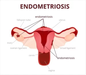
The uterus is a female reproductive organ located between the bladder and the rectum, in the pelvic area. The uterus has three layers: the inner lining (endometrium); the middle muscular layer (myometrium); and the outer layer (perimetrium). The uterus is connected to the fallopian tubes, the cervix and (via the cervix) the vagina.

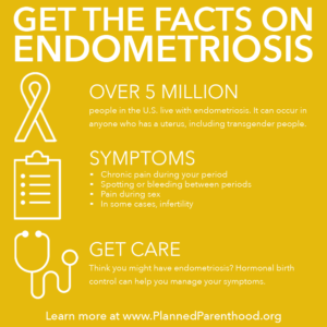
Endometriosis is a painful, chronic disease that affects at least 6.3 million women and girls in the U.S., 1 million in Canada, and millions more worldwide. It occurs when tissue like that which lines the uterus (tissue called the endometrium) is found outside the uterus.  The symptoms occur during menses and the endometrial tissue outside the uterus will be painful like the uterus cramps due to the period.

What Causes Endometriosis?

The cause of endometriosis is unknown. The retrograde menstruation theory (transtubal migration theory) suggests that during menstruation some of the menstrual tissue backs up through the fallopian tubes, implants in the abdomen, and grows.  Some experts believe that all women experience some menstrual tissue backup and that an immune system problem or a hormonal problem allows this tissue to grow in the women who develop endometriosis.

Another theory suggests that endometrial tissue is distributed from the uterus to other parts of the body through the lymph system or through the blood system. A genetic theory suggests that it may be carried in the genes in certain families or that some families may have predisposing factors to endometriosis.

Surgical transplantation has also been cited in many cases where endometriosis is found in abdominal scars, although it has also been found in such scars when accidental implantation seems unlikely.

Another theory suggests that remnants of tissue from when the woman was an embryo may later develop into endometriosis, or that some adult tissues retain the ability they had in the embryo stage to transform reproductive tissue in certain circumstances.

Research by the Endometriosis Association revealed a startling link between dioxin (TCCD) exposure and the development of endometriosis. Dioxin is a toxic chemical byproduct of pesticide manufacturing, bleached pulp and paper products, and medical and municipal waste incineration. The EA discovered a colony of rhesus monkeys that had developed endometriosis after exposure to dioxin. 79% of the monkeys exposed to dioxin developed endometriosis, and, in addition, the more dioxin exposure, the more severe the endometriosis.

Endometriosis tissus found outside of the uterus:

  • Outside and back of your uterus.
  • Fallopian tubes.
  • Ovaries.
  • Vagina.
  • Peritoneum (the lining of your abdomen and pelvis).
  • Bladder and ureters.
  • Intestines.
  • Rectum.
  • Diaphragm (a muscle near the bottom of your chest that plays an important role in breathing).
  • Less commonly they are found in the lung, arm, thigh, and other locations.

This misplaced tissue develops into growths or lesions which respond to the menstrual cycle in the same way that the tissue of the uterine lining does: each month the tissue builds up, breaks down, and sheds. Menstrual blood flows from the uterus and out of the body through the vagina, but the blood and tissue shed from endometrial growths has no way of leaving the body. This results in internal bleeding, breakdown of the blood and tissue from the lesions, and inflammation — and can cause pain, infertility, scar tissue formation, adhesions, and bowel problems.

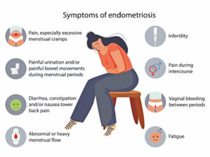
Pain before and during period: Pain with sex, Infertility, Fatigue, Painful urination during periods, Painful bowel movements during periods and Other Gastrointestinal upsets such as diarrhea, constipation, nausea.

In addition, many women with endometriosis suffer from:

  • Allergies
  • Chemical sensitivities
  • Frequent yeast infections

Diagnosis:

Diagnosis is considered uncertain until proven by laparoscopy, a minor surgical procedure done under anesthesia. A laparoscopy usually shows the location, size, and extent of the growths. This helps the doctor and patient make better treatment choices.


QUOTE FOR MONDAY:

“U.S. adult women age 18 and older (33%) are more likely than men (27%) to wake up every night and more likely to stay up late every night (31% vs. 20%).

More than half of women ages 25-44 with children get less than 7 hours of sleep per night, compared to 38% of men in this group.

Men in this group also get 30 minutes more sleep per night than the women.

Within this group, twice as many women are woken every night to care for others compared to men.

Nearly a quarter of women ages 18-24 lose sleep due to premenstrual syndrome (PMS), while more than half of women ages 45-54 lose sleep due to perimenopause or menopause.

Women (51%) are more likely than men (42%) to attribute their sleep issues to anxiety and depression.”

SleepFoundation.org

(https://www.sleepfoundation.org/sleep-news/the-sleep-gender-gap-nighttime-disparities-between-women-and-men)

How good are you sleeping?


How well do you sleep? 

When you go to bed do you wake up to the slightest noise (car going by the house, a dog barking outside, the heat going on or the cat fight outside)?  I know I do and have for years; unfortunately the slightest noise wakes me up, even a pin dropping to the floor. I probably live on 5 to 6 hours of sleep a day.  Some people even feel when they are scrambling to meet the countless demands of their day, cutting back on sleep might seem like the only answer. Who can afford to spend so much time sleeping, anyway? The truth is you can’t afford not to. Even minimal sleep loss takes a toll on your mood, energy, and ability to handle stress. By understanding your nightly sleep needs and what you can do to bounce back from chronic sleep loss, you can finally get on a healthy sleep schedule or pattern.

Sleep deprivation

Many of us try to sleep as little as possible. There are so many things that seem more interesting or important than getting a few more hours of sleep, but just as exercise and nutrition are essential for optimal health and happiness, so is sleep. The quality of your sleep directly affects the quality of your waking life, including your mental sharpness, productivity, emotional balance, creativity, physical vitality, and even your weight. No other activity delivers so many benefits with so little effort!

According to the National Institutes of Health, the average adult sleeps less than seven hours per night. In today’s fast-paced society, six or seven hours of sleep may sound pretty good. In reality, though, it’s a recipe for chronic sleep deprivation.

Remember sleep deprivation can affect you like a drunk.  This could be many symptoms like fatigue, lethargy, lack of motivation, moodiness and irritability, reduced creativity and problem-solving skills, inability to cope with stress, reduced immunity; frequent colds and infections, concentration and memory problems, weight gain, impaired motor skills, increased risk of accidents, difficulty making decisions to even increased risk of diabetes, heart disease, and other health problems.

 On your average sleep schedule there is a big difference between the amount of sleep you allow yourself to get so you can make it through each day compared to the amount you need to function optimally. Just because you’re able to operate on seven hours of sleep doesn’t mean you wouldn’t feel a lot better and get more done if you spent an extra hour or two in bed.

While sleep requirements vary slightly from person to person, most healthy adults need between seven and a half to nine hours of sleep per night to function at their best. Children and teens need even more. And despite the notion that our sleep needs decrease with age, older people still need at least seven and a half to eight hours of sleep. Since older adults often have trouble sleeping this long at night, daytime naps can help fill in the gap.

The best way to figure out if you’re meeting your sleep needs is to evaluate how you feel as you go about your day. If you’re logging enough hours, you’ll feel energetic and alert all day long, from the moment you wake up until your regular bedtime.

Consistency doing the following measures is the key to prevent sleep deprivation: Settle short-term sleep debt with an extra hour or two per night, Keep a sleep diary, Take a sleep vacation to pay off a long-term sleep debt, finally you must make sleep a priority.

Understanding sleep

Sleep isn’t exactly a time when your body and brain shut off. While you rest, your brain stays busy, overseeing a wide variety of biological maintenance that keeps your body running in top condition, preparing you for the day ahead. Without enough hours of restorative sleep, you won’t be able to work, learn, create, and communicate at a level even close to your true potential. Regularly skimp on “service” and you’re headed for a major mental and physical breakdown.

The good news is that you don’t have to choose between health and productivity. As you start getting the sleep you need, your energy and efficiency will go up. In fact, you’re likely to find that you actually get more done during the day than when you were skimping on shuteye.

It’s not just the number of hours in bed that’s important—it’s the quality of those hours of sleep. If you’re giving yourself plenty of time for sleep, but you’re still having trouble waking up in the morning or staying alert all day, you may not be spending enough time in the different stages of sleep.

Each stage of sleep in the sleep cycle offers benefits to the sleeper. However, deep sleep (Stage N3) and REM sleep are particularly important.  Learn more about the stages of sleep tomorrow and maybe it will help you strive for better health!

QUOTE FOR FRIDAY:

“Prothrombin deficiency is a disorder caused by a lack of a protein in the blood called prothrombin. It leads to problems with blood clotting (coagulation). Prothrombin is also known as factor II (factor two).

When you bleed, a series of reactions take place in the body that helps blood clots form. This process is called the coagulation cascade. It involves special proteins called coagulation, or clotting, factors. You may have a higher chance of excess bleeding if one or more of these factors are missing or are not functioning like they should.

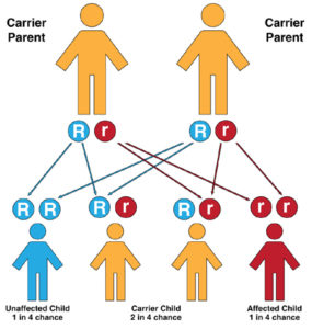
Prothrombin, or factor II, is one such coagulation factor. Prothrombin deficiency runs in families (inherited) and is very rare. Both parents must have the gene to pass the disorder on to their children. A family history of a bleeding disorder can be a risk factor.

Prothrombin deficiency can also be due to another condition or use of certain medicines. This is called acquired prothrombin deficiency.”

Mount Sinai  (https://www.mountsinai.org/health-library/diseases-conditions/factor-ii-deficiency)


Bleeding Disorder Month – Prothrombin Deficiency

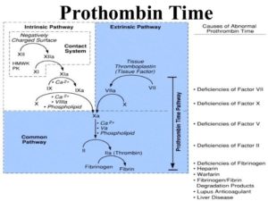
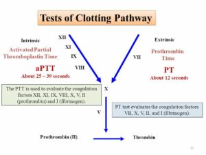
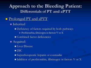
Prolonged PT and aPTT. Inherited. Deficiency of factors required by both pathways. Prothrombin, fibrinogen or factors V or X. Combined factor deficiencies. Acquired: Liver Disease. DIC. Supratherapeutic heparin or coumadin. Inhibitor of prothrombin, fibrinogen or factors V or X.


Prothrombin deficiency is a bleeding disorder that slows the blood clotting process. People with this condition often experience prolonged bleeding following an injury, surgery, or having a tooth pulled. … Women with prothrombin deficiency can have prolonged and sometimes abnormally heavy menstrual bleeding.  This blood clotting factor II deficiency.  Remember we have blood clotting factors that have to be present for the blood clotting factor but if one is lacking then bleeding can occur.

Practice Essentials

Clotting factor II, or prothrombin, is a vitamin K–dependent proenzyme that functions in the blood coagulation cascade. Factor II deficiency is a rare, inherited or acquired bleeding disorder disorder with an estimated incidence of one case per 2 million population. [1]

Inherited factor II deficiency is an autosomal recessive disorder that can manifest as hypoprothrombinemia, a decrease in the overall synthesis of prothrombin; or as dysprothrombinemia, the synthesis of dysfunctional prothrombin. [2, 3, 4] Homozygous individuals are generally asymptomatic and have functional prothrombin levels of 2-25%. However, symptomatic individuals may experience one or more of the following [1] :

  • Easy bruising
  • Epistaxis
  • Soft-tissue hemorrhage
  • Excessive postoperative bleeding
  • Menorrhagia
  • Muscle hematomas
  • Hemarthrosis
  • Intracranial bleeding

In true hypoprothrombinemia, immunologic assays correlate well with functional assays in that both reveal low prothrombin values. Heterozygous patients are generally asymptomatic and have prothrombin levels of 50% or greater on both immunologic and functional assays.

In dysprothrombinemia, only the functional assay for prothrombin returns significantly reduced values, whereas the immunologic assay reveals normal values. Acquired factor II deficiency can be caused by severe liver disease, vitamin K deficiency, anticoagulant drugs (eg, warfarin), or the presence of an antibody directed against the protein. [5]

Aside from the prothrombin deficiencies, another disorder of prothrombin is the prothrombin 20210a mutation. First reported in 1996 as a familial cause of venous thromboembolism, the prothrombin 20210a mutation results in increased levels of plasma prothrombin and a concurrent increased risk for the development of thrombosis. [6]

Prothrombin 20210a has an estimated prevalence of 2% in whites. [7, 8] The mutation is more prevalent in those of southern European descent than in those of northern European descent, and it is rarely seen in Asians or Africans. [7] A study of patients in Turkey revealed the presence of the prothrombin 20210a mutation in 0.7% of subjects. [9]

Individuals carrying the prothrombin 20210a mutation have a 2- to 3-fold increased risk for developing thrombosis. [6, 10] One case-control study found evidence of an increased risk of developing an ischemic cerebrovascular event in men aged younger than 60 years with the prothrombin 20210a mutation. [11] A study of cancer patients in the Netherlands found that the presence of the prothrombin 20210a mutation in these patients may increase the risk of venous thrombosis to a level greater than that attributable to the malignancy alone. [12]

The prothrombin 20210a mutation can be identified without DNA analysis and should be considered in any patient experiencing a thrombotic event without other risk factors. Treatment with oral anticoagulants is useful in preventing recurrence in patients with the mutation who have already experienced a thrombotic event. Additionally, women who are known to carry the mutation may want to avoid oral contraceptives because of the additional risk of thrombosis.

Laboratory studies for factor II deficiency include coagulation studies and clotting factor assays (see Workup). Coagulation study results are as follows:

  • Prothrombin time (PT) is prolonged
  • Activated partial thromboplastin time (aPTT) is prolonged
  • Bleeding time is within reference range

Treatment of factor II deficiency is aimed at restoring circulating factor II to levels sufficient for hemostasis. Levels greater than 30% of normal are usually adequate. Treatment measures include fresh frozen plasma (FFP), prothrombin complex concentrates (PCCs), and vitamin K. Additionally, in patients with acquired factor II deficiency, the underlying cause should be found and treated.

Frequency

Both congenital and acquired factor II deficiencies are rare. The prevalence of congenital factor II deficiency is approximately 1 per 1 to 2 million population. [41]

Mortality/Morbidity

Congenital factor II deficiency is a lifelong bleeding disorder. Death can result because of massive hemorrhage from relatively minor accidents or trauma. Hemorrhage can also occur as a result of surgery if precautions are not taken. Intracranial bleeding is another serious sequela of this disorder. Rarely, hemarthroses can occur. [2]

Myocardial infarction is a rare complication in young people, with coronary thrombosis due to hypercoagulable states being one cause. A heterozygote prothrombin gene mutation (G-20210-A) and protein S deficiency were described in a 19-year-old with a myocardial infarction with normal coronary arteries. [42]

Race- sex-, and age-related demographics

Factor II deficiency has no known racial or ethnic predilection. Males and females are affected equally. Patients with severe congenital factor II deficiency present early in life, whereas those with less severe forms can present at any age. Acquired forms can be observed in all age groups.


Bleeding Disorder Awareness Month – Von Willebrand Disease

It arises from a deficiency in the quality or quantity of von Willebrand factor (vWF), a multimeric protein that is required for platelet adhesion.

Von Willebrand disease (VWD) is a genetic disorder caused by missing or defective von Willebrand factor (VWF), a clotting protein. VWF binds factor VIII, a key clotting protein, and platelets in blood vessel walls, which help form a platelet plug during the clotting process. The condition is named after Finnish physician Erik von Willebrand, a who first described it in the 1920s.

VWD is the most common bleeding disorder, affecting up to 1% of the US population. It is carried on chromosome 12 and occurs equally in men and women.

Symptoms

People with VWD experience frequent nosebleeds, easy bruising and excessive bleeding during and after invasive procedures, such as tooth extractions and surgery. Women often experience menorrhagia, heavy menstrual periods that last longer than average, and hemorrhaging after childbirth.

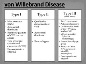
There are three main types of VWD based on qualitative or quantitative defects in VWF. A fourth type, acquired VWD, is not hereditary.

  • Type 1 VWD is found in 60%-80% of patients. People with type 1 VWD have a quantitative deficiency of VWF. Levels of VWF in the blood range from 20%-50% of normal. The symptoms are usually mild.
  • Type 2 VWD is found in 15%-30% of patients. People with type 2 VWD have a qualitative deficiency in their VWF. Type 2 is broken down into four subtypes: type 2A, type 2B, type 2M and type 2N, depending on the presence and behavior of multimers, molecular chains of VWF. Symptoms are mild to moderate.
  • Type 3 VWD is found in 5%-10% of patients. People with type 3 VWD have a quantitative deficiency of VWF. Symptoms are typically severe, and include spontaneous bleeding episodes, often into their joints and muscles.
  • Acquired VWD. This type of VWD in adults results after a diagnosis of an autoimmune disease, such as lupus, or from heart disease or some types of cancer. It can also occur after taking certain medications.

Diagnosis

The best place for patients with bleeding disorders to be diagnosed and treated is at one of the federally-funded hemophilia treatment centers (HTCs) that are spread throughout the country. HTCs provide comprehensive care from skilled hematologists and other professional staff, including nurses, physical therapists, social workers and sometimes dentists, dieticians and other healthcare providers.

A medical health history is important to help determine if other relatives have been diagnosed with a bleeding disorder or have experienced symptoms. Tests that evaluate clotting time and a patient’s ability to form a clot may be ordered. A clotting factor test, called an assay, and tests measuring platelet function also may be performed. The VWF antigen test measures the amount of VWF in blood plasma. Patients with VWD typically have <50% of normal VWF in their plasma. After VWD is confirmed, a test to determine the exact type is performed.

It should be noted that diagnostic testing to confirm VWD may have to be repeated because levels of VWF fluctuate. VWF can rise due to stress, exercise, the use of oral contraceptives, pregnancy and hyperthyroidism.

Treatment

Treatment for VWD depends on the diagnosis and severity. The mainstay of treatment is DDAVP (desmopressin acetate), the synthetic version of a natural hormone vasopressin,. It stimulates the release of VWF from cells, which also increases FVIII.DDAVP comes in two forms: injectable and nasal spray. Because DDAVP is an antidiuretic, causing the body to retain water, fluid restrictions are important so patients don’t develop hyponatremia, reduced sodium in the bloodstream.

There are a few clotting factor concentrates that are rich in VWF, and are recommended for patients with VWD. These therapies are given by intravenous infusion. In December 2015, the US Food and Drug Administration (FDA) approved Baxalta’s Vonvendi®, the first recombinant VWF product. Unlike other products, it contains VWF only, not VWF and factor VIII. It is approved to treat on-demand and for control of bleeding in adults 18 and older.

Aminocaproic acid and tranexamic acid are antifibrinolytics agents that prevent the breakdown of blood clots. These drugs are often recommended before dental procedures, to treat nose and mouth bleeds, and for menorrhagia.  Antifibrinolytics are taken orally, as a tablet or liquid. MASAC recommends that a dose of clotting factor be taken first to form a clot, then aminocaproic acid, to preserve the clot and keep it from being prematurely broken down.

QUOTE FOR WEDNESDAY:

“Your kidneys, each just the size of a computer mouse, filter all the blood in your body every 30 minutes. They work hard to remove wastes, toxins, and excess fluid. They also help control blood pressure, stimulate production of red blood cells, keep your bones healthy, and regulate blood chemicals that are essential to life.

Kidneys that function properly are critical for maintaining good health, however, more than one in seven American adults are estimated to have chronic kidney disease (CKD).”

Centers for Disease Control and Prevention – CDC

(https://www.cdc.gov/kidneydisease/basics.html#:~:text=disease (CKD).-,About Chronic Kidney Disease,as heart disease and stroke.)