QUOTE FOR FRIDAY:

“Prothrombin deficiency is a disorder caused by a lack of a protein in the blood called prothrombin. It leads to problems with blood clotting (coagulation). Prothrombin is also known as factor II (factor two).

When you bleed, a series of reactions take place in the body that helps blood clots form. This process is called the coagulation cascade. It involves special proteins called coagulation, or clotting, factors. You may have a higher chance of excess bleeding if one or more of these factors are missing or are not functioning like they should.

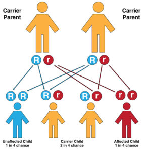
Prothrombin, or factor II, is one such coagulation factor. Prothrombin deficiency runs in families (inherited) and is very rare. Both parents must have the gene to pass the disorder on to their children. A family history of a bleeding disorder can be a risk factor.

Prothrombin deficiency can also be due to another condition or use of certain medicines. This is called acquired prothrombin deficiency.”

Mount Sinai  (https://www.mountsinai.org/health-library/diseases-conditions/factor-ii-deficiency)


Bleeding Disorder Month – Prothrombin Deficiency

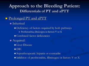
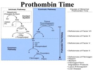
Prolonged PT and aPTT. Inherited. Deficiency of factors required by both pathways. Prothrombin, fibrinogen or factors V or X. Combined factor deficiencies. Acquired: Liver Disease. DIC. Supratherapeutic heparin or coumadin. Inhibitor of prothrombin, fibrinogen or factors V or X.


Prothrombin deficiency is a bleeding disorder that slows the blood clotting process. People with this condition often experience prolonged bleeding following an injury, surgery, or having a tooth pulled. … Women with prothrombin deficiency can have prolonged and sometimes abnormally heavy menstrual bleeding.  This blood clotting factor II deficiency.  Remember we have blood clotting factors that have to be present for the blood clotting factor but if one is lacking then bleeding can occur.

Practice Essentials

Clotting factor II, or prothrombin, is a vitamin K–dependent proenzyme that functions in the blood coagulation cascade. Factor II deficiency is a rare, inherited or acquired bleeding disorder disorder with an estimated incidence of one case per 2 million population. [1]

Inherited factor II deficiency is an autosomal recessive disorder that can manifest as hypoprothrombinemia, a decrease in the overall synthesis of prothrombin; or as dysprothrombinemia, the synthesis of dysfunctional prothrombin. [2, 3, 4] Homozygous individuals are generally asymptomatic and have functional prothrombin levels of 2-25%. However, symptomatic individuals may experience one or more of the following [1] :

  • Easy bruising
  • Epistaxis
  • Soft-tissue hemorrhage
  • Excessive postoperative bleeding
  • Menorrhagia
  • Muscle hematomas
  • Hemarthrosis
  • Intracranial bleeding

In true hypoprothrombinemia, immunologic assays correlate well with functional assays in that both reveal low prothrombin values. Heterozygous patients are generally asymptomatic and have prothrombin levels of 50% or greater on both immunologic and functional assays.

In dysprothrombinemia, only the functional assay for prothrombin returns significantly reduced values, whereas the immunologic assay reveals normal values. Acquired factor II deficiency can be caused by severe liver disease, vitamin K deficiency, anticoagulant drugs (eg, warfarin), or the presence of an antibody directed against the protein. [5]

Aside from the prothrombin deficiencies, another disorder of prothrombin is the prothrombin 20210a mutation. First reported in 1996 as a familial cause of venous thromboembolism, the prothrombin 20210a mutation results in increased levels of plasma prothrombin and a concurrent increased risk for the development of thrombosis. [6]

Prothrombin 20210a has an estimated prevalence of 2% in whites. [7, 8] The mutation is more prevalent in those of southern European descent than in those of northern European descent, and it is rarely seen in Asians or Africans. [7] A study of patients in Turkey revealed the presence of the prothrombin 20210a mutation in 0.7% of subjects. [9]

Individuals carrying the prothrombin 20210a mutation have a 2- to 3-fold increased risk for developing thrombosis. [6, 10] One case-control study found evidence of an increased risk of developing an ischemic cerebrovascular event in men aged younger than 60 years with the prothrombin 20210a mutation. [11] A study of cancer patients in the Netherlands found that the presence of the prothrombin 20210a mutation in these patients may increase the risk of venous thrombosis to a level greater than that attributable to the malignancy alone. [12]

The prothrombin 20210a mutation can be identified without DNA analysis and should be considered in any patient experiencing a thrombotic event without other risk factors. Treatment with oral anticoagulants is useful in preventing recurrence in patients with the mutation who have already experienced a thrombotic event. Additionally, women who are known to carry the mutation may want to avoid oral contraceptives because of the additional risk of thrombosis.

Laboratory studies for factor II deficiency include coagulation studies and clotting factor assays (see Workup). Coagulation study results are as follows:

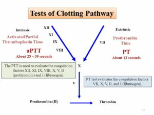
  • Prothrombin time (PT) is prolonged
  • Activated partial thromboplastin time (aPTT) is prolonged
  • Bleeding time is within reference range

Treatment of factor II deficiency is aimed at restoring circulating factor II to levels sufficient for hemostasis. Levels greater than 30% of normal are usually adequate. Treatment measures include fresh frozen plasma (FFP), prothrombin complex concentrates (PCCs), and vitamin K. Additionally, in patients with acquired factor II deficiency, the underlying cause should be found and treated.

Frequency

Both congenital and acquired factor II deficiencies are rare. The prevalence of congenital factor II deficiency is approximately 1 per 1 to 2 million population. [41]

Mortality/Morbidity

Congenital factor II deficiency is a lifelong bleeding disorder. Death can result because of massive hemorrhage from relatively minor accidents or trauma. Hemorrhage can also occur as a result of surgery if precautions are not taken. Intracranial bleeding is another serious sequela of this disorder. Rarely, hemarthroses can occur. [2]

Myocardial infarction is a rare complication in young people, with coronary thrombosis due to hypercoagulable states being one cause. A heterozygote prothrombin gene mutation (G-20210-A) and protein S deficiency were described in a 19-year-old with a myocardial infarction with normal coronary arteries. [42]

Race- sex-, and age-related demographics

Factor II deficiency has no known racial or ethnic predilection. Males and females are affected equally. Patients with severe congenital factor II deficiency present early in life, whereas those with less severe forms can present at any age. Acquired forms can be observed in all age groups.


Bleeding Disorder Awareness Month – Von Willebrand Disease

It arises from a deficiency in the quality or quantity of von Willebrand factor (vWF), a multimeric protein that is required for platelet adhesion.

Von Willebrand disease (VWD) is a genetic disorder caused by missing or defective von Willebrand factor (VWF), a clotting protein. VWF binds factor VIII, a key clotting protein, and platelets in blood vessel walls, which help form a platelet plug during the clotting process. The condition is named after Finnish physician Erik von Willebrand, a who first described it in the 1920s.

VWD is the most common bleeding disorder, affecting up to 1% of the US population. It is carried on chromosome 12 and occurs equally in men and women.

Symptoms

People with VWD experience frequent nosebleeds, easy bruising and excessive bleeding during and after invasive procedures, such as tooth extractions and surgery. Women often experience menorrhagia, heavy menstrual periods that last longer than average, and hemorrhaging after childbirth.

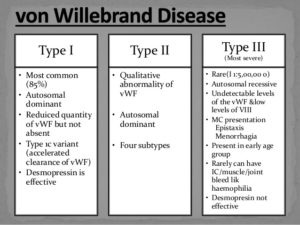
There are three main types of VWD based on qualitative or quantitative defects in VWF. A fourth type, acquired VWD, is not hereditary.

  • Type 1 VWD is found in 60%-80% of patients. People with type 1 VWD have a quantitative deficiency of VWF. Levels of VWF in the blood range from 20%-50% of normal. The symptoms are usually mild.
  • Type 2 VWD is found in 15%-30% of patients. People with type 2 VWD have a qualitative deficiency in their VWF. Type 2 is broken down into four subtypes: type 2A, type 2B, type 2M and type 2N, depending on the presence and behavior of multimers, molecular chains of VWF. Symptoms are mild to moderate.
  • Type 3 VWD is found in 5%-10% of patients. People with type 3 VWD have a quantitative deficiency of VWF. Symptoms are typically severe, and include spontaneous bleeding episodes, often into their joints and muscles.
  • Acquired VWD. This type of VWD in adults results after a diagnosis of an autoimmune disease, such as lupus, or from heart disease or some types of cancer. It can also occur after taking certain medications.

Diagnosis

The best place for patients with bleeding disorders to be diagnosed and treated is at one of the federally-funded hemophilia treatment centers (HTCs) that are spread throughout the country. HTCs provide comprehensive care from skilled hematologists and other professional staff, including nurses, physical therapists, social workers and sometimes dentists, dieticians and other healthcare providers.

A medical health history is important to help determine if other relatives have been diagnosed with a bleeding disorder or have experienced symptoms. Tests that evaluate clotting time and a patient’s ability to form a clot may be ordered. A clotting factor test, called an assay, and tests measuring platelet function also may be performed. The VWF antigen test measures the amount of VWF in blood plasma. Patients with VWD typically have <50% of normal VWF in their plasma. After VWD is confirmed, a test to determine the exact type is performed.

It should be noted that diagnostic testing to confirm VWD may have to be repeated because levels of VWF fluctuate. VWF can rise due to stress, exercise, the use of oral contraceptives, pregnancy and hyperthyroidism.

Treatment

Treatment for VWD depends on the diagnosis and severity. The mainstay of treatment is DDAVP (desmopressin acetate), the synthetic version of a natural hormone vasopressin,. It stimulates the release of VWF from cells, which also increases FVIII.DDAVP comes in two forms: injectable and nasal spray. Because DDAVP is an antidiuretic, causing the body to retain water, fluid restrictions are important so patients don’t develop hyponatremia, reduced sodium in the bloodstream.

There are a few clotting factor concentrates that are rich in VWF, and are recommended for patients with VWD. These therapies are given by intravenous infusion. In December 2015, the US Food and Drug Administration (FDA) approved Baxalta’s Vonvendi®, the first recombinant VWF product. Unlike other products, it contains VWF only, not VWF and factor VIII. It is approved to treat on-demand and for control of bleeding in adults 18 and older.

Aminocaproic acid and tranexamic acid are antifibrinolytics agents that prevent the breakdown of blood clots. These drugs are often recommended before dental procedures, to treat nose and mouth bleeds, and for menorrhagia.  Antifibrinolytics are taken orally, as a tablet or liquid. MASAC recommends that a dose of clotting factor be taken first to form a clot, then aminocaproic acid, to preserve the clot and keep it from being prematurely broken down.

QUOTE FOR WEDNESDAY:

“Your kidneys, each just the size of a computer mouse, filter all the blood in your body every 30 minutes. They work hard to remove wastes, toxins, and excess fluid. They also help control blood pressure, stimulate production of red blood cells, keep your bones healthy, and regulate blood chemicals that are essential to life.

Kidneys that function properly are critical for maintaining good health, however, more than one in seven American adults are estimated to have chronic kidney disease (CKD).”

Centers for Disease Control and Prevention – CDC

(https://www.cdc.gov/kidneydisease/basics.html#:~:text=disease (CKD).-,About Chronic Kidney Disease,as heart disease and stroke.)

Part III March is kidney month – Chronic Kidney Failure


Chronic Renal or Kidney Disease (CRF):

In giving a short and easily understandable definition Chronic kidney disease happens when your kidneys no longer filter your blood the way they should, so wastes (toxins, usually end products of an acid) build up in your blood. This has probably been going on for years, and it may keep getting worse over time. Just like a car engine damaged but still using the car without getting the engine repaired sooner or later in time the engine no longer functions the same with any organ of the body getting damaged by some long term condition. If your disease gets worse and worse over time, you could have kidney failure or some multi organ failure, depending on the condition causing this.

Regarding Chronic Kidney Failure the causes can be:

** Diabetes (uncontrolled diabetes (Type 1 or 2) for many years.

** High blood pressure for many years.

These are the top 2 causes of most chronic kidney disease. Controlling these diseases can help slow or stop the damage to the individual’s kidneys who has one of these, if not both.

Other common causes of chronic renal failure (CRF) include:

-recurring pyelonephritis (kidney infection)

-polycystic kidney disease (multiple cysts in the kidneys

-autoimmune disorders such as systemic lupus erythematosus.

-hardening of the arteries, which can damage blood vessels in the kidney.

-A narrowed or blocked renal artery. A renal artery carries blood to the kidneys. Know this for starters, each of your kidneys has about a million tiny filters, called nephrons. The nephron is the tiny filtering structure in your kidneys. Each of your kidneys contain more than a million tiny filtering nephrons that help clean your blood removing toxins dumping them into your urinary bladder so you can evacuate them though urine (urea, urine; get it). Your nephrons play a vital role to our essential daily living. If over a long time you have a renal artery blocked the nephrons stop their function and die.

Remember the nephrons help all humans do the following if there kidneys or one kidney is functioning properly:

-Remove excess water, wastes (like urea, ammonia, etc.) & other substances from your blood.

-Return substances like sodium, potassium or phosphorus to the body whenever any of these substances run llow in your body or do the opposite if they run high to evacuate them through voiding dumping the sodium or phosphorus or potassium in the urinary bladder through the tube from the kidneys to the urinary bladder called ureters.

**If nephrons are damaged by the high sugar content or high blood pressure in the kidneys, they stop working. For a while, healthy nephrons can take on the extra work or overload. But if the damage continues, more and more nephrons shut down. After a certain point, the nephrons that are left cannot filter your blood well enough to keep you’re blood filtered properly to keep you healthy. Just like running from a bear in the street chancing you. We can run only so long but sooner or later we will run out of energy and not be able to run anymore, same concept for the kidney nephrons when they run out of enough energy due to the kidneys not properly working.**

The symptoms can be:

Urinate less than normal.

Have swelling and weight gain from fluid buildup in your tissues. This is called edema.

Feel very tired or sleepy.

Not feel hungry, or you may lose weight without trying.

Often feel sick to your stomach (nauseated) or vomit.

Have trouble sleeping.

Have headaches or trouble thinking clearly.

So what will you do GO TO A DOCTOR OR CALL 911 AND GO TO THE ER:

Your doctor will do blood and urine tests to help find out how well your kidneys are working. These tests can show signs of kidney disease and anemia. (You can get anemia from having damaged kidneys.) You may have other tests to help rule out other problems that could cause your symptoms.

To diagnose chronic renal failure is pretty much the same tests that are listed above on acute renal failure plus:

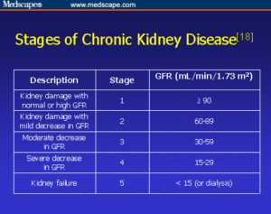
Chronic kidney disease is also called chronic renal failure or chronic renal insufficiency.There are five stages of kidney disease, from kidney damage with normal GFR to kidney failure.  So GFR will help the MD rule out acute versus chronic to give the MD direction on Rx.Your doctor will ask questions about any past kidney problems. He or she will also ask whether you have a family history of kidney disease and what medicines you take, both prescription and over-the-counter drugs.

Diagnosis & Treatment (Rx) for Chronic Kidney (Renal) Failure (CRF):

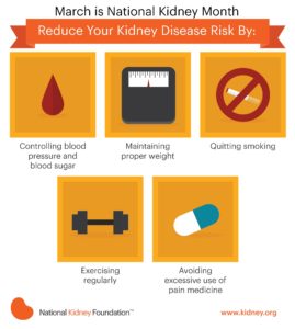
There are things you can do to slow or stop the damage to your kidneys. Taking medicines and making some lifestyle changes can help you manage your disease, prevent further damage to the kidneys, if their functioning at all and make you possibly feel better.

Kidney disease is a complex problem. You will probably need to take a number of medicines and have many tests. To stay as healthy as possible, take your medicines just the way your doctor says to and work closely with your doctor.

Go to all your appointments for the MD to see a increase in function or decrease in function of your kidney or kidneys you have still functioning to a level. To do that you can’t just go every 6 months especially when first diagnosed with it or with a collapse of an exacerbation of kidney failure in a worse level that brought on new symptoms that brought you to the ER.

Lifestyle changes are an important part of your treatment. Taking these steps can help slow down kidney disease and reduce your symptoms. These steps may also help with high blood pressure, diabetes, and other problems that make kidney disease worse or made the kidney disease happen with the secondary diagnosis you had originally for years (ex. Hypertension or Diabetes if not both especially is uncontrolled)

Very hard, never a complete 100 % resolution. It is like emphysema done by smokers the damage is done or like a heart attack the area of the infarction=damage is already done to the heart muscle.

Scared now, understandable but unfortunately the damage is done, so its get the organ to its optimal level of functioning or replace the damaged kidney (s) through a transplant of one.  You need to know this, do it if you want to live LONGER.  Remember fear is fear itself, the fear run it over and deal with what you have and make your life longer & better!  It’s all up to you.  HANDSOME I know you can do it , I am there for you as a good friend and professional RN and I would love you to last longer John!! XO

You may have a test done that lets your doctor look at a picture of your kidneys, such as an ultrasound or CT (Cat Scan of the kidneys). These tests can help your doctor measure the size of your kidneys, estimate blood flow to the kidneys, and see if urine flow is blocked. In some cases, your doctor may take a tiny sample of kidney tissue (biopsy) to help find out what caused your kidney disease.

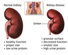
Chronic kidney disease is caused by damage of the kidneys whether the cause of it be primary a Renal or Kidney problem or a secondary, another disease or disorder that affects the kidneys in doing their job, like hyperglycemia related to a individual with uncontrolled diabetes, for instance.

Chronic kidney disease may seem to have come on suddenly. But it has been happening bit by bit for many years as a result of damage to your kidneys.

One way to measure how well your kidneys are working is to figure out your glomelular filtration rate (GFR). The GFR is usually calculated using results from your blood creatinine test. Then the stage of kidney disease is figured out using the GFR (glomelular filtration rate). There are five stages of kidney disease, from kidney damage with normal GFR to kidney failure.

Your doctor will do tests that measure the amount of urea (BUN) and creatinine in your blood. These tests can help measure how well your kidneys are filtering your blood. As your kidney function gets worse, the amount of nitrogen (shown by the BUN test) and creatinine in your blood increases. The level of creatinine in your blood is used to find out the glomerular filtration rate (GFR). The GFR is used to show how much kidney function you still have. The GFR is also used to find out the stage of your kidney disease your in if you have it and its to guide decisions about treatment. *
QUOTE FOR TUESDAY:

Acute kidney injury (AKI) is where your kidneys suddenly stop working properly. It can range from minor loss of kidney function to complete kidney failure.

AKI normally happens as a complication of another serious illness. It’s not the result of a physical blow to the kidneys, as the name might suggest.

This type of kidney damage is usually seen in older people who are unwell with other conditions and the kidneys are also affected.

It’s essential that AKI is detected early and treated promptly.

National Health Services-NHS (https://www.nhs.uk/conditions/acute-kidney-injury/)

Part II March is kidney month – Acute Kidney Damage

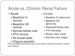
ARF VERSUS CRF1

Acute Renal (Kidney) Failure:

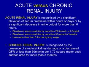
Kidney failure occurs when the kidneys lose their ability to function. To treat kidney failure effectively, it is important to know whether kidney disease has developed suddenly (acute) or over the long term (chronic). Many conditions, diseases, and medicines can create situations that lead to acute and chronic kidney disease. Acute kidney injury, also called acute renal failure, is more commonly reversible than chronic kidney failure since the chronic condition has lasted longer in the body affecting systems for several months to years (some decades). Acute Renal Failure is new to the body as opposed to chronic; making it higher odds this can be treated and cured.

When acute kidney injury (ARF) occurs, the kidneys are unable to remove waste products and excess fluids, which then build up in the body and upset the body’s normal chemical balance.*

The most common causes of acute kidney injury are:

-dehydration

-blood loss from major surgery or injury

-medicines such as nonsteroidal anti-inflammatory drugs (NSAIDs), antibiotics, or the dyes (contrast agents) used in X-ray tests.

Symptoms depend on the cause of acute renal failure and can include:

    • -Little or no urine output.
    • -Dizziness upon standing.
    • -Swelling, especially of the legs and feet.
    • -Loss of appetite, nausea, and vomiting.
    • -Feeling confused, anxious and restless, or sleepy.
    • -Pain in the flank, which is felt just below the rib cage and above the waist on one or both sides of the back.*

If your signs and symptoms suggest that you have acute kidney failure, your doctor may recommend certain tests and procedures to verify your diagnosis. These may include:

  • Urine output measurements. The amount of urine you excrete in a day may help your doctor determine the cause of your kidney failure.
  • Urine tests. Analyzing a sample of your urine, a procedure called urinalysis, may reveal abnormalities that suggest kidney failure.
  • Blood tests. A sample of your blood may reveal rapidly rising levels of urea and creatinine — two substances used to measure kidney function.  Most cases of acute kidney injury occur in people who are already in the hospital for other reasons. In these people, acute kidney injury is usually diagnosed when routine tests show a sudden increase in creatinine and blood urea nitrogen (BUN) levels.   **A buildup of these waste products in the blood points to a loss of kidney function!**
  • With a patient just coming in the first time to an MD with no history of renal disease but has factors or symptoms indicating possible kidney failure involvement don’t be surprised if the doctor orders glomerular filtration rate (GFR); which is a test used to check how well the kidneys are working. Specifically, it estimates how much blood passes through the glomeruli each minute. Glomeruli are tiny filters in the kidneys that filter waste from the blood.
  • Imaging tests. Imaging tests such as ultrasound and computerized tomography may be used to help your doctor see your kidneys.  Also commonly done is an ultrasound of the kidneys which may help determine whether kidney problems are acute or chronic. Normal-sized kidneys may be present in either condition, but when both kidneys are smaller than normal, chronic kidney disease is usually the problem.  This helps rule out acute from chronic.correcting the cause and supporting the kidneys with dialysis until proper functioning is restored.
  • Removing a sample of kidney tissue for testing. In some situations, your doctor may recommend a kidney biopsy to remove a small sample of kidney tissue for lab testing. Your doctor inserts a needle through your skin and into your kidney to remove the sample.

TREATMENT FOR ARF (reversible in most cases):

Treatment for acute kidney failure typically requires a hospital stay. Most people with acute kidney failure are already hospitalized. How long you’ll stay in the hospital depends on the reason for your acute kidney failure and how quickly your kidneys recover.

In some cases, you may be able to recover at home.

Treating the underlying cause of your AKF:

Treatment for acute kidney failure (AKF) involves identifying the illness or injury that originally damaged your kidneys. Your treatment options depend on what’s causing your kidney failure.

Learn tomorrow Chronic Kidney Failure (CKF).


QUOTE FOR MONDAY:

“The body takes nutrients from food and converts them to energy. After the body has taken the food components that it needs, waste products are left behind in the bowel and in the blood.

The kidney and urinary systems help the body to eliminate liquid waste called urea, and to keep chemicals, such as potassium and sodium, and water in balance. Urea is produced when foods containing protein, such as meat, poultry, and certain vegetables, are broken down in the body. Urea is carried in the bloodstream to the kidneys, where it is removed along with water and other wastes in the form of urine.”

John Hopkins Medicine (https://www.hopkinsmedicine.org/health/wellness-and-prevention/anatomy-of-the-urinary-system)

Part I March is Kidney Month – The importance of kidneys and know its functions to understand kidney failure.

kidney 3

    kidney failure

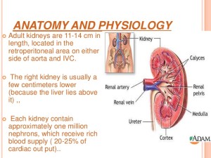
The kidneys are important organs with many functions in the body, including producing hormones, absorbing minerals, and filtering blood and producing urine. While they are important and kidney failure can be fatal, a human only needs one healthy kidney to survive.

The kidneys are two bean-shaped organs that extract waste from blood, balance body fluids, form urine, and aid in other important functions of the body.

They reside against the back muscles in the upper abdominal cavity. They sit opposite each other on either side of the spine. The right kidney sits a little bit lower than the left to accommodate the liver.

When it comes to components of the urinary system, the kidneys are multi-functional powerhouses of activity, for if the kidneys aren’t working, meaning they don’t filter toxic wastes out of our blood stream (with other functions it does) than the waste products don’t get dumped into the urinary bladder from the renal tubes, called right and left ureters. In human anatomy, the ureters are tubes made of smooth muscle fibers that propel urine from the kidneys to the urinary bladder. If the kidneys are not working they are not filtering our blood (same principle as filtering beer to make it to perfection, the kidneys do it for our blood to be able to have the cells do their function to the optimal levels with keeping toxins out of the body in preventing many blood problems with more due to acidosis (toxin build up). In the adult, the ureters are usually 25–30 cm (10–12 in) long and ~3–4 mm in diameter.

The kidneys have multiple functions! Some of the core actions of a healthy kidney or kidneys of a human body include:

  • Waste excretion: There are many things your body doesn’t want inside of it. The kidneys filter out toxins, excess salts, and urea (a toxin), a nitrogen-based waste created by cell metabolism.
  • * Urea is an organic chemical compound and is essentially the waste produced by the body after metabolizing protein. Naturally the compound urea is produced when the liver breaks down protein or amino acids, and ammonia, the kidneys then transfer the urea from the blood to the urine, when they do filtering of the blood.  Urea is a byproduct of protein metabolism, the ending result. Extra nitrogen is expelled from the body through urea because it is extremely soluble (solid); it is a very efficient process. The average person excretes about 30 grams of urea a day, mostly through urine but a small amount is also secreted in perspiration. Synthetic versions of the chemical compound can be created in liquid or solid form and is often an ingredient found in fertilizers, animal food, and diuretics, just to name a few . Urea is what gives our urine the color yellow.         In the gastrointestinal tract, blood proteins are broken down into ammonia (could be due to high protein eating to drugs with actual conditions); and goes to the liver converting it to Urea. It is then released into the blood stream where the kidney’s take it up and eliminate it. Urea is then eliminated by the kidney’s, but not produced by it.  Urea is synthesized in the liver and transported through the blood to the kidneys for removal.
  •  A Healthy Kidney or Kidneys functions in the human body doing:
  • Water level balancing: As the kidneys are key in the chemical breakdown of urine, they react to changes in the body’s water level throughout the day. As water intake decreases, the kidneys adjust accordingly and leave water in the body instead of helping excrete it which aides in electrolyte balancing in the blood with keeping the body hydrated properly.
  • Blood pressure regulation: The kidneys need constant pressure to filter the blood. When it drops too low, the kidneys increase the pressure. One way is by producing a blood vessel-constricting protein (angiotensin) that also signals the body to retain sodium and water. Both the constriction and retention help restore normal blood pressure.
  • Red blood cell regulation: When the kidneys don’t get enough oxygen, they send out a distress call in the form of erythropoietin, a hormone that stimulates the bone marrow to produce more oxygen-carrying red blood cells.
  • Acid regulation: As cells metabolize, they produce acids. Foods we eat can either increase the acid in our body or neutralize it. If the body is to function properly, it needs to keep a healthy balance of these chemicals. The kidneys do that, too.Because of all of the vital functions the kidneys perform and the toxins they encounter, the kidneys are susceptible to various problems.
  • Acute kidney failure is a condition in which the kidneys suddenly lose their ability to function properly. This can occur for many reasons, including:

  • Infection
  • Blood-clotting disorders
  • Decreased blood flow caused by low blood pressure
  • Autoimmune kidney disorders
  • Urinary tract infections
  • Complications from pregnancy
  • Most people are born with two kidneys, but many people can live on just one. Kidney transplant surgeries with live donors are common medical procedures today. *
  • Chronic kidney failure – same as acute in that the kidney (s) loses its function. 
  • DehydrationDiseases and conditions that commonly cause chronic kidney disease include:
  • Type 1 or type 2 diabetes.
  • High blood pressure
  • Glomerulonephritis (gloe-mer-u-lo-nuh-FRY-tis), an inflammation of the kidney’s filtering units (glomeruli)
  • Interstitial nephritis, an inflammation of the kidney’s tubules and surrounding structures
  • Polycystic kidney disease
  • Prolonged obstruction of the urinary tract, from conditions such as enlarged prostate, kidney stones and some cancers
  • Vesicoureteral (ves-ih-koe-yoo-REE-ter-ul) reflux, a condition that causes urine to back up into your kidneys
  • Recurrent kidney infection, also called pyelonephritis (pie-uh-lo-nuh-FRY-tis)
  • A chronic condition caused the failure to happen called a secondary diagnosis.
  • Learn more about Acute  Chronic kidney failure this week coming up. 

QUOTE FOR THE WEEKEND:

“Anemia is a problem of not having enough healthy red blood cells or hemoglobin to carry oxygen to the body’s tissues. Hemoglobin is a protein found in red cells that carries oxygen from the lungs to all other organs in the body. Having anemia can cause tiredness, weakness and shortness of breath.

There are many forms of anemia. Each has its own cause. Anemia can be short term or long term. It can range from mild to severe. Anemia can be a warning sign of serious illness.”

MAYO Clinic (https://www.mayoclinic.org/diseases-conditions/anemia/symptoms-causes/syc-20351360)