Part I Thyroid Awareness Month-Learn the A&P of the Thyroid Gland and Understand how the organ works.

thyroid-awareness-month1 thyroid-awareness-month-goiter6

There is an alarming number of people in America that have issues with their thyroid, in fact it’s a huge number, around 59 million people suffer from a thyroid problem. A thyroid handles your metabolism and is a gland located in the neck area. It can have huge negative affects on your health if it is not treated properly. Many people aren’t even aware that they have any symptoms that are connected with a thyroid; but before going into the problems lets first talk about what the thyroid is.

The Anatomy & Physiology of the Thyroid Gland:

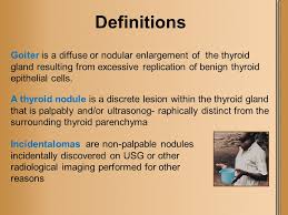
The thyroid is a small gland, measuring about 2 inches (5 centimeters) across, that lies just under the skin below the Adam’s apple in the neck. The two halves (lobes) of the gland are connected in the middle (called the isthmus), giving the thyroid gland the butterfly shapes organ, sort of looking like a bow tie. Normally, the thyroid gland cannot be seen and can barely be felt. If it becomes enlarged, doctors can feel it easily, and a prominent bulge (goiter) may appear below or to the sides of the Adam’s apple.

How the Thyroid Gland Works:

  • The thyroid is part of the endocrine system, which is made up of glands that produce, store, and release hormones into the bloodstream so the hormones can reach the body’s cells. The thyroid gland uses iodine from the foods you eat to make two main hormones:
  • Triiodothyronine (T3)
  • Thyroxine (T4)

T4, the major hormone produced by the thyroid gland, has only a slight effect, if any, on speeding up the body’s metabolic rate. When this occurs instead, T4 is converted into T3, the more active hormone. The conversion of T4 to T3 occurs in the liver and other tissues. Now yes T3 is made by the thyroid but if too much in the body it is converted to T4, more inactivating or decreasing your T3 levels to prevent metabolism too high in the body causing metabolic problems.  Many factors control the conversion of T4 to T3, including the body’s needs from moment to moment and the presence or absence of illnesses.

The thyroid gland secretes thyroid hormones, which control the speed at which the body’s chemical functions proceed (metabolic rate).  This is the vital function of this gland.

Thyroid hormones influence the metabolic rate in two ways:

1. By stimulating almost every tissue in the body to produce proteins

2. The stimulating other areas of the body.  This depends on all how much hormone the thyroid is releasing in the blood stream.  This in turn will decrease or increase the amount of oxygen that cells use.  Which all depends upon how much of the 2 major hormones the thyroid secretes as the factor.  If the thyroid is doing low hormone release this will decrease metabolism in the body or if the organ is in high hormone release it will increase metabolism in the body.

Thyroid hormones affect many vital body functions through either increasing or decreasing metabolism.  This would all be effected on the rate of thyroid hormone releasing from the pituitary and how much T3 or T4 is being released from the thyroid, Did you know in turn this will effect the following areas of the body:

A. the heart rate  B. the rate at which calories are burned  C. skin maintenance  D. growth  E. heat production

F. fertility  E. digestion  G. breathing H. CNS and Peripheral Nervous System I. Body Wt. & Temp J. Cholesterol and much more!

If metabolism is increased so will all these functions listed above regarding its activity; or metabolism that is decreased so will all these functions listed above.

In a nut shell the thyroid gland is the gland that releases hormones that plays an impact on metaboloism = the way your body uses energy. The thyroid’s hormones regulate vital body functions.

Two Thyroid Hormones made and released by this organ that impact your metabolism=Triiodothyronine-also known as T3 and T4-also known as thyroxine.

It is important that T3 and T4 levels are neither too high nor too low but within normal range of therapeutic levels. Two glands in the brain—the hypothalamus and the pituitary communicate to maintain T3 and T4 balance.

The Hypothalamus:

A structure deep in your brain, acts as your body’s smart control coordinating center. Its main function is to keep your body in a stable state called homeostasis. It does its job by directly influencing your autonomic nervous system or by managing hormones. and one of the organs it affects is the THYROID.

The pituitary gland:

This organ sometimes called the “master” gland of the endocrine system because it controls the functions of many of the other endocrine glands. The pituitary gland is no larger than a pea, and is located at the base of the brain. The gland is attached to the hypothalamus by nerve fibers and blood vessels (a part of the brain that affects the pituitary gland). The pituitary gland itself consists of 2 major structures:

  • Anterior lobe – this is where TSH is produced and released.
  • Posterior lobe

This is how the hypothalamus and the pituitary gland work:

The Hypothalamus produces and releases Thyroid Releasing Hormone when T3 & T4 are low in the blood.             The Pituitary produces and releases Thyroid Stimulating Hormone TSH to  tell your thyroid how much thyroid hormone it needs to make.

The hypothalamus senses low circulating levels of thyroid hormones (T3 & T4) and responds by releasing thyrotropin-releasing hormone (TRH). The TRH stimulates the pituitary telling it to produce thyroid-stimulating hormone (TSH) and causing the following effect in the body:

  • When T3 and T4 levels are low in the blood, the pituitary gland releases more of TSH to tell the thyroid gland to produce more thyroid hormones.
  • If T3 and T4 levels are high, the pituitary gland releases less TSH to the thyroid gland to slow production of these hormones.  The inhibiting release of TRH and TSH through a negative feedback loop.

T3 and T4 travel in your bloodstream to reach almost every cell in the body. The hormones regulate the speed with which the cells/metabolism work. For example, T3 and T4 regulate your heart rate and how fast your intestines process food. So if T3 and T4 levels are low, your heart rate may be slower than normal, and you may have constipation/weight gain. If T3 and T4 levels are high, you may have a rapid heart rate and diarrhea/weight loss.

Did you know the third hormone produced by the thyroid gland is called calcitonin. Calcitonin is made by C-cells. It is involved in and metabolism of the bone only.


QUOTE FOR FRIDAY:

“The term “goiter” simply refers to the abnormal enlargement of the thyroid gland. It is important to know that the presence of a goiter does not necessarily mean that the thyroid gland is malfunctioning. A goiter can occur in a gland that is producing too much hormone (hyperthyroidism), too little hormone (hypothyroidism), or the correct amount of hormone (euthyroidism). A goiter indicates there is a condition present which is causing the thyroid to grow abnormally.  One of the most common causes of goiter formation worldwide is iodine deficiency. While this was a very frequent cause of goiter in the United States many years ago, it is no longer commonly observed. The primary activity of the thyroid gland is to concentrate iodine from the blood to make thyroid hormone. The gland cannot make enough thyroid hormone if it does not have enough iodine. Therefore, with iodine deficiency the individual will become hypothyroid. Consequently, the pituitary gland in the brain senses the thyroid hormone level is too low and sends a signal to the thyroid. This signal is called thyroid stimulating hormone (TSH). As the name implies, this hormone stimulates the thyroid to produce thyroid hormone and to grow in size. This abnormal growth in size produces what is termed a “goiter.” Thus, iodine deficiency is one cause of goiter development. Wherever iodine deficiency is common, goiter will be common. It remains a common cause of goiters in other parts of the world.”

American Thyroid Association (https://www.thyroid.org/goiter/)

Nodules & Thyroid goiter

thyroid awareness month goiter6thyroid awareness goiter1thyroid awareness month goiter4  thyroidawarenessmonthGoiter2

Goiter in the thyroid

A goiter is simply an enlarged thyroid gland. Multiple conditions can lead to goiter, including hypothyroidism, hyperthyroidism, excessive iodine intake, or thyroid tumors. Goiter is a non-specific finding that warrants medical evaluation.

History: The doctor will take a detailed history, evaluating both past and present medical problems. If the patient is younger than 20 or older than 70 years, there is increased likelihood that a nodule is cancerous. Similarly, the nodule is more likely to be cancerous if there is any history of radiation exposure, difficulty swallowing, or a change in the voice. It was actually customary to apply radiation to the head and neck in the 1950s to treat acne! Significant radiation exposures include the Chernobyl and Fukushima disasters. Although women tend to have more thyroid nodules than men, the nodules found in men are more likely to be cancerous. Despite its value, the history cannot differentiate benign from malignant nodules. Thus, many patients with risk factors uncovered in the history will have benign lesions. Others without risk factors for malignant nodules may still have thyroid cancer.

Physical examination: The physician should determine if there is one nodule or many nodules, and what the remainder of the gland feels like. The probability of cancer is higher if the nodule is fixed to the surrounding tissue (unmovable). In addition, the physical exam should search for any abnormal lymph nodes nearby that may suggest the spread of cancer. In addition to evaluating the thyroid, the physician should identify any signs of gland malfunction, such as thyroid hormone overproduction (hyperthyroidism) or underproduction (hypothyroidism).

Blood tests: Initially, blood tests should be done to assess thyroid function. These tests include:

  • The free T4 and thyroid stimulating hormone (TSH) levels. Elevated levels of the thyroid hormones T4 or T3 in the context of suppressed TSH suggests hyperthyroidism
  • Reduced T4 or T3 in the context of high TSH suggests hypothyroidism
  • Antibody titers to thyroperoxidase or thyroglobulin may be useful to diagnose autoimmune thyroiditis
  • (for example, Hashimoto’s thyroiditis).
  • If surgery is likely to be considered for treatment, it is strongly recommended that the physician als determine the level of thyroglobin. Produced only in the thyroid hormone in the blood. Thyroglobulin carries thyroid hormone in the blood. Thyroglobulin levels should fall quickly within 48 hours in the thyroid gland is completely remobed. If thyroglobulin levels start to climb.
  • Ultrasonography: A physician may order an ultrasound examination of the thyroid to:
  • Detect nodules that are not easily felt
  • Determine the number of nodules and their sizes
  • Determine if a nodule is solid or cystic
  • Assist obtaining tissue for diagnosis from the thyroid with a fine needle aspirate (FNA)Radionuclide scanning: Radionuclide scanning with radioactive chemicals is another imaging technique a physician may use to evaluate a thyroid nodule. The normal thyroid gland accumulates iodine from the blood and uses it to make thyroid hormones. Thus, when radioactive iodine (123-iodine) is administered orally or intravenously to an individual, it accumulates in the thyroid and causes the gland to “light up” when imaged by a nuclear camera (a type of Geiger counter). The rate of accumulation gives an indication of how the thyroid gland and any nodules are functioning. A “hot spot” appears if a part of the gland or a nodule is producing too much hormone. Non-functioning or hypo-functioning nodules appear as “cold spots” on scanning. A cold or non-functioning nodule carries a higher risk of cancer than a normal or hyper-functioning nodule. Cancerous nodules are more likely to be cold, because cancer cells are immature and don’t accumulate the iodine as well as normal thyroid tissue. However, cold spots can also be caused by cysts. This makes the ultrasound a much better tool for determining the need to do an FNA.
  • Fine needle aspiration: Fine needle aspiration (FNA) of a nodule is a type of biopsy and the most common, direct way to determine what types of cells are present. The needle used is very thin. The procedure is simple and can be done in an outpatient office, and anesthetic is injected into tissues traversed by the needle. FNA is possible if the nodule is easily felt. If the nodule is more difficult to feel, fine needle aspiration can be performed with ultrasound guidance. The needle is inserted into the thyroid or nodule to withdraw cells. Usually, several samples are taken to maximize the chance of detecting abnormal cells. These cells are examined microscopically by a pathologist to determine if cancer cells are present. The value of FNA depends upon the experience of the physician performing the FNA and the pathologist reading the specimen. Diagnoses that can be made from FNA include:
  • Despite its value, the ultrasound cannot determine whether a nodule is benign or cancerous.
  • Benign thyroid tissue (non-cancerous) can be consistent with Hashimoto’s thyroiditis, a colloid nodule, or a thyroid cyst. This result is reported from approximately 60% of biopsies.
  • Cancerous tissue (malignant) can be consistent with diagnosis of papillary, follicular, or medullary cancer. This result is reported from approximately 5% of biopsies. The majority of these are papillary cancers.
  • Suspicious biopsy can show a follicular adenoma. Though usually benign, up to 20% of these nodules are found ultimately to be cancerous.
  • Non-diagnostic results usually arise because insufficient cells were obtained. Upon repeat biopsy, up to 50% of these cases can be distinguished as benign, cancerous, or suspicious.

One of the most difficult problems for the pathologist is to be confident that a follicular adenoma – usually a benign nodule – is not a follicular cell carcinoma or cancer. In these cases, it is up to the physician and the patient to weigh the option of surgery on a case-by-case basis, with less reliance on the pathologist’s interpretation of the biopsy. It is also important to remember that there is a small risk (3%) that a benign nodule diagnosed by FNA may still be cancerous. Thus, even benign nodules should be followed closely by the patient and physician. Another biopsy may be necessary, especially if the nodule is growing. Most thyroid cancers are not very aggressive; that is, they do not spread rapidly. The exception is poorly differentiated (anaplastic) carcinoma, which spreads rapidly and is difficult to treat.

QUOTE FOR THURSDAY:

“Participants testify to how great a month off from alcohol can be; they sleep better, have more energy, some lose weight and save money, and others notice improvements in their skin and hair.

More recently, research from the Royal Free Hospital in London has shown that there are positive physiological effects to be gained from taking part in Dry January. The study, performed on moderate drinkers (those drinking around the levels of recommended limits), has shown people experiencing improvements in concentration and sleep patterns, as well as having reduced cholesterol and lower glucose levels, lower blood pressure, weight loss overall, and losing 40% of their liver fat.”

National Library of Medicine NIH

Try the dry free alcohol challenge for one month & learn how its good for the body.

Harvard Health Publishing states the following:

“If you’d like to cut down your alcohol consumption or start the new year with a clean slate, join in the Dry January challenge by choosing not to drink beer, wine, or spirits for one month. Dry January began in 2012 as a public health initiative from Alcohol Change UK, a British charity. Now millions take part in this health challenge every year.

While past observational studies suggested a link between drinking a moderate amount of alcohol and health benefits for some people, more recent research has questioned whether any amount of alcohol improves health outcomes. And heavier drinking or long-term drinking can increase physical and mental problems, especially among older adults. Heart and liver damage, a higher cancer risk, a weakened immune system, memory issues, and mood disorders are common issues.

Yet, cutting out alcohol for even a month can make a noticeable difference in your health. Regular drinkers who abstained from alcohol for 30 days slept better, had more energy, and lost weight, according to a study in BMJ Open. They also lowered their blood pressure and cholesterol levels and reduced cancer-related proteins in their blood.

Tips for a successful Dry January

A month may seem like a long time, but most people can be successful. Still, you may need assistance to stay dry in January. Here are some tips:

  • Find a substitute non-alcoholic drink. For social situations, or when you crave a cocktail after a long day, reach for alcohol-free beverages like sparkling water, soda, or mocktails (non-alcoholic cocktails.)
    Non-alcoholic beer or wine also is an option, but some brands still contain up to 0.5% alcohol by volume, so check the label. “Sugar is often added to these beverages to improve the taste, so try to choose ones that are low in sugar,” says Dawn Sugarman, a research psychologist at Harvard-affiliated McLean Hospital in the division of alcohol, drugs, and addiction.
  • Avoid temptations. Keep alcohol out of your house. When you are invited to someone’s home, bring your non-alcoholic drinks with you.
  • Create a support group. Let friends and family know about your intentions and encourage them to keep you accountable. Better yet, enlist someone to do the challenge with you.
  • Use the Try Dry app. This free app from the UK helps you track your drinking, set personal goals, and offers motivational information like calories and money saved from not drinking. It’s aimed at cutting back on or cutting out alcohol, depending on your choices.
  • Don’t give up. If you slip up, don’t feel guilty. Just begin again the next day.

Check your feelings

Sugarman recommends people also use Dry January to reflect on their drinking habits. It’s common for people to lose their alcohol cravings and realize drinking need not occupy such an ample space in their lives. If this is you, consider continuing for another 30 days, or just embrace your new attitude toward drinking where it’s an occasional indulgence.

If you struggle during the month, or give up after a week or so, you may need extra help cutting back. Talk to your doctor about getting the help you need.

The Rethinking Drinking site created by the National Institute on Alcohol Abuse and Alcoholism (NIAAA) is also an excellent resource. For the record, NIAAA recommends limiting alcohol to two daily drinks or less for men and no more than one drink a day for women.

Be aware of problems that might crop up

Dry January can reveal signs of potential alcohol problems, including symptoms of alcohol withdrawal ranging from mild to serious, depending on how much you usually drink.

  • Mild symptoms include anxiety, shaky hands, headache, nausea, vomiting, sweating, and insomnia.
  • Severe symptoms often kick in within two or three days after you stop drinking. They can include hallucinations, delirium, racing heart rate, and fever.

“If you suffer alcohol withdrawal symptoms at any time, you should seek immediate medical help,” says Sugarman.”

QUOTE FOR WEDNESDAY:

“Glaucoma medication, in the form of eye drops or pills, is the most common early treatment for glaucoma. Medications work by lowering eye pressure, either by slowing the production of aqueous humor fluid or by improving the drainage of fluid from the eye.

Prior vision loss from glaucoma is not reversible with treatment or surgery.

UCLA Health (https://www.uclahealth.org/medical-services/ophthalmology/non-surgical-treatments)

Part III Glaucoma National Awareness: The treatments of Glaucoma!

If you are diagnosed with glaucoma, it is important to set a regular schedule of examinations with your eye doctor to monitor your condition and make sure that your prescribed treatment is effectively maintaining a safe eye pressure.

Treatments

The Treatment of Glaucoma: The damage caused by glaucoma can’t be reversed. But treatment and regular checkups can help slow or prevent vision loss, especially in you catch the disease in its early stage. The goal of glaucoma treatment is to lower pressure in your eye (intraocular pressure). Depending on your situation, your options may include eyedrops, laser treatment or surgery. 

Remember The damage caused by glaucoma can’t be reversed. But treatment and regular checkups can help slow or prevent vision loss, especially in you catch the disease in its early stage. The goal of glaucoma treatment is to lower pressure in your eye (intraocular pressure). Depending on your situation, your options may include eyedrops, laser treatment or surgery.

Eyedrops

Glaucoma treatment often starts with prescription eyedrops. These can help decrease eye pressure by improving how fluid drains from your eye or by decreasing the amount of fluid your eye makes. Depending on how low your eye pressure needs to be, more than one of the eyedrops below may need to be prescribed.

Prescription eyedrop medications include:

  • Prostaglandins. These increase the outflow of the fluid in your eye (aqueous humor), thereby reducing your eye pressure. Medicines in this category include latanoprost (Xalatan), travoprost (Travatan Z), tafluprost (Zioptan), bimatoprost (Lumigan) and latanoprostene bunod (Vyzulta). Possible side effects include mild reddening and stinging of the eyes, darkening of the iris, darkening of the pigment of the eyelashes or eyelid skin, and blurred vision. This class of drug is prescribed for once-a-day use.
  • Beta blockers. These reduce the production of fluid in your eye, thereby lowering the pressure in your eye (intraocular pressure). Examples include timolol (Betimol, Istalol, Timoptic) and betaxolol (Betoptic). Possible side effects include difficulty breathing, slowed heart rate, lower blood pressure, impotence and fatigue. This class of drug can be prescribed for once- or twice-daily use depending on your condition.
  • Alpha-adrenergic agonists. These reduce the production of aqueous humor and increase outflow of the fluid in your eye. Examples include apraclonidine (Iopidine) and brimonidine (Alphagan P, Qoliana). Possible side effects include an irregular heart rate, high blood pressure, fatigue, red, itchy or swollen eyes, and dry mouth. This class of drug is usually prescribed for twice-daily use but sometimes can be prescribed for use three times a day.
  • Carbonic anhydrase inhibitors. These medicines reduce the production of fluid in your eye. Examples include dorzolamide (Trusopt) and brinzolamide (Azopt). Possible side effects include a metallic taste, frequent urination, and tingling in the fingers and toes. This class of drug is usually prescribed for twice-daily use but sometimes can be prescribed for use three times a day.
  • Rho kinase inhibitor. This medicine lowers eye pressure by suppressing the rho kinase enzymes responsible for fluid increase. It is available as netarsudil (Rhopressa) and is prescribed for once-a-day use. Possible side effects include eye redness, eye discomfort and deposits forming on the cornea.
  • Miotic or cholinergic agents. These increase the outflow of fluid from your eye. An example is pilocarpine (Isopto Carpine). Side effects include headache, eye ache, smaller pupils, possible blurred or dim vision, and nearsightedness. This class of medicine is usually prescribed to be used up to four times a day. Because of potential side effects and the need for frequent daily use, these medications are not prescribed very often anymore.

Because some of the eyedrop medicine is absorbed into your bloodstream, you may experience some side effects unrelated to your eyes. To minimize this absorption, close your eyes for one to two minutes after putting the drops in. You may also press lightly at the corner of your eyes near your nose to close the tear duct for one or two minutes. Wipe off any unused drops from your eyelid.

If you have been prescribed multiple eyedrops or you need to use artificial tears, space them out so that you are waiting at least five minutes in between types of drops.

Oral medications

If eyedrops alone don’t bring your eye pressure down to the desired level, your doctor may also prescribe an oral medication, usually a carbonic anhydrase inhibitor. Possible side effects include frequent urination, tingling in the fingers and toes, depression, stomach upset, and kidney stones.

Surgery and other therapies

Other treatment options include laser therapy and various surgical procedures. The following techniques are intended to improve the drainage of fluid within the eye, thereby lowering pressure:

  • Laser therapy. Laser trabeculoplasty (truh-BEK-u-low-plas-tee) is an option if you have open-angle glaucoma. It’s done in your doctor’s office. Your doctor uses a small laser beam to open clogged channels in the trabecular meshwork. It may take a few weeks before the full effect of this procedure becomes apparent.
  • Filtering surgery. With a surgical procedure called a trabeculectomy (truh-bek-u-LEK-tuh-me), your surgeon creates an opening in the white of the eye (sclera) and removes part of the trabecular meshwork.
  • Drainage tubes. In this procedure, your eye surgeon inserts a small tube shunt in your eye to drain away excess fluid to lower your eye pressure.
  • Minimally invasive glaucoma surgery (MIGS). Your doctor may suggest a MIGS procedure to lower your eye pressure. These procedures generally require less immediate postoperative care and have less risk than trabeculectomy or installing a drainage device. They are often combined with cataract surgery. There are a number of MIGS techniques available, and your doctor will discuss which procedure may be right for you.

After your procedure, you’ll need to see your doctor for follow-up exams. And you may eventually need to undergo additional procedures if your eye pressure begins to rise again or other changes occur in your eye.


QUOTE FOR TUESDAY:

“Glaucoma is the silent thief of sight. Glaucoma has no symptoms in its early stages. In fact, half the people with glaucoma do not know they have it! Having regular eye exams can help your ophthalmologist find this disease before you lose vision. Your ophthalmologist can tell you how often you should be examined.”

American Academy of Ophthalmology (https://www.aao.org/eye-health/diseases/what-is-glaucoma)

Part II Glaucoma National Awareness: Other types of Glaucoma & The key to preventing glaucoma.

glaucoma3   glaucoma2                                        Glaucoma-Table

Continuation of Other Types of Glaucoma:

Normal-Tension Glaucoma (NTG)

Also called low-tension or normal-pressure glaucoma. In normal-tension glaucoma the optic nerve is damaged even though the eye pressure is not very high. We still don’t know why some people’s optic nerves are damaged even though they have almost normal pressure levels.

Congenital Glaucoma

This type of glaucoma occurs in babies when there is incorrect or incomplete development of the eye’s drainage canals during the prenatal period. This is a rare condition that may be inherited. When uncomplicated, microsurgery can often correct the structural defects. Other cases are treated with medication and surgery.

Secondary Glaucomas

Sometimes glaucoma is caused by another medical condition — this is called secondary glaucoma.

Neovascular glaucoma

Treatments: Medicines, laser treatment, surgery

Neovascular glaucoma happens when the eye makes extra blood vessels that cover the part of your eye where fluid would normally drain. It’s usually caused by another medical condition, like diabetes or high blood pressure.

If you have neovascular glaucoma, you may notice:

  • Pain or redness in your eye
  • Vision loss

This type of glaucoma can be hard to treat. Doctors need to treat the underlying cause (like diabetes or high blood pressure) and use glaucoma treatments to lower the eye pressure that results from it.

Pigmentary glaucoma

Treatments: Medicines, laser treatment, surgery

Pigment dispersion syndrome happens when the pigment (color) from your iris (the colored part of your eye) flakes off. The loose pigment may block fluid from draining out of your eye, which can increase your eye pressure and cause pigmentary glaucoma.

Young, white men who are near-sighted are more likely to have pigment dispersion syndrome than others. If you have this condition, you may have blurry vision or see rainbow-colored rings around lights, especially when you exercise.

Doctors can treat pigmentary glaucoma by lowering eye pressure, but there currently isn’t a way to prevent pigment from detaching from the iris.

Exfoliation glaucoma

Treatments: Medicines, laser treatment, surgery

Exfoliation glaucoma (sometimes called pseudoexfoliation) is a type of open-angle glaucoma that happens in some people with exfoliation syndrome, a condition that causes extra material to detach from parts of the eye and block fluid from draining.

Recent research shows that genetics may play a role in exfoliation glaucoma. You are at higher risk if someone else in your family has exfoliation glaucoma.

This type of glaucoma can progress faster than primary open-angle glaucoma, and often causes higher eye pressure. This means that it’s especially important for people who are at risk to get eye exams regularly.

Uveitic glaucoma

Treatments: Medicines, surgery

Uveitic glaucoma can happen in people who have uveitis, a condition that causes inflammation (irritation and swelling) in the eye. About 2 in 10 people with uveitis will develop uveitic glaucoma.

Experts aren’t sure how uveitis causes uveitic glaucoma, but they think that it may happen because uveitis can cause inflammation and scar tissue in the middle of the eye. This may damage or block the part of the eye where fluid drains out, causing high eye pressure and leading to uveitic glaucoma.

In some cases, the medicines that treat uveitis may also cause uveitic glaucoma, or make it worse. This is because corticosteroid medicines may cause increased eye pressure as a side effect.

The KEY is take the steps to help PREVENT Glaucoma:

These self-care steps can help you detect glaucoma in its early stages, which is important in preventing vision loss or slowing its progress.

  • Get regular dilated eye examinations. Regular comprehensive eye exams can help detect glaucoma in its early stages, before significant damage occurs. As a general rule, the American Academy of Ophthalmology recommends having a comprehensive eye exam every five to 10 years if you’re under 40 years old; every two to four years if you’re 40 to 54 years old; every one to three years if you’re 55 to 64 years old; and every one to two years if you’re older than 65. If you’re at risk of glaucoma, you’ll need more frequent screening. Ask your doctor to recommend the right screening schedule for you.
  • Know your family’s eye health history. Glaucoma tends to run in families. If you’re at increased risk, you may need more frequent screening.
  • Exercise safely. Regular, moderate exercise may help prevent glaucoma by reducing eye pressure. Talk with your doctor about an appropriate exercise program.
  • Take prescribed eyedrops regularly. Glaucoma eyedrops can significantly reduce the risk that high eye pressure will progress to glaucoma. To be effective, eyedrops prescribed by your doctor need to be used regularly even if you have no symptoms.
  • Wear eye protection. Serious eye injuries can lead to glaucoma. Wear eye protection when using power tools or playing high-speed racket sports in enclosed courts.


QUOTE FOR MONDAY:

“There are different types of glaucoma.

Glaucoma is a group of eye diseases that can cause vision loss and blindness by damaging a nerve in the back of your eye called the optic nerve.

The symptoms can start so slowly that you may not notice them. The only way to find out if you have glaucoma is to get a comprehensive dilated eye exam.

There’s no cure for glaucoma unfortunately, but early treatment can often stop the damage and protect your vision.”

National Eye Institute (https://www.nei.nih.gov/learn-about-eye-health/eye-conditions-and-diseases/glaucoma#:~:text=Glaucoma%20is%20a%20group%20of,a%20comprehensive%20dilated%20eye%20exam.)