Part II Glaucoma National Awareness: Other types of Glaucoma & The key to preventing glaucoma.

glaucoma3   glaucoma2                                        Glaucoma-Table

Continuation of Other Types of Glaucoma:

Normal-Tension Glaucoma (NTG)

Also called low-tension or normal-pressure glaucoma. In normal-tension glaucoma the optic nerve is damaged even though the eye pressure is not very high. We still don’t know why some people’s optic nerves are damaged even though they have almost normal pressure levels.
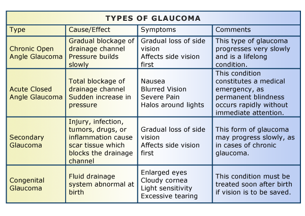
Congenital Glaucoma

This type of glaucoma occurs in babies when there is incorrect or incomplete development of the eye’s drainage canals during the prenatal period. This is a rare condition that may be inherited. When uncomplicated, microsurgery can often correct the structural defects. Other cases are treated with medication and surgery.

Secondary Glaucomas

Sometimes glaucoma is caused by another medical condition — this is called secondary glaucoma.

Neovascular glaucoma

Treatments: Medicines, laser treatment, surgery

Neovascular glaucoma happens when the eye makes extra blood vessels that cover the part of your eye where fluid would normally drain. It’s usually caused by another medical condition, like diabetes or high blood pressure.

If you have neovascular glaucoma, you may notice:

  • Pain or redness in your eye
  • Vision loss

This type of glaucoma can be hard to treat. Doctors need to treat the underlying cause (like diabetes or high blood pressure) and use glaucoma treatments to lower the eye pressure that results from it.

Pigmentary glaucoma

Treatments: Medicines, laser treatment, surgery

Pigment dispersion syndrome happens when the pigment (color) from your iris (the colored part of your eye) flakes off. The loose pigment may block fluid from draining out of your eye, which can increase your eye pressure and cause pigmentary glaucoma.

Young, white men who are near-sighted are more likely to have pigment dispersion syndrome than others. If you have this condition, you may have blurry vision or see rainbow-colored rings around lights, especially when you exercise.

Doctors can treat pigmentary glaucoma by lowering eye pressure, but there currently isn’t a way to prevent pigment from detaching from the iris.

Exfoliation glaucoma

Treatments: Medicines, laser treatment, surgery

Exfoliation glaucoma (sometimes called pseudoexfoliation) is a type of open-angle glaucoma that happens in some people with exfoliation syndrome, a condition that causes extra material to detach from parts of the eye and block fluid from draining.

Recent research shows that genetics may play a role in exfoliation glaucoma. You are at higher risk if someone else in your family has exfoliation glaucoma.

This type of glaucoma can progress faster than primary open-angle glaucoma, and often causes higher eye pressure. This means that it’s especially important for people who are at risk to get eye exams regularly.

Uveitic glaucoma

Treatments: Medicines, surgery

Uveitic glaucoma can happen in people who have uveitis, a condition that causes inflammation (irritation and swelling) in the eye. About 2 in 10 people with uveitis will develop uveitic glaucoma.

Experts aren’t sure how uveitis causes uveitic glaucoma, but they think that it may happen because uveitis can cause inflammation and scar tissue in the middle of the eye. This may damage or block the part of the eye where fluid drains out, causing high eye pressure and leading to uveitic glaucoma.

In some cases, the medicines that treat uveitis may also cause uveitic glaucoma, or make it worse. This is because corticosteroid medicines may cause increased eye pressure as a side effect.

The KEY is take the steps to help PREVENT Glaucoma:

These self-care steps can help you detect glaucoma in its early stages, which is important in preventing vision loss or slowing its progress.

  • Get regular dilated eye examinations. Regular comprehensive eye exams can help detect glaucoma in its early stages, before significant damage occurs. As a general rule, the American Academy of Ophthalmology recommends having a comprehensive eye exam every five to 10 years if you’re under 40 years old; every two to four years if you’re 40 to 54 years old; every one to three years if you’re 55 to 64 years old; and every one to two years if you’re older than 65. If you’re at risk of glaucoma, you’ll need more frequent screening. Ask your doctor to recommend the right screening schedule for you.
  • Know your family’s eye health history. Glaucoma tends to run in families. If you’re at increased risk, you may need more frequent screening.
  • Exercise safely. Regular, moderate exercise may help prevent glaucoma by reducing eye pressure. Talk with your doctor about an appropriate exercise program.
  • Take prescribed eyedrops regularly. Glaucoma eyedrops can significantly reduce the risk that high eye pressure will progress to glaucoma. To be effective, eyedrops prescribed by your doctor need to be used regularly even if you have no symptoms.
  • Wear eye protection. Serious eye injuries can lead to glaucoma. Wear eye protection when using power tools or playing high-speed racket sports in enclosed courts.


QUOTE FOR MONDAY:

“There are different types of glaucoma.

Glaucoma is a group of eye diseases that can cause vision loss and blindness by damaging a nerve in the back of your eye called the optic nerve.

The symptoms can start so slowly that you may not notice them. The only way to find out if you have glaucoma is to get a comprehensive dilated eye exam.

There’s no cure for glaucoma unfortunately, but early treatment can often stop the damage and protect your vision.”

National Eye Institute (https://www.nei.nih.gov/learn-about-eye-health/eye-conditions-and-diseases/glaucoma#:~:text=Glaucoma%20is%20a%20group%20of,a%20comprehensive%20dilated%20eye%20exam.)

Part I National Glaucoma Awareness: What this is, the two major types and their symptoms.

   glaucoma2                      

 Glaucoma-Table

A common eye condition in which the fluid pressure inside the eye rises to a level higher than healthy for that eye. If untreated, it may damage the optic nerve, causing the loss of vision or even blindness. The elderly, African-Americans, and people with family histories of the disease are at greatest risk.

Glaucoma is a multi-factorial, complex eye disease with specific characteristics such as optic nerve damage and visual field loss. While increased pressure inside the eye (called intraocular pressure or IOP) is usually present, even patients with normal range IOP can develop glaucoma.

There is no specific level of elevated eye pressure that definitely leads to glaucoma; conversely, there is no lower level of IOP that will absolutely eliminate a person’s risk of developing glaucoma. That is why early diagnosis and treatment of glaucoma is the key to preventing vision loss.

Eye pressure is measured in millimeters of mercury (mm Hg). Normal eye pressure ranges from 12-22 mm Hg, and eye pressure of greater than 22 mm Hg is considered higher than normal. When the IOP is higher than normal but the person does not show signs of glaucoma, this is referred to as ocular hypertension.

High eye pressure alone does not cause glaucoma. However, it is a significant risk factor. Individuals diagnosed with high eye pressure should have regular comprehensive eye examinations by an eyecare professional to check for signs of the onset of glaucoma.

A person with elevated IOP is referred to as a glaucoma suspect, because of the concern that the elevated eye pressure might lead to glaucoma. The term glaucoma suspect is also used to describe those who have other findings that could potentially, now or in the future, indicate glaucoma. For example, a suspicious optic nerve, or even a strong family history of glaucoma, could put someone in the category of a glaucoma suspect.

Vision loss from glaucoma occurs when the eye pressure is too high for the specific individual and damages the optic nerve. Any resultant damage cannot be reversed. The peripheral (side) vision is usually affected first. The changes in vision may be so gradual that they are not noticed until a lot of vision loss has already occurred.

In time, if the glaucoma is not treated, central vision will also be decreased and then lost; this is how visual impairment from glaucoma is most often noticed. The good news is that glaucoma can be managed if detected early, and with medical and/or surgical treatment, most people with glaucoma will not lose their sight.

Most common signs and symptoms of Glaucoma:

There are several forms of glaucoma; the two most common forms are primary open-angle glaucoma (POAG) and angle-closure glaucoma (ACG). Open-angle glaucoma is often called “the sneak thief of sight” because it has no symptoms until significant vision loss has occurred.

2 Commonly Known Types of Glaucoma:

1-Open Angle Glaucoma 2-Angle Closure Glaucoma


1-Open-Angle Glaucoma

Open-angle glaucoma, the most common form of glaucoma, accounting for at least 90% of all glaucoma cases:

  • Is caused by the slow clogging of the drainage canals, resulting in increased eye pressure
  • Has a wide and open angle between the iris and cornea
  • Develops slowly and is a lifelong condition
  • Has symptoms and damage that are not noticed.

“Open-angle” means that the angle where the iris meets the cornea is as wide and open as it should be. Open-angle glaucoma is also called primary or chronic glaucoma. It is the most common type of glaucoma, affecting about three million Americans.

Symptoms of Open-Angle Glaucoma

There are typically no early warning signs or symptoms of open-angle glaucoma. It develops slowly and sometimes without noticeable sight loss for many years.

Most people who have open-angle glaucoma feel fine and do not notice a change in their vision at first because the initial loss of vision is of side or peripheral vision, and the visual acuity or sharpness of vision is maintained until late in the disease.

By the time a patient is aware of vision loss, the disease is usually quite advanced. Vision loss from glaucoma is not reversible with treatment, even with surgery.

Because open-angle glaucoma has few warning signs or symptoms before damage has occurred, it is important to see a doctor for regular eye examinations. If glaucoma is detected during an eye exam, your eye doctor can prescribe a preventative treatment to help protect your vision.

In open-angle glaucoma, the angle in your eye where the iris meets the cornea is as wide and open as it should be, but the eye’s drainage canals become clogged over time, causing an increase in internal eye pressure and subsequent damage to the optic nerve. It is the most common type of glaucoma, affecting about four million Americans, many of whom do not know they have the disease.

You are at increased risk of glaucoma if your parents or siblings have the disease, if you are African-American or Latino, and possibly if you are diabetic or have cardiovascular disease. The risk of glaucoma also increases with age.

2-Angle-Closure Glaucoma

Angle-closure glaucoma, a less common form of glaucoma:

  • Is caused by blocked drainage canals, resulting in a sudden rise in intraocular pressure
  • Has a closed or narrow angle between the iris and cornea
  • Develops very quickly
  • Has symptoms and damage that are usually very noticeable
  • Demands immediate medical attention.

It is also called acute glaucoma or narrow-angle glaucoma. Unlike open-angle glaucoma, angle-closure glaucoma is a result of the angle between the iris and cornea closing.

Symptoms of Angle-Closure Glaucoma

  • Hazy or blurred vision
  • The appearance of rainbow-colored circles around bright lights
  • Severe eye and head pain
  • Nausea or vomiting (accompanying severe eye pain)
  • Sudden sight loss

Angle-closure glaucoma is caused by blocked drainage canals in the eye, resulting in a sudden rise in intraocular pressure. This is a much more rare form of glaucoma, which develops very quickly and demands immediate medical attention

In contrast with open-angle glaucoma, symptoms of acute angle-closure glaucoma are very noticeable and damage occurs quickly. If you experience any of these symptoms, seek immediate care from an ophthalmologist.

If you are diagnosed with glaucoma, it is important to set a regular schedule of examinations with your eye doctor to monitor your condition and make sure that your prescribed treatment is effectively maintaining a safe eye pressure.

Tomorrow stay tune for part II on glaucoma awareness giving other types of glaucoma, how its diagnosed & the risks.

QUOTE FOR THE WEEKEND:

“January marks the 53rd anniversary of National Blood Donor Month – a time to recognize the importance of giving blood and platelets while celebrating the lifesaving impact of those who roll up a sleeve to help patients in need. It is also a time to encourage new and lapsed donors to resolve to give blood during one of the most difficult times to maintain a sufficient blood supply – the winter months.

This year’s National Blood Donor Month comes as the nation’s blood supply has dipped to concerning levels and could force hospitals to delay essential blood and platelet transfusions. Blood donors of all blood types – particularly type O blood, the blood group hospitals need most – are needed now to give blood or platelets to help meet daily hospital demands.  Donors are urged to schedule an appointment now

The American Red Cross shelters, feeds and provides comfort to victims of disasters; supplies about 40% of the nation’s blood; teaches skills that save lives; distributes international humanitarian aid; and supports veterans, military members and their families. The Red Cross is a nonprofit organization that depends on volunteers and the generosity of the American public to deliver its mission.

American Red Cross (https://www.redcross.org/about-us/news-and-events/news/2021/help-save-lives-during-national-blood-donor-month.html)

National Blood Donor Month

January is National Blood Donor Month, a time to celebrate generous volunteer donors and raise awareness for the need for blood donations.  The purpose is to raise awareness about the vital importance of blood donation. Every drop counts and every donor is a hero.

It began in 1970 when President Richard Nixon made an historic proclamation. During this special month, we encourage all those impacted by the lifesaving power of blood donation to come together to honor and celebrate the incredible generosity of America’s voluntary blood donors.

According to the American Red Cross, winter is “one of the most difficult times of year to collect enough blood products to meet patient needs.” That’s because of, among other things, busy holiday schedules and bad weather often resulting in canceled blood drives. Furthermore, seasonal illnesses such as the flu force potential donors to forgo their blood donations.

That’s just one of the reasons that National Blood Donor Month, which has taken place each January since 1970, is such an important observance. Donating blood saves many lives and improves health for many people. According to the World Health Organization, “blood is the most precious gift that anyone can give to another person — the gift of life. A decision to donate your blood can save a life.”

Did you know these facts? Seek below:

1. About 36,000 units of red blood cells and 7,000 units of platelets are needed every single day in the U.S.

2. A single-car accident victim may need up to 100 pints of blood to survive.

3. 38% of our population is eligible to donate, but less than 10% actually do.

4. A single donation from a single patient can help more than one person.

5. The most requested blood type by hospitals is type O. This kind of blood can be transfused to patients of all blood types, so it’s always in great demand and very short supply. Only 7% of people in the U.S. have type O.

6. About 6.8 million people donate blood every year in the U.S.

7. Donating blood is a simple, safe process. All you have to do is register, take a mini-medical history test, donate, and then accept free refreshments like water, Gatorade, granola bars, etc.

Give blood. Don’t wait for a disaster. Someone needs blood now! Contact the American Red Cross or your local blood bank for more information.


QUOTE FOR FRIDAY:

“Cervical cancer is highly preventable and highly curable if caught early. Nearly all cervical cancers could be prevented by HPV vaccination, routine cervical cancer screening (So it is important for people with a cervix to have regular screening tests starting in their 20s.), and appropriate follow-up treatment when needed. Condoms, which prevent some sexually transmitted diseases, can decrease the risk of HPV transmission. Condoms can assist in prevention. HOWEVER, they do not completely prevent it. Therefore, exposure to HPV is still possible in areas that are not covered by the condom. You can get HPV by having vaginal, anal, or oral sex with someone who has the virus. It is most commonly spread during vaginal or anal sex. It also spreads through close skin-to-skin touching during sex. A person with HPV can pass the infection to someone even when they have no signs or symptoms.”

National Cancer Institute
(https://www.cancer.gov/types/cervical/causes-risk-prevention)

Part IV Cervical Cancer Awareness Month – Prevention methods and Treatments for cervical cancer!


There is steps you can take early in life and steps you can take through out your life to prevent cervical cancer:

1. Three HPV vaccines—9-valent HPV vaccine (Gardasil 9, 9vHPV), quadrivalent HPV vaccine (Gardasil, 4vHPV), and bivalent HPV vaccine (Cervarix, 2vHPV)—have been licensed by the U.S. Food and Drug Administration (FDA). All three HPV vaccines protect against HPV types 16 and 18 that cause most HPV cancers.

Gardasil 9 is an HPV vaccine approved by the U.S. Food and Drug Administration and can be used for both girls and boys. This vaccine can prevent most cases of cervical cancer if the vaccine is given before girls or women are exposed to the virus. This vaccine can also prevent vaginal and vulvar cancer.

2. For cancer prevention as well as overall health and wellness through out your life, many experts recommend a plant-based diet that consists primarily of fruits, vegetables, beans and whole grains. These foods contain a variety of beneficial chemical compounds that can be easily incorporated into daily meals. Some examples include:

  • Flavonoids – These chemical compounds, which are believed to provide protection against cancer, are found in apples, asparagus, black beans, broccoli, brussels sprouts, cabbage, cranberries, garlic, lettuce, lima beans, onions, soy and spinach.
  • Folate – This water-soluble B vitamin has been found to reduce the risk of cervical cancer in women who have HPV. Foods that are rich in folate include avocados, chickpeas, lentils, orange juice, romaine lettuce and strawberries.
  • Carotenoids – These valuable sources of vitamin A are found in most fruits, vegetables and beans, and particularly in orange foods such as carrots, sweet potatoes, pumpkin and squash.

While a healthy diet is an important component of an overall cancer prevention plan, it should not be the only component.

Treatment of cervical cancer:

Cervical cancer treatments include surgery, chemotherapy, and radiation therapy. If your doctor says that you have cervical cancer, ask to be referred to a gynecologic oncologist—a doctor who has been trained to treat cancers of a woman’s reproductive system.

Different treatments may be provided by different doctors on your medical team.

  • Gynecologic oncologists are doctors who have been trained to treat cancers of a woman’s reproductive system.
  • Surgeons are doctors who perform operations.
  • Medical oncologists are doctors who treat cancer with medicine.
  • Radiation oncologists are doctors who treat cancer with radiation.

You always go with MD specialist in treating any cancer starting with a oncologist who will work you up with further specialists if needed.


QUOTE FOR THURSDAY:

“Doctors assign the stage of the cancer by evaluating the tumor and whether the cancer has spread to other parts of the body. Staging is based on the results of a physical exam, imaging scans, and biopsies.

After someone is diagnosed with cervical cancer, doctors will try to figure out if it has spread, and if so, how far. This process is called staging. The stage of a cancer describes the extent of the cancer in the body. It helps determine how serious the cancer is and how best to treat it. The stage is one of the most important factors in deciding how to treat the cancer and determining how successful treatment might be.  Cervical cancer stage ranges from stages I (1) through IV (4).  The lower the better.

To determine the cancer’s stage after a cervical cancer diagnosis, doctors try to answer these questions:

  • How far has the cancer grown into the cervix?
  • Has the cancer reached nearby structures?
  • Has the cancer spread to the nearby lymph nodes or to distant organs?”

American Cancer Society (https://www.cancer.org/cancer/types/cervical-cancer/detection-diagnosis-staging/staged.html)

Part III Cervical Cancer Awareness Month – Diagnosing & Staging this cancer.


HOW TO GET DIAGNOSE FOR CERVICAL CANCER:

Key Points to remember about cervical cancer:

  • Cervical cancer is a disease in which malignant (cancer) cells form in the cervix.
  • Screening for cervical cancer using the Pap test has decreased the number of new cases of cervical cancer and the number of deaths due to cervical cancer since 1950.
  • Human papillomavirus (HPV) infection is the major risk factor for cervical cancer.

1-Screening is looking for cancer before a person has any symptoms. This can help find cancer at an early stage. When abnormal tissue or cancer is found early, it may be easier to treat. By the time symptoms appear, cancer may have begun to spread.

Know this about screening:

  • Tests are used to screen for different types of cancer when a person does not have symptoms.
  • Studies show that screening for cervical cancer helps decrease the number of deaths from the disease.
  • A Pap test is commonly used to screen for cervical cancer.
  • After certain positive Pap test results, an HPV test may be done.
  • An HPV test may be done with or without a Pap test to screen for cervical cancer.
  • Samples for an HPV test may be self-collected.
  • Screening tests for cervical cancer are being studied in clinical trials.

Scientists are trying to better understand which people are more likely to get certain types of cancer.

Cervical dysplasia occurs more often in women who are in their 20s and 30s. Death from cervical cancer is rare in women younger than 30 years and in women of any age who have regular screenings with the Pap test. The Pap test is used to detect cancer and changes that may lead to cancer. The chance of death from cervical cancer increases with age. In recent years, deaths from cervical cancer have been slightly higher in Black women younger than 50 years than in White women younger than 50 years. Deaths from cervical cancer are almost twice as likely in Black women older than 60 years than in White women older than 60 years.

Human papillomavirus (HPV) infection is the major risk factor for cervical cancer.

2.If cervical cancer is suspected, your doctor is likely to start with a thorough examination of your cervix. A special magnifying instrument (colposcope) is used to check for abnormal cells.

During the colposcopic examination, your doctor is likely to take a sample of cervical cells (biopsy) for laboratory testing. To obtain tissue, your doctor may use:

  • Punch biopsy, which involves using a sharp tool to pinch off small samples of cervical tissue.
  • Endocervical curettage, which uses a small, spoon-shaped instrument (curet) or a thin brush to scrape a tissue sample from the cervix

If the punch biopsy or endocervical curettage is worrisome, your doctor may perform one of the following tests:

  • Electrical wire loop, which uses a thin, low-voltage electrified wire to obtain a small tissue sample. Generally this is done under local anesthesia in the office.
  • Cone biopsy (conization), which is a procedure that allows your doctor to obtain deeper layers of cervical cells for laboratory testing. A cone biopsy may be done in a hospital under general anesthesia.

Staging the cervical cancer:

Cervical Cancer. Carcinoma of Cervix. Malignant neoplasm arising from cells in the cervix uteri. Vaginal bleeding. Vector diagram

If your doctor determines that you have cervical cancer, you’ll have further tests to determine the extent (stage) of your cancer. Your cancer’s stage is a key factor in deciding on your treatment.

Staging exams include:

  • Imaging tests. Tests such as X-ray, CT, MRI and positron emission tomography (PET) help your doctor determine whether your cancer has spread beyond your cervix.
  • Visual examination of your bladder and rectum. Your doctor may use special scopes to see inside your bladder and rectum.

stay tune for Part III in treatment of Cervical Cancer

QUOTE FOR WEDNESDAY:

“Cancer is a disease in which cells in the body grow out of control. Cancer is always named for the part of the body where it starts, even if it spreads to other body parts later.When the body’s immune system can’t get rid of an HPV infection with oncogenic HPV types, it can linger over time and turn normal cells into abnormal cells and then cancer. In general, HPV is thought to be responsible for more than 90% of anal and cervical cancers, about 70% of vaginal and vulvar cancers, and 60% of penile cancers. HPV is also very common in men, and often has no symptoms. About 10% of women with HPV infection on their cervix will develop long-lasting HPV infections that put them at risk for cervical cancer.All HPV vaccines protect against at least HPV types 16 and 18, which cause the greatest risk of cervical cancer. Each year in the United States, about 11,500 new cases of cervical cancer are diagnosed and about 4,000 women die of this cancer.”
Center for Disease Control and Prevention – CDC