QUOTE FOR FRIDAY:

“Seizures are unpredictable. When a person has a seizure, it is usually not in a doctor’s office or other medical setting where health care providers can observe what is happening, so diagnosing seizures is a challenge. Accurate diagnosis depends on taking a careful medical history and using brain imaging and other tests to assess abnormal patterns of electrical activity in the brain. Proper diagnosis of seizures and epilepsy is essential for effective treatment. Diagnostic tests can help determine if and where a lesion in the brain is causing seizures. . In the majority of cases, there may be no cause that can be discovered for epilepsy or in some cases there are actual causes.”

John Hopkins Medicine (https://www.hopkinsmedicine.org/health/conditions-and-diseases/epilepsy)

Part II National Epilepsy Awareness Month-Causes, how its diagnosed, and what to know before, during,, and after the seizure!


Possible causes of Epilepsy:

Their epilepsy that is diagnosed with a IDIOPATHIC cause – meaning unknown cause and the patient could grow out of it in childhood in some cases (not all) depending on the type of seizure disorder and if the child doesn’t grow out of it the condition becomes chronic (for life).

Genetic influence (heredity). Some types of epilepsy run in families. In these instances, it’s likely that there’s a genetic influence. Researchers have linked some types of epilepsy to specific genes. But some people have genetic epilepsy that isn’t hereditary. Genetic changes can occur in a child without being passed down from a parent.For most people, genes are only part of the cause of epilepsy. Certain genes may make a person more sensitive to environmental conditions that trigger seizures.

  • A genetic tendency, passed down from one or both parents (inherited).
  • A genetic tendency that is not inherited, but is a new change in the person’s genes.

Although heredity has been known since antiquity to cause epilepsy, the progress to date in identifying the genetic basis of epilepsy has been limited primarily to the discovery of single gene mutations that cause epilepsy in relatively rare families. For the more common types of epilepsy, heredity plays a subtler role, and it is thought that a combination of mutations in multiple genes likely determine an individual’s susceptibility to seizures, as well as the responsiveness to antiepileptic medications.

Epilepsy can be caused by genetic factors (inherited) or acquired (a etiology—cause) , although in most cases it arises in part from both. The neurology and neurological sciences of Stanford Epilepsy Center Dr. Robert S. Fischer Ph D. presents in the article Genetic Causes of Epilepsy.

He also presents in this article our genes are the instruction set for building the human body. Genes reside on chromosomes.

Going to the basics is every person has 46 chromosomes, carrying a total of about 30,000 genes. We get half our chromosomes from our mother and half from our father. While genes determine the structure of our body, they also control the excitability of our brain cells. Defective genes can make hyperexcitable brain cells, which are prone to seizures.

In recent years, several epilepsy conditions have been linked to mutations in genes, but the matter is complicated by the fact that different genes may be involved in different circumstances.

In general, the most common epilepsy conditions, including partial seizures, seem to be more acquired than genetic.

Gene testing will soon be able to identify predispositions to epilepsy, allowing doctors to help a patient get treatment and to assist with family counseling. One day, doctors may simply be able to swap a patient’s cheek, test his or her genes, and predict response to various epilepsy medicines, eliminating much of the trial and error in medication choice that goes on today. Eventually, we may even be able to repair or replace defective genes that predispose a person to epilepsy, a process called gene therapy.

Lastly, Dr. Robert Fischer Ph D presented in his article, that I found very interesting, the general population has about a 1% risk of developing epilepsy.  Meanwhile, children of mothers with epilepsy have a 3 to 9% risk of inheriting this disease, while children of fathers have a 1.5 to 3% risk of inheritence. Still, the actual risk is upon the specific type of epilepsy. For example, partial seizures are less likely to run in families than are generalized seizures. In any event, with the usual forms of epilepsy, even if a parent does have the condition, there is more than a 90% chance that their child will not. So most epilepsies are acquired than inherited.

Clearly, genes determine a great deal of who we are, including our possible risk for epilepsy but slim versus a actual cause. But what happens to us in life and what we do is still the larger part of the risk for epilepsy.

A person given this diagnosis in the 1970’s, or before  and even up to the early 1990’s was quiet about ever letting people know about this since in the 1970’s and back with lack of knowledge, information to the public and definitely technology versus now.  Epilepsy is much more an accepted disease in the overall community compared to 20-25 years ago and back.  Heck in the 1970’s and back these patients when having a seizure episode were characterized as “Freaks”.  This was due to ignorance and lack of information to society/community but due to the past 20 to 25 years with the computer used more as a must in our lives with media, television and even our government they all have made it possible for society everywhere in the world to learn and understand diseases with acceptance in wanting to help those, particularly the US, but we still need a healthier America. It will take time to get there with the many multicultural lives that all live in the U.S. which practice differently on how important a healthy diet is with exercise balanced with rest.  Also including stress well controlled is not always in America on their top priority list in living.  Stress can even be a catalyst for a seizure but not the cause.

For a person diagnosed with or without a cause of epilepsy these steps in learning about the disease with higher technology and continuous research with medications over the years has allowed them to be able to live a completely healthy life doing the same things other people do without the disease but only if the patient is UNDER COMPLETE CONTROL  which includes being COMPLIANT with your Rx; this does exist in America.

Compliant meaning taking their medications everyday as ordered by their neurologist with yearly or sooner follow-up visits with blood levels of the anti-seizure medications there on.  This is the only way one with chronic epilepsy is guaranteed that living this way MAY stop the seizures from occurring (inactive epilepsy you can call it — meaning you’ll always have the disease but can put the seizure activity in a remission by medications preventing the seizure.)

Other Etiologies or Causes of Epilepsy:

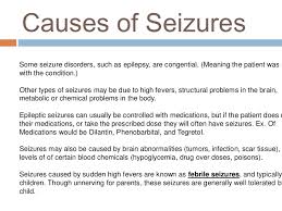
Epilepsy has no identifiable cause in about half the people with the condition. In the other half, the condition may be traced to various factors, including:

  • Low oxygen during birth.
  • Head trauma. Head trauma as  a result of a car accident or other traumatic injury can cause epilepsy.
  • Factors in the brain. Brain tumors can cause epilepsy. Epilepsy also may be caused by the way blood vessels form in the brain. People with blood vessel conditions such as arteriovenous malformations and cavernous malformations can have seizures. And in adults older than age 35, stroke is a leading cause of epilepsy.
  • Infections. Meningitis, HIV, viral encephalitis and some parasitic infections can cause epilepsy.
  • Injury before birth. Before they’re born, babies are sensitive to brain damage that could be caused by several factors. They might include an infection in the mother, poor nutrition or not enough oxygen. This brain damage can result in epilepsy or cerebral palsy.

Head trauma/Degenerative Disease like Alzheimer’s or Creutfeldz-Jacob or Huntington’s Chorea or Multiple Sclerosis or Pick’s Disease. There is also tumors or genetic disease or Stroke or Infections or Febrile seizures.

Different epilepsies are due to many different underlying causes. The causes can be complex, and sometimes hard to identify. A person might start having seizures because they have one or more of the following.

  • A structural (sometimes called ‘symptomatic’) change in the brain, such as the brain not developing properly.
  • A stroke or a tumour. A brain scan, such as Magnetic Resonance Imaging (MRI), may show this.

Some researchers now believe that the chance of developing epilepsy is probably always genetic to some extent, in that any person who starts having seizures has always had some level of genetic likelihood to do so. This level can range from high to low and anywhere in between.

Even if seizures start after a brain injury or other structural change, this may be due to both the structural change and the person’s genetic tendency to seizures, combined. This makes sense if we consider that many people might have a similar brain injury, but not all of them develop epilepsy afterwards.

Metabolic and Systemic Causes of Seizures:

a.) Electrolyte Imbalance=In the blood having acidosis, heavy metal poisoning, Hypocalcemia (low Ca+) , Hypocapnea (low carbon dioxide), Hypoglycemia (low glucose), Hypoxia (low oxygen), Sodium-Potassium imbalance, and than Systemic  diseases (liver, renal failure, etc…).  Then their is also toxemia of pregnancy, and water intoxication.

b.) Infections like meningitis, encephalitis, brain abcess.  Structural changes due to genetic conditions such as tuberous sclerosis, or neurofibromatosis, which can cause growths affecting the brain.

Tuberous sclerosis  – a genetic condition that causes growths in organs including the brain. Tuberous sclerosis can cause epilepsy.

Neurofibromatosis  – a genetic condition that causes benign tumours to grow on the covering of nerves. Neurofibromatosis can cause epilepsy.

c.) Withdrawal of sedative-hypnotic drugs=Alcohol, Antiepileptic drugs, Barbiturates, Benzodiazepines.

d.) Iatrogenic drug overdose=Theopylline, Penicillin.

How Epilepsy is Diagnosed:

The purpose for intial visits is for the Neurologist to determine if the patient is having a seizure or something else and to determine what diagnotic tooling tests to start with to help the doctor to find out the problem.  Apart from the description of the seizure, there are other things that can help to explain why your seizures have happened. Your medical history and any other medical conditions will also be considered as part of your diagnosis.

If you have a seizure you may not remember what has happened. It can be helpful to have a description of what happened from someone who saw your seizure, to pass on to your GP or specialist.

Here are some questions that may help you or someone who witnessed your seizure to record useful information about what happened:

Before the seizure

  • Did anything trigger (set off) the seizure – for example, did you feel tired, hungry, or unwell?
  • Did you have any warning that the seizure was going to happen?
  • Did your mood change – for example, were you excited, anxious or quiet?
  • Did you make any sound, such as crying out or mumbling?
  • Did you notice any unusual sensations, such as an odd smell or taste, or a rising feeling in your stomach?
  • Where were you and what were you doing before the seizure?
  • TIME the seizure when it started and ended to tell the MD if not in the hospital!!

During the seizure

  • Did you appear to be ‘blank’ or stare into space?
  • Did you lose consciousness or become confused?
  • Did you do anything unusual such as mumble, wander about or fiddle with your clothing?
  • Did your colour change (become pale or flushed) and if so, where (face or lips)?
  • Did your breathing change (for example, become noisy or look difficult)?
  • Did any part of your body move, jerk or twitch?
  • Did you fall down, or go stiff or floppy?
  • Did you wet yourself?
  • Did you bite your tongue or cheek?

After the seizure

  • How did you feel after the seizure – did you feel tired, worn out or need to sleep?
  • How long was it before you were able to carry on as normal?
  • Did you notice anything else?

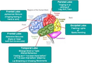
For F/U (follow up) visits is for the neurologist to see how well your seizures are under control by taking drug blood levels of the anti seizure medication your taking to make sure the medication is in a therapeutic drug level and if not he or she will make dose changes in the med(s) your on.  Possible do a EEG (electroencephalogram); the only test to decipher if you have spikes in your brain waves indicating you had a seizure determining from which lobe of the brain is having the seizures (a 26 lead to wires on the brain, which is painless).  Go to the expert for keeping you on the right track.  Its just like based on the principle why a person gets a check up on there car by seeing the mechanic (the car’s doctor) who fixes it.  The expert,  the Neurologist,  fix your seizures or get them under control.


QUOTE FOR THURSDAY:

“-New cases of epilepsy are most common among children, especially during the first year of life.
-The rate of new cases of epilepsy gradually goes down until about age 10 and then becomes stable.
-After age 55, the rate of new cases of epilepsy starts to increase, as people develop strokes, brain tumors, or Alzheimer’s disease, which all can cause epilepsy.”

PART I National Epilepsy Awareness Month-Learn what it is & know the facts!

       epilepsy2

What is epilepsy?

 Most people with epilepsy are otherwise healthy; as long as it’s well controlled like most other diseases.  A seizure is a physical manifestation of paroxysmal and abnormal electrical firing of neurons in the brain.  Think of it as numerous voltage (hyperexcitability of neurons) going throughout the brain meaning brain waves going in all directions with the brain saying its too much activity going through my organ and can’t think normally; instead the brain goes through a shock.   In simpler terms the brain is  getting too much brain wave excitability for the organ to register in what to do causing the brain to go into a type of a seizure.

When the seizure occurs there is a decrease in oxygen since the brain isn’t capable to send messages during the seizure.  The problem it too much electrical stimulation is happening in the brain causing the type of seizure to come on.  If the seizure continues to repeat one right after another the person is in status epilepticus and if the seizures do not stop the person can lead to a neuronal death;  like John Travolta’s son who died of this for example.

The term seizure disorder may refer to any number of conditions that result  in such a paroxysmal electrical discharge.  These conditions could be metabolic or structural in nature.

For example, if a metabolic condition this could be “Canavan disease” which is primarily a disease of demyelination.  Your myelin sheath that protects and insulates the nerves is being destroyed and can cause a seizure as one of the symptoms.

*Another example being metabolic is thought to be caused by brain acetate deficiency resulting from a defect of Nacetylaspartic acid (NAA) catabolism (meaning breakdown is occurring).  Accumulation of NAA, a compound thought to be responsible for maintaining cerebral fluid balance, can lead to cerebral edema and neurological injury, like a seizure as one symptoms of the disease.

*Sometimes there is a known cause and than there is just idiopathic, unknown cause for the epilepsy which if starts in childhood can resolve by the child growing out it, like in petite mal seizures but other times that is not the case and goes into motor/focal or grand mal that is permanent so the individual needs Rx for life.

Remember, not all seizures are due to epilepsy. Other conditions that can look like epilepsy include fainting, or very low blood sugar in some people being treated for diabetes.

Remember, etiology (the cause) of Epilepsy can be generally a sign of underlying pathology involving the brain–knowing the cause.  To find this out diagnostic tooling be a neurologist who specializes in epilepsy is the best resource to go to.  The epilepsy may be the first sign of a nervous system disease (ex. Brain tumor), or it may be a sign of a systemic or metabolic derangement.  Where the treatment may be able to resolve the seizure symptom completely where this wasn’t a seizure disorder or epilepsy but just a symptom due to another disorder that may be 100% cured, like a operable tumor removed surgically from the brain.

Facts and Statistics on Seizures:

  • Most seizures happen suddenly without warning, last a short time (a few seconds or minutes) and stop by themselves.
  • Seizures can be different for each person.
  • Just knowing that someone has epilepsy does not tell you what their epilepsy is like, or what seizures they have.
  • Calling seizures ‘major’ or ‘minor’ does not tell you what happens to the person during the seizure. The names of seizures used on this page describe what happens during the seizure.
  • Some people have more than one type of seizure, or their seizures may not fit clearly into the types described on this page. But even if someone’s seizures are unique, they usually follow the same pattern each time they happen.
  • Not all seizures involve convulsions (jerking or shaking movements). Some people seem vacant, wander around or are confused during a seizure.
  • Some people have seizures when they are awake, called ‘awake seizures’. Some people have seizures while they are asleep, called ‘asleep seizures’ (or ‘nocturnal seizures’). The names ‘awake’ and ‘asleep’ do not explain the type of seizures, only when they happen.
  • Injuries can happen during seizures, but many people don’t hurt themselves and don’t need to go to hospital or see a doctor.

Check out Part II tomorrow!


QUOTE FOR WEDNESDAY:

“Lung cancer diagnosis often starts with an imaging test to look at the lungs. If you have symptoms that worry you, a healthcare professional might start with an X-ray. If you smoke or used to smoke, you might have an imaging test to look for signs of lung cancer before you develop symptoms.  People with an increased risk of lung cancer may consider yearly lung cancer screening using low-dose CT scans. Lung cancer screening is generally offered to people 50 and older who smoked heavily for many years. Screening also is offered to people who have quit smoking in the past 15 years. Discuss your lung cancer risk with your healthcare professional. Together you can decide whether lung cancer screening is right for you.  Treatment for lung cancer usually begins with surgery to remove the cancer. If the cancer is very large or has spread to other parts of the body, surgery may not be possible. Treatment might start with medicine and radiation instead. Your healthcare team considers many factors when creating a treatment plan.”

MAYO CLINIC (https://www.mayoclinic.org/diseases-conditions/lung-cancer/diagnosis-treatment/drc-20374627)

Part III Lung Cancer Awareness Month – diagnosing and treatment options for Lung Cancer!

For many people, the first sign that they may have lung cancer is the appearance of a suspicious spot on a chest x-ray or a CT scan. But an image alone is not enough to tell you whether you have cancer and, if so, what type of cancer it is.

Most people who come to us for a lung cancer diagnosis first meet with a surgeon. He or she will work with pathologists, radiologists, and other lung cancer specialists to determine the specific type of lung cancer you have and how advanced it is. These findings help your disease management team develop the most successful treatment plan for you.

The first step is for your doctor to get a tissue sample using one of several biopsy methods. Then a pathologist — a type of doctor who specializes in diagnosing disease —who focuses on lung cancer studies the tissue under a microscope to determine whether you have lung cancer and, if so, what type. He or she will be able to tell this by looking closely at the cancer cells’ shape and other features.

Knowing which type of lung cancer you have will help your doctors to stage the tumor accurately and to begin identifying the best treatment approach. Understanding what type of cancer you have is also important because each type responds differently to certain chemotherapy drugs.

Testing healthy people for lung cancer

Several organizations recommend people with an increased risk of lung cancer consider annual computerized tomography (CT) scans to look for lung cancer. If you’re 55 or older and smoke or used to smoke, talk with your doctor about the benefits and risks of lung cancer screening.

 Some studies show lung cancer screening saves lives by finding cancer earlier, when it may be treated more successfully. But other studies find that lung cancer screening often reveals more benign conditions that may require invasive testing and expose people to unnecessary risks and worry.

Tests to diagnose lung cancer

If there’s reason to think that you may have lung cancer, your doctor can order a number of tests to look for cancerous cells and to rule out other conditions. In order to diagnose lung cancer, your doctor may recommend:

  • Imaging tests. An X-ray image of your lungs may reveal an abnormal mass or nodule. A CT scan can reveal small lesions in your lungs that might not be detected on an X-ray.
  • Sputum cytology. If you have a cough and are producing sputum, looking at the sputum under the microscope can sometimes reveal the presence of lung cancer cells.
  • Tissue sample (biopsy). A sample of abnormal cells may be removed in a procedure called a biopsy.Your doctor can perform a biopsy in a number of ways, including bronchoscopy, in which your doctor examines abnormal areas of your lungs using a lighted tube that’s passed down your throat and into your lungs; mediastinoscopy, in which an incision is made at the base of your neck and surgical tools are inserted behind your breastbone to take tissue samples from lymph nodes; and needle biopsy, in which your doctor uses X-ray or CT images to guide a needle through your chest wall and into the lung tissue to collect suspicious cells.A biopsy sample may also be taken from lymph nodes or other areas where cancer has spread, such as your liver.

Treatment Options for Lung Cancer Patients

Depending on the type of lung cancer you have and what stage it has progressed to, the treatment options will vary. From aggressive chemotherapy and radiation regimens to surgery and immunotherapy, two patients’ lung cancer journeys can look very different from each other.  Treatment options for lung cancer may involve cutting-edge targeted therapies and immunotherapies. Patients may also be eligible to enroll in clinical trials, where they’ll have early access to the latest innovations. comprehensive palliative care and integrative care.

QUOTE FOR MONDAY:

“There are many cancers that affect the lungs, but we usually use the term “lung cancer” for two main kinds: non-small cell lung cancer and small cell lung cancer. Non-small cell lung cancer (NSCLC) is the most common type of lung cancer. It accounts for over 80% of lung cancer cases. Common types include adenocarcinoma and squamous cell carcinoma. Adenosquamous carcinoma and sarcomatoid carcinoma are two less common types of NSCLC. Small cell lung cancer (SCLC) grows more quickly and is harder to treat than NSCLC. It’s often found as a relatively small lung tumor that’s already spread to other parts of your body. Specific types of SCLC include small cell carcinoma (also called oat cell carcinoma) and combined small cell carcinoma.  Cancer is usually staged based on the size of the initial tumor, how far or deep into the surrounding tissue it goes, and whether it’s spread to lymph nodes or other organs. Each type of cancer has its own guidelines for staging but ranges from stage 0 to stage 5.”

Cleveland Clinic (https://my.clevelandclinic.org/health/diseases/4375-lung-cancer)

Part II Lung Cancer Awareness Month -Staging of NonSmall and Small Cell Lung Cancer and How Staging Works!

What is staging and why is it important?

Understanding if and where lung cancer has spread (the stage) is important to determining what options are available for treatment. Imaging tests, biopsies and laboratory tests help to determine staging.

Non-Small Cell Lung Cancer

Non-small cell lung cancer is one of several cancers staged using the TNM system. The cancer is staged according to the size of the tumor (T), the extent to which the cancer has spread to the lymph nodes (N), and the extent to which the cancer has spread beyond the lymph nodes, or metastasis (M).

How Does The TNM Staging System Work?

The TNM staging system:

  • Was created by merging the staging systems of the American Joint Committee on Cancer (AJCC) http://www.cancerstaging.org/ and the International Union Against Cancer (UICC) http://www.uicc.org/ in 1987
  • Is one of the most commonly used cancer staging systems
  • Standardizes cancer staging internationally

T is for Tumor

How big is the tumor? Where is it located? Has it spread to nearby tissue?

TX The primary tumor cannot be assessed, or the presence of a tumor was only proven by the finding of cancer cells in sputum or bronchial washings but not seen in imaging tests or bronchoscopy.
T0 No evidence of a primary tumor.
Tis “In situ” – cancer is only in the area where the tumor started and has not spread to nearby tissues.
T1 The tumor is less than 3 cm (just slightly over 1 inch), has not spread to the membranes that surround the lungs (visceral pleura), and does not affect the air tunes (bronchi) that brand out on either side from the windpipe (trachea).
T1a The tumor is less than 2 cm.
T1b The tumor is larger than 2 cm but less than 3 cm.
T2 The tumor is larger than 3 cm but less than 7 cm or involves the main air tubes (bronchus) that brand out from the windpipe (trachea) or the membranes that surround the lungs (visceral pleura). The tumor may partially block the airways but has not caused the entire lung to collapse (atelectasis) or to develop pneumonia).
T2a The tumor is larger than 3 cm but less than or equal to 5 cm.
T2b The tumor is larger than 5 cm but less than or equal to 7 cm.
T3 The tumor is more than 7 cm or touches an area near the lung (such as the chest wall or diaphragm, or sac surrounding the heart (pericardium) or has grown into the main air tubes (bronchus) that brand out from the windpipe (trachea) but not the area where the windpipe divides or has caused one lunch to collapse (atelectasis) or pneumonia in an entire lung or there is a separate tumor(s) in the same lobe.
T4 The tumor is of any size and has spread to the area between the lungs (mediastinum), heart, trachea, esophagus, backbone or the place where the windpipe (trachea) branches or there is a separate tumor(s) in a different lobe of the same lung.

N is for Lymph Node

Has the cancer spread to the lymph nodes in and around the lungs? For more information on the lymph system and lymph nodes, see Lymph System

NX Regional lymph nodes cannot be assessed.
N0 No cancer found in lymph nodes.
N1 Cancer has spread to lymph nodes within the lung or to the area where the air pipes (bronchus) that branch out from the windpipe enter the lung, but only on the same side of the lung as the tumor (ipsilateral).
N2 Cancer has spread to lymph nodes near where the windpipe (trachea) branches into the left and right air tubes (bronchi) or near the area in the center of the lung (mediastinum) but only on the same side of the lung as the tumor.
N3 Cancer has spread to lymph nodes found on the opposite side of the lung as the tumor (contralateral) or lymph nodes in the neck.

M is for Metastasis

Has the cancer spread to other parts of the body?

MX Cancer spread cannot be assessed
M0 Cancer has not spread.
M1 Cancer has spread.
M1a Cancer has spread: separate tumor(s) in a lobe in the opposite lung from the primary tumor (contralateral), or malignant nodules in the membrane that surround the lung (pleura) or malignant excess fluid (effusion) in the pleura or membrane that surround the hear (pericardium).
M1b Cancer has spread to distant part of the body such as brain, kidney, bone.

Stages

After the Tumor (T), Lymph Nodes (N) and Metastasis (M) have been determined, the cancer is then staged accordingly:

Overall Stage T N M
Stage 0 Tis (in situ) N0 M0
Stage IA T1a, b N0 M0
Stage IB T2a N0 M0
Stage IIA T1a, b
T2a
T2b
N1
N1
N0
M0
M0
M0
Stage IIB T2b
T3
N1
N0
M0
M0
Stage IIIA T1, T2
T3
T4
N2, N1
N2, N0
N1
M0
M0
M0
Stage IIIB T4
Any T
N2
N3
M0
M0
Stage IV Any T Any N M1a, b

Small Cell Lung Cancer

Small cell lung cancer is most often staged as either limited-stage or extensive-stage.

Limited-Stage

Indicates that the cancer has not spread beyond one lung and the lymph nodes near that lung.

Extensive-Stage

The cancer is in both lungs or has spread to other areas of the body.

Source:

International Association for the Study of Lung Cancer. Goldstraw P, ed. Staging Handbook in Thoracic Oncology. Orange Park: Editorial Rx Press; 2009.

QUOTE FOR THE WEEKEND:

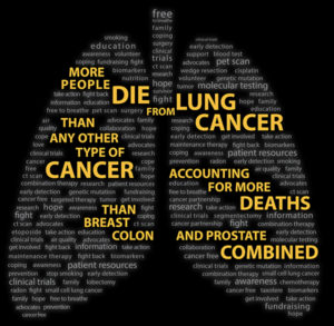
“LUNG FORCE is uniting people across the country to stand together against lung cancer, the leading cause of cancer deaths in the U.S.  Lung cancer happens when cells in the lung change (or mutate). Most often, this is because of exposure to dangerous chemicals that we breathe. But lung cancer can also happen in people with no known exposure to toxic substances. Unlike normal cells, cancer cells grow uncontrollably and cluster together to form a tumor, destroying healthy lung tissue around them. Symptoms usually do not appear until cancer cells spread to other parts of the body and prevent other organs from functioning properly. At this point, it is harder to treat lung cancer.   Smoking poses the greatest risk, but there are others such as exposure to radon and air pollution.Screening high-risk individuals has the potential to drama-tically improve lung cancer survival rates. There are multiple types of lung cancer. Knowing this information can help inform treatment options.”

American Lung Association (https://www.lung.org/lung-health-diseases/lung-disease-lookup/lung-cancer/basics)

Part I Lung Cancer Awareness Month


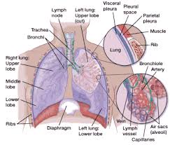
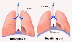
When you breathe in, air enters through your mouth and nose and goes into your lungs through the trachea (windpipe). The trachea divides into tubes called the bronchi (singular, bronchus), which enter the lungs and divide into smaller branches called the bronchioles. At the end of the bronchioles are tiny air sacs known as alveoli.

Many tiny blood vessels run through the alveoli. They absorb oxygen from the inhaled air into your bloodstream and pass carbon dioxide (a waste product from the body) into the alveoli. This is expelled from the body when you exhale. Taking in oxygen and getting rid of carbon dioxide are your lungs’ main functions.

A thin lining called the pleura surrounds the lungs. The pleura protects your lungs and helps them slide back and forth as they expand and contract during breathing. The space inside the chest that contains the lungs is called the pleural space (or pleural cavity).

Below the lungs, a thin, dome-shaped muscle called the diaphragm separates the chest from the abdomen. When you breathe, the diaphragm moves up and down, forcing air in and out of the lungs.

LUNG CANCER

There are 3 types of lungs cancer.  The two most common types of lung cancer that exist are 1 non-small cell lung cancer (NSCLC), which is the most common, and 2 small cell lung cancer (SCLC), an aggressive cancer that occurs in just over 10 percent of all lung cancer cases.

The third group is 3 lung carcinoid tumors (also known as lung carcinoids) are a type of lung cancer, which is a cancer that starts in the lungs. Cancer starts when cells begin to grow out of control. Cells in nearly any part of the body can become cancer, and can spread to other areas of the body.

Lung carcinoid tumors are uncommon and tend to grow slower than other types of lung cancers. They are made up of special kinds of cells called neuroendocrine cells.

Lung Cancer Symptoms

Both major types of lung cancer have similar symptoms. These symptoms often include a cough that doesn’t go away and shortness of breath.

Sometimes lung cancer does not cause any signs or symptoms. It may be found during a chest X-ray done for another condition. Signs and symptoms may be caused by lung cancer or by other conditions. Check with your doctor if you have any of the following:

  • Chest discomfort or pain
  • A cough that doesn’t go away or gets worse over time
  • Trouble breathing
  • Wheezing
  • Blood in sputum (mucus coughed up from the lungs)
  • Hoarseness
  • Loss of appetite
  • Weight loss for no known reason
  • Tiredness/lethargy
  • Trouble swallowing
  • Swelling in the face and/or veins in the neck

For both conditions, early detection through a low-dose computed topography (CT) scan is especially critical. Identifying lung cancer in its earliest stages even before you have symptoms can reduce the risk of death by 20 percent, according to recent studies.

Non-small cell lung Cancer (NSCLC)

Non-small cell lung cancer (NSCLC) is the most common type of cancer in lung tissues. Your risk of developing this disease increases if you are a longtime or former smoker, have been exposed to passive smoke, or have had environmental or occupational exposure to radon, asbestos, uranium, and other substances. The primary types of NSCLC are named for the type of cells found in the cancer:

  • Squamous-cell carcinoma (also called epidermoid carcinoma)
  • Adenocarcinoma
  • Large-cell carcinoma
  • Adenosquamous carcinoma
  • Undifferentiatiated carcinoma

Small Cell Lung Cancer (SCLC)

In small cell lung cancer (SCLC), small cancerous cells arise in the airway, usually in a central location. This is an aggressive cancer that spreads quickly throughout the body through the blood and lymphatic (node) systems. Typically occurring in people who smoke or who used to smoke, SCLC accounts for just over 10 percent of all lung cancers.