QUOTE FOR FRIDAY:

“Gaucher disease can affect anyone, occurring in up to 1 in 40,000 live births in the general population.

Gaucher disease is more common among Jewish people of Ashkenazi (Eastern European) descent, occurring in approximately 1 in 450 within this population. As many as 1 in 10 may carry the mutated gene responsible for Gaucher disease.

Gaucher (go-SHAY) disease is the result of a buildup of certain fatty substances in certain organs, particularly your spleen and liver. This causes these organs to enlarge and can affect their function.  The fatty substances also can build up in bone tissue, weakening the bone and increasing the risk of fractures.  If the bone marrow is affected, it can interfere with your blood’s ability to clot.  An enzyme that breaks down these fatty substances doesn’t work properly in people with Gaucher disease.  Treatment often includes enzyme replacement therapy.”

National Gaucher Foundation (https://www.mayoclinic.org/diseases-conditions/gauchers-disease/symptoms-causes/syc-20355546)


Gaucher (go-SHAY) disease

Gaucher (go-SHAY) disease is the result of a buildup of certain fatty substances in certain organs, particularly your spleen and liver. This causes these organs to enlarge and can affect their function.

The fatty substances also can build up in bone tissue, weakening the bone and increasing the risk of fractures. If the bone marrow is affected, it can interfere with your blood’s ability to clot.

An enzyme that breaks down these fatty substances doesn’t work properly in people with Gaucher disease. Treatment often includes enzyme replacement therapy.

An inherited disorder, Gaucher disease is most common in Jewish people of Eastern and Central European descent (Ashkenazi). Symptoms can appear at any age.

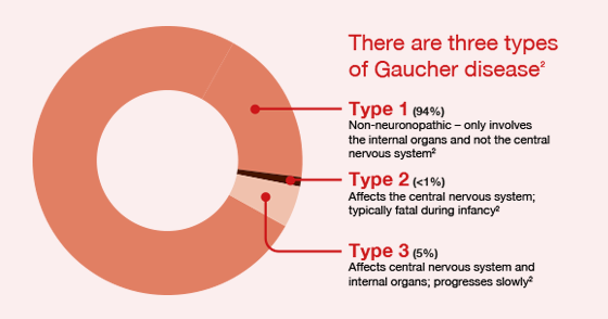
Types of Gaucher Disease:

Scientists divide Gaucher disease into 3 different types based on the presence or absence of early-onset brain involvement, including:

  • Gaucher disease type 1: Gaucher disease type 1 is the most common form of the disease in western countries, making up roughly 95 percent of patients there. Symptoms include spleen and liver enlargement, bone problems, and fatigue. Brain development is normal.
  • Gaucher disease type 2: This type of Gaucher disease is rare and involves severe neurological (brain stem) abnormalities. It is usually fatal within the first 2 years, and it is currently untreatable because of the severe, irreversible brain damage.
  • Gaucher disease type 3: This type of Gaucher disease is rare in the United States and Europe; however, it is the most common form of the disease worldwide. Gaucher disease type 3 has a severity between types 1 and 2, causing the same symptoms as type 1 plus some neurological involvement. While patients typically have a shortened lifespan, some can live into their 50s with treatment.

Symptoms

There are different types of Gaucher disease, and signs and symptoms of disease vary widely, even within the same type. Type 1 is by far the most common.

Siblings, even identical twins, with the disease can have different levels of severity. Some people who have Gaucher disease have only mild or no symptoms.

Most people who have Gaucher disease have varying degrees of the following problems:

  • Abdominal complaints. Because the liver and especially the spleen can enlarge dramatically, the abdomen can become painfully distended.
  • Skeletal abnormalities. Gaucher disease can weaken bone, increasing the risk of painful fractures. It can also interfere with the blood supply to your bones, which can cause portions of the bone to die.
  • Blood disorders. A decrease in healthy red blood cells (anemia) can result in severe fatigue. Gaucher disease also affects the cells responsible for clotting, which can cause easy bruising and nosebleeds.

More rarely, Gaucher disease affects the brain, which can cause abnormal eye movements, muscle rigidity, swallowing difficulties and seizures. One rare subtype of Gaucher disease begins in infancy and typically results in death by 2 years of age.

Causes

Gaucher disease is passed along in an inheritance pattern called autosomal recessive. Both parents must be carriers of a Gaucher changed (mutated) gene for their child to inherit the condition.

Risk factors

People of Eastern and Central European Jewish (Ashkenazi) ancestry are at higher risk of developing the most common variety of Gaucher disease.

Complications

Gaucher disease can result in:

  • Delays in growth and puberty in children
  • Gynecological and obstetric problems
  • Parkinson’s disease
  • Cancers such as myeloma, leukemia and lymphoma

When to see a doctor

If you or your child has the signs and symptoms associated with Gaucher disease, make an appointment with your doctor ASAP.

QUOTE FOR THURSDAY:*

“If you’ve been diagnosed with ovarian cancer, your cancer care team will discuss your treatment options with you. It’s important that you think carefully about each of your choices. Weigh the benefits of each treatment option against the possible risks and side effects.  Typically, treatment plans are based on the type of ovarian cancer, its stage, and any special situations. Most women with ovarian cancer will have some type of surgery to remove the tumor. Depending on the type of ovarian cancer and how advanced it is, you might need other types of treatment as well, either before or after surgery, or sometimes both. Typically the range of treatments are from surgery to chemo therapy, hormone therapy, targeted temperature therapy and/or immunotherapy depending on the oncologist team on the case.”

American Cancer Society (https://www.cancer.org/cancer/types/ovarian-cancer/treating.html)


QUOTE FOR WEDNESDAY:

“Ovarian cancer is the second most common gynecologic cancer in the United States. Ovarian cancer causes more deaths than any other cancer of the female reproductive system.  In 2020, the latest year for which incidence data are available, in the United States, 18,518 new cases of Ovarian cancer were reported among women, and 13,438 women died of this cancer. For every 100,000 women, 9 new Ovarian cancer cases were reported and 6 women died of this cancer.

Cancer is the second leading cause of death in the United States, exceeded only by heart disease. One of every five deaths in the United States is due to cancer.

Centers for Disease Control and Prevention – CDC  (https://gis.cdc.gov/Cancer/USCS/#/AtAGlance/)

QUOTE FOR TUESDAY:

“In Lower Manhattan, the plane crashes—which resulted in the collapse of the Twin Towers—created massive dust clouds that filled the air and left hundreds of highly populated city blocks covered with ash, debris, and harmful particles, including asbestos, silica, metals, concrete, and glass. Fires within the debris pile and the collapse of 7 WTC burned through the end of December 2001 with continued flare-ups in 2002, releasing carcinogenic combustion by-products. These contaminants remained in Lower Manhattan and parts of Brooklyn for an undetermined amount of time after 9/11.”

Center for Disease Control and Prevention – CDC (https://www.cdc.gov/wtc/exhibition/toxins-and-health-impacts.html)

QUOTE FOR WEEKEND:

“Duchenne muscular dystrophy (DMD) is a genetic disorder characterized by progressive muscle degeneration and weakness due to the alterations of a protein called dystrophin that helps keep muscle cells intact. DMD is one of four conditions known as dystrophinopathies.  It is the most common type of muscular dystrophy.

DMD symptom onset is in early childhood, usually between ages 2 and 3. The disease primarily affects boys, but in rare cases it can affect girls.

In Europe and North America, the prevalence of DMD is approximately 6 per 100,000 individuals.DMD was first described by the French neurologist Guillaume Benjamin Amand Duchenne in the 1860s, but until the 1980s, little was known about the cause of any kind of muscular dystrophy. In 1986, MDA-supported researchers identified a particular gene on the X chromosome that, when flawed (mutated), leads to DMD. In 1987, the protein associated with this gene was identified and named dystrophin

Muscular Dystrophy Association – MDA (https://www.mda.org/disease/duchenne-muscular-dystrophy)


Duchenne Muscular Dystrophy

The most common form of muscular dystrophy – Duchenne muscular dystrophy – typically affects young boys, but other variations can strike in adulthood.

Currently, there is no cure for muscular dystrophy, but certain physical and medical treatments can improve symptoms and slow the disease’s progression.

is a group of diseases that make muscles weaker and less flexible over time. It is caused by a problem in the genes that control how the body keeps muscles healthy. For some people, the disease starts early in childhood. Others don’t have any symptoms until they are teenagers or middle-aged adults.

How muscular dystrophy affects you or your child depends on the kind. Most people’s condition will get worse over time, and some people may lose the ability to walk, talk, or care for themselves. But that doesn’t happen to everyone. Other people can live for many years with mild symptoms.

There are more than 30 kinds of muscular dystrophy, and each is different based on:

  • The genes that cause it
  • The muscles it affects
  • The age when symptoms first appear
  • How quickly the disease gets worse

People usually get one of nine major forms of the disease:

  • Duchenne muscular dystrophy (DMD) is the most common form. It mainly affects boys, and starts between ages 3 and 5.
  • Becker muscular dystrophy is like Duchenne, except milder. It also affects boys but the symptoms start later — between ages 11 and 25.
  • Myotonic muscular dystrophy is the most common form in adults. People who have it can’t relax their muscles after they contract. It can affect both men and women, and it usually starts when people are in their 20s.
  • Congenital muscular dystrophy starts at birth or shortly afterwards.
  • Limb-Girdle muscular dystrophy often starts in a person’s teens or 20s.
  • Facioscapulohumeral muscular dystrophy affects the muscles of the face, shoulders, and upper arms. It can affect anyone from teenagers to adults in their 40s.
  • Distal muscular dystrophy affects the muscles of the arms, legs, hands, and feet. It usually comes on later in life, between ages 40 and 60.
  • Oculopharyngeal muscular dystrophy starts in a person’s 40s or 50s. It causes weakness in the muscles of the face, neck, and shoulders, and droopy eyelids (ptosis), followed by difficulty swallowing (dysphagia).
  • Emery-Dreifuss muscular dystrophy affects mainly boys, usually starting around age 10. People with this form often have heart problems along with muscle weakness.
There are many treatments that can help keep muscles strong and flexible, and scientists are looking for new ones, too.The important thing is to get the treatment you need and find support.

QUOTE FOR FRIDAY:

“Cervical cancer screening tests are usually done during a pelvic exam, which takes only a few minutes. During the exam, you lie on your back on an exam table, bend your knees, and put your feet into supports at the end of the table. The health care provider uses a speculum to gently open your vagina to see the cervix. A soft, narrow brush or tiny spatula is used to collect a small sample of cells from your cervix.

Cervical cancer is highly preventable and highly curable if caught early. Nearly all cervical cancers could be prevented by HPV vaccination, routine cervical cancer screening, and appropriate follow-up treatment when needed.

HPV commonly caused by multiple sex partners and unprotected sex. People who become sexually active at a young age, especially before age 18, or have multiple sexual partners are more likely to become infected with a high-risk type of HPV.”

National Cancer Institute (https://www.cancer.gov/types/cervical/causes-risk-prevention)


Know the diagnosing tools used in determining Cervical Cancer!


HOW TO GET DIAGNOSE FOR CERVICAL CANCER:

Key Points to remember about cervical cancer:

  • Cervical cancer is a disease in which malignant (cancer) cells form in the cervix.
  • Screening for cervical cancer using the Pap test has decreased the number of new cases of cervical cancer and the number of deaths due to cervical cancer since 1950.
  • Human papillomavirus (HPV) infection is the major risk factor for cervical cancer.

1-Screening is looking for cancer before a person has any symptoms. This can help find cancer at an early stage. When abnormal tissue or cancer is found early, it may be easier to treat. By the time symptoms appear, cancer may have begun to spread.

Know this about screening:

  • Tests are used to screen for different types of cancer when a person does not have symptoms.
  • Studies show that screening for cervical cancer helps decrease the number of deaths from the disease.
  • A Pap test is commonly used to screen for cervical cancer.
  • After certain positive Pap test results, an HPV test may be done.
  • An HPV test may be done with or without a Pap test to screen for cervical cancer.
  • Samples for an HPV test may be self-collected.
  • Screening tests for cervical cancer are being studied in clinical trials.

Scientists are trying to better understand which people are more likely to get certain types of cancer.

Cervical dysplasia occurs more often in women who are in their 20s and 30s. Death from cervical cancer is rare in women younger than 30 years and in women of any age who have regular screenings with the Pap test. The Pap test is used to detect cancer and changes that may lead to cancer. The chance of death from cervical cancer increases with age. In recent years, deaths from cervical cancer have been slightly higher in Black women younger than 50 years than in White women younger than 50 years. Deaths from cervical cancer are almost twice as likely in Black women older than 60 years than in White women older than 60 years.

Human papillomavirus (HPV) infection is the major risk factor for cervical cancer.

2.If cervical cancer is suspected, your doctor is likely to start with a thorough examination of your cervix. A special magnifying instrument (colposcope) is used to check for abnormal cells.

During the colposcopic examination, your doctor is likely to take a sample of cervical cells (biopsy) for laboratory testing. To obtain tissue, your doctor may use:

  • Punch biopsy, which involves using a sharp tool to pinch off small samples of cervical tissue.
  • Endocervical curettage, which uses a small, spoon-shaped instrument (curet) or a thin brush to scrape a tissue sample from the cervix

If the punch biopsy or endocervical curettage is worrisome, your doctor may perform one of the following tests:

  • Electrical wire loop, which uses a thin, low-voltage electrified wire to obtain a small tissue sample. Generally this is done under local anesthesia in the office.
  • Cone biopsy (conization), which is a procedure that allows your doctor to obtain deeper layers of cervical cells for laboratory testing. A cone biopsy may be done in a hospital under general anesthesia.

Staging the cervical cancer:

Cervical Cancer. Carcinoma of Cervix. Malignant neoplasm arising from cells in the cervix uteri. Vaginal bleeding. Vector diagram

If your doctor determines that you have cervical cancer, you’ll have further tests to determine the extent (stage) of your cancer. Your cancer’s stage is a key factor in deciding on your treatment.

Staging exams include:

  • Imaging tests. Tests such as X-ray, CT, MRI and positron emission tomography (PET) help your doctor determine whether your cancer has spread beyond your cervix.
  • Visual examination of your bladder and rectum. Your doctor may use special scopes to see inside your bladder and rectum.