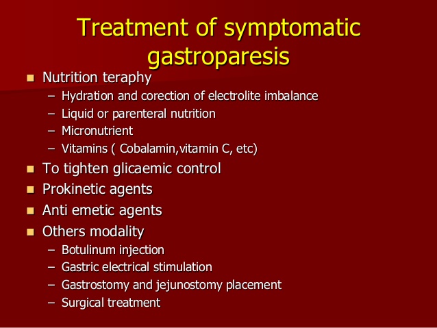
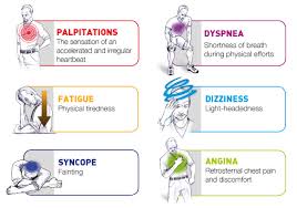
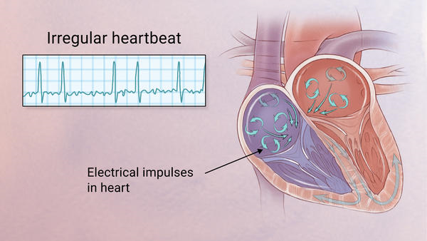
QUOTE FOR TUESDAY:

“More and more US adults are dealing with stress, which can lead to mental health problems.  In August 2022, more than 32% of US adults reported having symptoms of depression or anxiety in the last 2 weeks.  Quick activities to improve your emotional well being!

  • Be active—Take a dance break! Lift weights. Do push-ups or sit-ups. Or kick around a soccer ball for a few minutes.
  • Close your eyes, take deep breaths, stretch, or meditate.
  • Write three things you are grateful for.
  • Check in with yourself—take time to ask yourself how you are feeling.
  • Laugh! Think of someone who makes you laugh or the last time you laughed so hard you cried.
  • Find an inspiring song or quote and write it down (or screenshot it) so you have it nearby.”

Centers for Disease Control and Prevention (https://www.cdc.gov/emotional-wellbeing/features/reduce-stress.htm)

QUOTE FOR THE WEEKEND:

“Atrial fibrillation is the most common type of abnormal heart rhythm. Nearly 4 million emergency room visits from 2007 to 2014 in the United States were for atrial fibrillation. An estimated 2 million people in the U.S. have A-fib Complications arising from afib vary from Cardumyopathy to CVA (stroke), Low B/P,  CHF and more.”.

Cleveland Clinic (https://my.clevelandclinic.org/health/diseases/24578-atrial-fibrillation-with-rvr)

Part II What is RVR (Rapid Ventricular Rate) & Atrial Fibrillation?

Symptoms:

 Some of the symptoms of this disease atrial fibrillation RVR include Chest Pain or Discomfort, heart palpitations (described as unnoticed skipped beats or skipped beats noticed from experienced dizziness or difficulty in breathing), shortness of breath when lying flat (orthopnea), shortness of breath (dyspnea after exertion) sudden onset of short breath during the night (also called paroxysmal nocturnal dyspnea) and gradual swelling of lower extremities. As a result of inadequate blood flow, some patients complain of light headedness or dizziness and may feel like they are about to faint, a condition referred to as presyncope and may actually lose consciousness (syncope). Some patients experience respiratory distress that results in them appearing blue. A close examination of jugular veins usually reveals elevated pressure in some patients (jugular venous distention). When some patients are subjected to lung examinations, crackles and rales may be observed pointing to possible lung edema.

Importance of proper diagnosis:

A good diagnosis of the symptoms shown by patients is important to ascertain that the patient is suffering from atrial fibrillation with RVR.  This is because some forms or irregular and rapid heart rates, tachyarrhythmia, are dangerous and must be ruled out as they are life threatening – such as ventricular tachycardia. Some patients are usually placed on continuous cardio respiratory monitoring, but an electrocardiogram ECG is vital for correct diagnosis.

 How is it diagnosed?

 Simple, a typical 12 lead electrocardiogram (ECG). This test shows cardiac rhythms which atrial fibrillation is. Rhythms are made up of types of waves that the ECG shows which are P waves, QRS waves, T waves and U waves.

The QRS complexes should be narrow, to signify that they are being initiated by normal conduction of atrial electrical activity through the Intra-ventricular conduction system, or heart conduction system. Wide QRS complexes could point to ventricular tachycardia, although wide complexes may also be an indication of disease processes in the Intra-ventricular conduction system. The R-R internal will also likely be irregular. Meaning measuring from each R section of the QRS rhythm. It is also important to find out if there are triggering causes for the tachycardia which include dehydration, Hypovolemia – a decrease in blood volume, and more specifically decrease in blood plasma volume. You can go ahead to eliminate Acute coronary syndrome – which refers to any diseases that are directly attributed to the obstruction of coronary arteries.

How A Fib is treated:

1-A Shock

This is known as cardioversion and is used typically either when an immediate result is required or used when the Afib is of relatively recent onset or only intermittent, and so has more chance of staying in normal rhythm. In cardioversion a small shock is given using defibrillation pads. It is done under light anesthesia therefore it doesn’t hurt. The Afib may return however.

2-Rate Control Drugs

The biggest problem in Afib with RVR is too fast a heart rate. In a rhythm control strategy we use drugs such as beta-blockers to slow the heart rate down. These drugs typically will leave the patient in AF. For many people with AF it turns out that a rate control strategy is preferred as it is considered less risky than the rhythm control drugs used to get rid of the AF while being just as effective. In Afib with RVR rate control drugs can often slow the heart rate down fairly quickly and improve symptoms.

b-Rhythm Control Drugs

These medications are generally more powerful than the rate control drugs and attempt to convert the Afib back in to a normal rhythm. They are often given after a shock treatment to try and help the heart stay in normal rhythm. These drugs are also commonly used in hospitalized Afib with RVR patients. The problem with these drugs is that they may have side effects and associated risks. Many patients simply cannot tolerate Afib even if the rate is controlled and therefore require rhythm control drugs. They may be safe and effective however if used in selected patients. In cases of Afib with RVR these medications may need to be used if patients cannot tolerate other rate control medications.

3-Ablation Procedures

Ablation procedures are minimally invasive procedures typically done through the groin. They are typically used in patients that have tried, or cannot tolerate medicines for control of AFib. Ablation is typically not used as an emergency treatment of Afib with RVR, rather it is used for stable patients in AF, or those with intermittent AFib that wish to remain in normal rhythm. In patients that have had persistent Afib for a long time these procedures are not likely to be successful in the long term.

b-Pacemaker

This is typically the last throw of the dice for AF control. In some patients, drugs can either not control the rate in AFib with RVR, or the drugs can simply not be tolerated. In these patients who have no other choice, and in whom it is determined the Afib is causing harmful effects, a procedure called AV node ablation and pacemaker is done. In a relatively minor procedure, a small burn is made to the connection that connects the top and bottom chambers of the heart. A pacemaker is then inserted. This prevents Afib with RVR as although the top chambers continue to fire at a fast rate, the pacemaker now controls the bottom chamber, in a nice regular way. The downside of course is that now although the patient cannot have Afib with RVR, they have a pacemaker.

Acute afib RVR patients are more likely to be converted to Normal Sinus Rhythm (the best rhythm you could be in) as opposed to patients with chronic afib. There are complete resolutions for both kind of afib but atrial fibrillation in RVR the heart can handle for only so long and remembering the engine of our body is the heart so take good care of it for if you don’t it could allow you to die.


QUOTE FOR FRIDAY:

“Atrial fibrillation with rapid ventricular rate (A-fib with RVR) is a type of irregular heart rhythm. With A-fib with RVR, your heart doesn’t have a normal signaling process telling your heart when to beat. Instead, signaling is disorganized and the parts of your heart beat out of sync. Medicines and procedures can help manage this condition.  They have a heart rate of 100 beats per minute or more.

When you have A-fib with RVR, it’s difficult for your heart to pump the amount of blood it should. That makes it hard for your body to get the oxygen-rich blood it needs to function.”

Cleveland Clinic (https://my.clevelandclinic.org/health/diseases/24578-atrial-fibrillation-with-rvr)


Part I What is RVR (Rapid Ventricular Rate) & Atrial Fibrillation?

afib afibAFIB Symptoms.

afib RVR
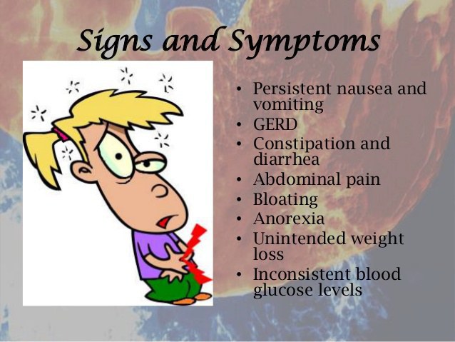
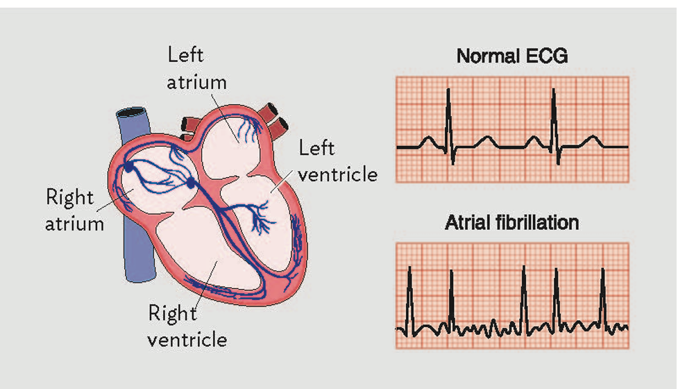
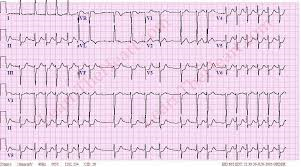
12Lead EKG with AFIB.


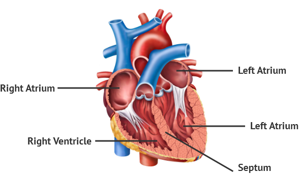
Working of the heart:

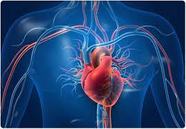
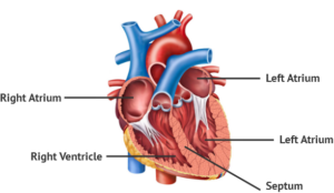
The anatomy to know in this discussion it you have 4 chambers and flow the blood through the heart.  Below is a picture of the heart showing vessels mainly carbon dioxide in blood=color blue & oxygenated vessels=color red.

  The Ventricles of the HEART

Our blood needs to be oxygenated and when oxygen is used up we have more de-oxygenated or high carbon dioxide (CO2) levels in the red blood cells in the blood stream that go to our lungs for oxygen from the right side of our heart, which is used up oxygen blood in our red blood cells to more carbon dioxide in the cell than oxygen.  So now those cells have to get replaced with oxygen and to that blood reaches the lungs from the right side of the heart pumping the blood, as its motility, to get to the lungs for more oxygen.

The RBC’s filled with more carbon dioxide are sent to the right side of the heart; first the right atrium to the right ventricle to the pulmonary artery to the lungs for more oxygen.

When we breath a oxygen / carbon dioxide process takes place at the bottom of the lungs.  We send oxygen down to our lungs and send out CO2 from our body to replace it with more oxygen.  This is how it works; when we inhale we send oxygen down to the lungs to replace the CO2 in our red blood cells (RBC’s).  We have to get rid of CO2 for replacement with oxygen.  When we exhale we send that gas, carbon dioxide, out of our blood stream and upon inhaling we send oxygen down to the lungs to replace the CO2 in our red blood cells (RBC’s).  That carbon dioxide blood in our RBC’s get sent out of the RBC’s leaving the membrane of the cell going into the lungs where a gas exchange takes place replaced with oxygen passing the RBC membrane into the RBC that’s in our blood at the bottom of the lungs that we get when we breath.  Those RBC’s that are now oxygenated are sent in the bloodstream to the left side of the heart where the heart pumps sending that oxygenated blood to all body tissues/organs to stay alive from the left atrium to left ventricle and sent out the aorta to all body parts.

When we do the process of  breathing it is what allows the exchange of gases to take place at the RBC’s.  Without oxygen we would have RBC starvation and we would be dead unless we where on a ventilator doing our breathing process.

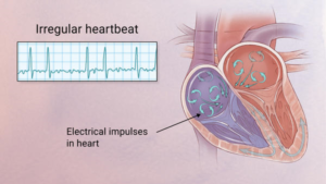
Knowing this also know we have areas of the heart that play an effect with cardiac impulses from our natural pacemaker of the heart SA node in the right atrium that sends cardiac impulsed all the way down the heart to send messages to the heart in doing beating and function that is not discussed in this since it would be a chapters.  Know this part the SA node is effected which when its now we are probably in a rhythm NSR normal sinus rhythm but when the SA node doesn’t work other areas in the atrium take over causing other rhythms and one could be atrial fibrillation or not.

So now with a quick overview of the anatomy of the 4 chambers of your heart and the process of breathing for 02 and CO2 and a quick touch of the firing system at the SA node lets go into atrial fibrillation also known as afib.

Understand in atrial fibrillation you show a line that squabbly with a pointed line.  The squabble line is

To easily identify atrial fibrillation with RVR, it is vital to understand the working of the heart.

The atrium or atria (plural) is the upper chamber of the heart, the heart has 2 atrium.  Now, in size compared to atriums you have the lower 2 chambers known as the ventricles and they are larger.

The atria function by gathering blood as it flows into the heart and shrinking to forward the blood into the ventricles. At the very moment, the ventricle must shrink to forward the blood to all parts of the body. This rhythm of blood flow creates a heart signature voice referred to as the Sinus rhythm. It is important that the sinus rhythm is synchronized so that the atrium does not send blood into the ventricle out of cue. To achieve this, an electric signal is generated to ensure the atrium contracts. When this signal short circuits (bypasses) the atrium, atrial fibrillation with RVR occurs, and the atrium is seen to vibrate just like jelly on a flat surface (see the second picture posted above to see the rhythm).

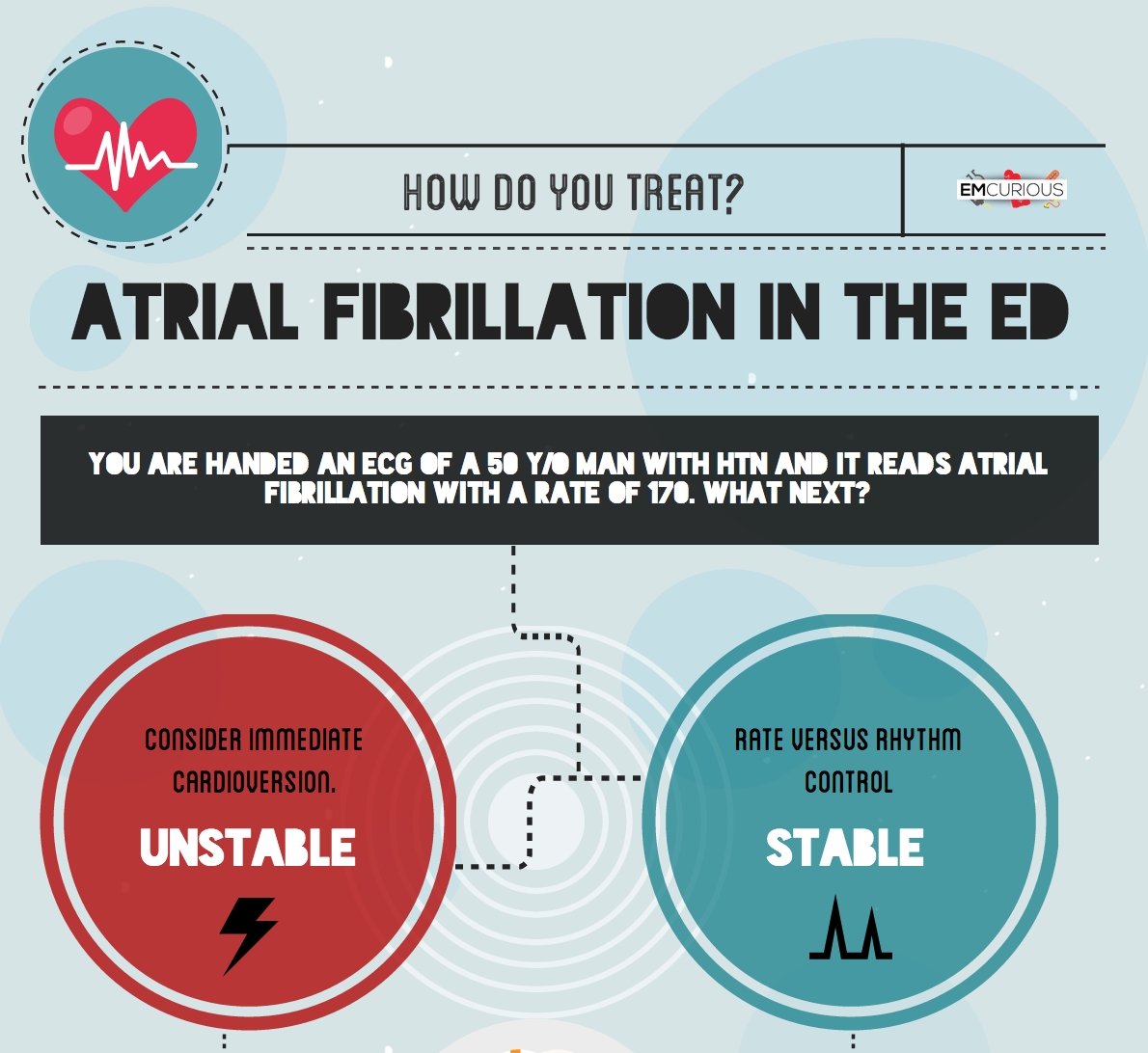
Atrial fib with RVR refers to atrial fibrillation with rapid ventricular rate. Usually the heart is like clockwork, the top (collecting) chambers beat then the bottom (main pumping) chambers sense this and also beat, and so on, in a nice regular fashion just like a clock ticking second after second. Usually the heart beats at about 60-80 beats per minute.

What is Atrial Fibrillation and RVR:

In atrial fibrillation the top chamber basically goes crazy often firing off over 400 beats per minute! Atrial fibrillation with RVR (Rapid Ventricular Response) is a heart condition caused by irregular electrical activity that results in irregular contractions of the 2 top heart chambers fibrillating. This means the heart (atriums), shakes with a rapid tremulous movement or makes fine irregular twitching movements, generally referred to as fibrillating in the upper chambers of the heart (atriums) causing little control in the heart output of blood by the heart making the lower chambers called the ventricles to take over.  Know when the atrial chambers can’t work the ventricles take over but know this they are pumping a slower than 400 beats a minute and could range from 100 and up to 200 if not controlled.

These bottom chambers don’t allow all those impulses through but it does let every second or third one through. This can give a heart rate of 100-180 beats per minute at rest, still too many beats, known as Afib with RVR, leading to symptoms and problems with heart function. Afib does not necessarily lead to Afib with RVR however, Afib can be rate controlled, sometimes naturally, sometimes using medications and sometimes requiring procedures.

In most people with AFib although symptoms can sometimes be unpleasant it is generally not harmful as long as the afib is controlled, meaning the heart in the afib rhythm with the pulse under 100. The main concern is stroke, but that can be treated with the use of blood thinning medications in people at risk. In Afib with RVR, basically the heart is beating too fast. Of course palpitations are the most common symptom. Other symptoms of AFib with RVR may include dizziness, lack of energy, exercise intolerance and shortness of breath. If Afib with RVR goes on for too long then this may result in heart failure and of course worsening of existing heart failure. Control of the heart rate in patients with Afib with RVR often causes these symptoms to improve, again meaning the HR is under 100 with the heart rhythm in afib.

A major indication of atrial fibrillation with RVR is a very rapid heartbeat rate, although some patients are known to have the condition without showing symptoms. Atrial fibrillation with RVR may occur when cardiac muscle cells overcome their intrinsic pacemaker’s signals and fire rapidly differently from their normal pattern spreading the abnormal activity to the ventricles. The rapid heart rate can strain the heart, developing a situation referred to as Tachycardia (meaning a pulse greater than 100). Atrial fibrillation with RVR can be detected from the various symptoms though it is important to remember that some patients have experienced the condition without symptoms.

Always remember the human body is like a car in the following:

The CAR’s heart is the engine! The CAR’s lungs are the transmission! One can’t live without the other or the car will die!  Without these part’s the car won’t start to run if the engine is damaged completely and it won’t be able to allow the car to slow down or speed up via the transmission!

The BODY’s engine is the heart! The BODY’s transmission is the LUNGS.  One can’t live without the other or the body will die!   Without them we will have no air exchange process and the tissues or organs or our body don’t get their food call oxygen and removing the toxic carbon dioxide!


QUOTE FOR THURSDAY:

“Fibrodysplasia ossificans progressiva is a disorder in which muscle tissue and connective tissue such as tendons and ligaments are gradually replaced by bone (ossified), forming bone outside the skeleton (extra-skeletal or heterotopic bone) that limits movement. This process generally becomes noticeable in early childhood, starting with the neck and shoulders and proceeding down the body and into the limbs.”

QUOTE FOR WEDNESDAY:

“An accurate diagnosis is necessary to treating gastroparesis, since the treatment depends on the cause. If your doctor diagnosed an underlying disease or condition that is causing the gastroparesis, the treatment will focus on correcting or reversing that condition; if there is no underlying cause or if it is not possible to treat it, then the goal of treatment is to promote gastric emptying and relieve your symptoms.

The first step is to stop taking medications that inhibit or delay gastric emptying.”

John Hopkins Medicine (https://www.hopkinsmedicine.org/health/conditions-and-diseases/gastroparesis/gastroparesis-treatment)

Part II Gastroparesis August Awareness Month

How is Gastroparesis Treated?
The treatment for gastroparesis in an individual depends on the severity of symptoms. Treatments are aimed at managing symptoms over a long-term.

Treatment approaches may involve one or a combination of:

  • dietary and lifestyle measures,
  • medications, and/or
  • procedures that may include surgery, such as
  • enteral nutrition,
  • parenteral nutrition,
  • gastric electrical stimulation (Enterra), or
  • other surgical procedures

Some people with gastroparesis have mild symptoms that come and go, which can be managed with dietary and lifestyle measures.

Others have moderate to more severe symptoms that additionally may be treated with medications to stimulate motility and/or reduce nausea and vomiting.

Some people have severe symptoms that are difficult to treat or do not respond to initial treatment approaches. They may require additional procedures to maintain nutrition and/or reduce symptoms.

Goals of Treatment
The goals of treatment are to manage and reduce symptoms, maintain quality of daily living, and minimize related problems such as:

  • Severe dehydration due to persistent vomiting
  • Bezoars (solid collections of food, fiber, or other material), which can cause nausea, vomiting, obstruction, or interfere with absorption of some medications in pill form
  • Difficulty managing blood glucose levels in people with diabetes
  • Malnutrition due to poor absorption of nutrients or a low calorie intake

Manage Risk and Benefit
No single treatment helps all persons with gastroparesis. All drugs and procedures have inherent risks, some more than others. Some of the risks are unavoidable, while others can be avoided and managed. For patients and families it is important to talk to the doctor or health care team about both benefit and risk.

As a patient, in the context of your personal illness status, consider:

  • How severe is your own condition – what effect is it having on your life
  • What is the possible benefit from the treatment suggested or prescribed to you
  • What are the chances that you will receive benefit from the treatment
  • How much benefit should you reasonably expect
  • What possible side effects or complications might there be from the treatment
  • What are the chances that you will experience a side effect or serious adverse event from the treatment
  • What can you do to reduce the chances of side effects or complications
  • How will you know when a side effect occurs
  • Exactly what should you do if a side effect or complication occurs

How to live with Gastroparesis:

Gastroparesis is a long-term condition that can impair quality of life and well-being. Living with gastroparesis affects not only those who suffer but also many others, especially family members and friends. It also touches on relationships in the classroom, in the workplace, or in social interactions.

It takes skills and strengths to deal with a challenging digestive condition like gastroparesis. It means being a kind of active researcher, always looking for what does and does not help, hurt, and work best.

It is important to understand the condition and to advocate for better health. If you or a friend or loved one has gastroparesis, it is also important to understand that you are not alone with this diagnosis.

QUOTE FOR TUESDAY:

“Gastroparesis is a condition that affects the normal spontaneous movement of the muscles (motility) in your stomach. Ordinarily, strong muscular contractions propel food through your digestive tract. But if you have gastroparesis, your stomach’s motility is slowed down or doesn’t work at all, preventing your stomach from emptying properly.”

MAYO CLINIC (https://www.mayoclinic.org/diseases-conditions/gastroparesis/symptoms-causes/)

Part I Gastroparesis August Awareness Month


Should focus attention on important health messages about gastroparesis diagnosis, treatment, and quality of life issues. The goals include improving understanding of gastroparesis to help patients and families manage the condition, and encouraging preventive strategies.

The number of people with gastroparesis appears to be rising. Yet gastroparesis is poorly understood. More community awareness is needed about the condition.

The more awareness for gastroparesis, the greater the ability to impact positive outcomes, such as additional research and improved patient care for the functional GI and motility disorders.

Gastroparesis is also called delayed gastric emptying. The term “gastric” refers to the stomach.

Normally, the stomach empties its contents in a controlled manner into the small intestines. In gastroparesis, the muscle contractions (motility) that move food along the digestive tract do not work properly and the stomach empties too slowly.

Gastroparesis is characterized by the presence of certain long-term symptoms together with delayed stomach emptying in the absence of any observable obstruction or blockage. The delayed stomach emptying is confirmed by a test.

Signs and Symptoms:

The signs and symptoms of gastroparesis may differ among persons with the condition. Symptoms usually occur during and after eating a meal.

Symptoms that are characteristic of gastroparesis include:

  • Nausea and/or vomiting
  • Retching (dry heaves)
  • Stomach fullness after a normal sized meal
  • Early fullness (satiety) – the inability to finish a meal

Diagnosing Gastroparesis:

The symptoms of gastroparesis are similar to those that occur in a number of other illnesses. When symptoms persist over time or keep coming back, it’s time to see a doctor to diagnose the problem. An accurate diagnosis is the starting point for effective treatment.

Diagnosis of gastroparesis begins with a doctor asking about symptoms and past medical and health experiences (history), and then performing a physical exam. Any medications that are being taken need to be disclosed.

Tests will likely be performed as part of the examination. These help to identify or rule out other conditions that might be causing symptoms. Tests also check for anything that may be blocking or obstructing stomach emptying. Examples of these tests include:

  • a blood test,
  • an upper endoscopy, which uses a flexible scope to look into the stomach,
  • an upper GI series that looks at the stomach on an x-ray, or
  • an ultrasound, which uses sound waves that create images to look for disease in the pancreas or gallbladder that may be causing symptoms.

If – after review of the symptoms, history, and examination – the doctor suspects gastroparesis, a test to measure how fast the stomach empties is required to confirm the diagnosis.

Slow gastric emptying alone does not correlate directly with a diagnosis of gastroparesis.

There are several different ways to measure the time it takes for food to empty from the stomach into the small intestine. These include scintigraphy, wireless motility capsule, or breath test. Your doctor will provide details of the one chosen.

Gastric Emptying Study (Scintigraphy)
The diagnostic test of choice for gastroparesis is a gastric emptying study (scintigraphy). The test is done in a hospital or specialty center.

It involves eating a bland meal of solid food that contains a small amount of radioative material so that it can be tracked inside the body. The abdomen is scanned over the next few hours to see how quickly the meal passes out of the stomach. A radiologist will interpret the study at periodic intervals after the meal.

A diagnosis of gastroparesis is confirmed when 10% or more of the meal is still in the stomach after 4 hours.

Other methods for measuring gastric emptying include a wireless motility capsule and a breath test.

Wireless Motility Capsule
The ingestible wireless motility capsule (SmartPill) is swallowed and transmits data to a small receiver that the patient carries. The data collected is interpreted by a radiologist. While taking the test, people can go about their daily routine. After a day or two, the disposable capsule is excreted naturally from the body.

Breath Test
The breath test involves eating a meal that contains a nonradioactive component that can be tracked and measured in the breath over a period of hours. The results can then be calculated to determine how quickly the stomach empties.

Stay tune for Part II tomorrow!