QUOTE FOR MONDAY:

“It all comes down to the pathophysiology.  Sepsis ultimately results from a complex interaction of pro-inflammatory, anti-inflammatory, activated complement system, and coagulation mediators that in association with detector and signaling markers, trigger a host response. Initiators (microbes, trauma, hypoxia, ischemia, toxins) cause local tissue damage, which release local pro- and anti-inflammatory markers. Proinflammatory signalers include TNF, IL-1, and IL-6, while anti-inflammatory markers include IL-4, IL-10, IL-11 and soluble TNF receptors. These are designed to function and contain at a local level. If the initiators overwhelm the local response, the mediators affect multiple systems in the body: dermal, cardiovascular, gastrointestinal, renal, neurologic, hematologic/coagulopathic, pulmonary, and endocrine.3,4 All of these mimics have a similar endgame: triggering a systemic reaction that looks just like sepsis.”

emDOCS (http://www.emdocs.net/mimics-of-sepsis/)

Part II Sepsis and SIRS (systemic inflammatory response syndrome)-Still a major problem in hospitals!

Patty Duke and sepsissepsismultihit

               sepsis inflammatory process

Part II talks to you about the multi-hit theory of SIRS with Inflammatory Cascade of SIRS and lastly the coagulation process in SIRS.   It also tells you an extensive amount of infectious and non-infectious causes of SIRS. Lastly the key antidote to SIRS.
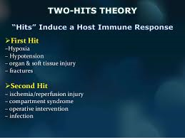
Multi-hit theory

A multi hit theory behind the progression of SIRS to organ dysfunction and possibly multiple organ dysfunction syndrome (MODS). In this theory, the event that initiates the SIRS cascade primes the pump. With each additional event, an altered or exaggerated response occurs, leading to progressive illness. The key to preventing the multiple hits is adequate identification of the ETIOLOGY or CAUSE of SIRS and appropriate resuscitation and therapy.

Inflammatory cascade

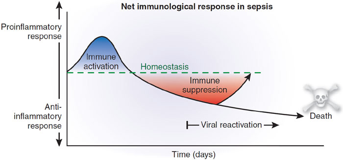
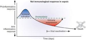
Trauma, inflammation, or infection leads to the activation of the inflammatory cascade. Initially, a pro-inflammatory activation occurs, but almost immediately thereafter a reactive suppressing anti-inflammatory response occurs. This SIRS usually manifests itself as increased systemic expression of both pro-inflammatory and anti-inflammatory species. When SIRS is mediated by an infectious insult, the inflammatory cascade is often initiated by endotoxin or exotoxin. Tissue macrophages, monocytes, mast cells, platelets, and endothelial cells are able to produce a multitude of cytokines. The cytokines tissue necrosis factor–alpha (TNF-α) and interleukin-1 (IL-1) are released first and initiate several cascades.

The release of certain factors without getting into medical specific terms they ending line induces the production of other pro-inflammatory cytokines, worsening the condition.

Some of these factors are the primary pro-inflammatory mediators. In research it suggests that glucocorticoids may function by inhibit-ing certain factors that have been shown to be released in large quantities within 1 hour of an insult and have both local and systemic effects. In studies they have shown that certain cytokines given individually produce no significant hemodynamic response but that they cause severe lung injury and hypotension. Others responsible for fever and the release of stress hormones (norepinephrine, vasopressin, activation of the renin-angiotensin-aldosterone system).

Other cytokines, stimulate the release of acute-phase reactants such as C-reactive protein (CRP) and pro-calcitonin.

The pro-inflammatory interleukins either function directly on tissue or work via secondary mediators to activate the coagulation cascade and the complement cascade and the release of nitric oxide, platelet-activating factor, prostaglandins, and leukotrienes.

High mobility group box 1 (HMGB1) is a protein present in the cytoplasm and nuclei in a majority of cell types. In response to infection or injury, as is seen with SIRS, HMGB1 is secreted by innate immune cells and/or released passively by damaged cells. Thus, elevated serum and tissue levels of HMGB1 would result from many of the causes of SIRS.

HMGB1 acts as a potent pro-inflammatory cytokine and is involved in delayed endotoxin lethality and sepsis.

Numerous pro-inflammatory polypeptides are found within the complement cascade. It is thought they are felt to contribute directly to the release of additional cytokines and to cause vasodilatation and increasing vascular permeability. Prostaglandins and leukotrienes incite endothelial damage, leading to multi-organ failure.

Polymorphonuclear cells (PMNs) from critically ill patients with SIRS have been shown to be more resistant to activation than PMNs from healthy donors, but, when stimulated, demonstrate an exaggerated micro-bicidal response (agents that kill microbes). This may represent an auto-protective mechanism in which the PMNs in the already inflamed host may avoid excessive inflammation, thus reducing the risk of further host cell injury and death.[4]

Coagulation

The correlation between inflammation and coagulation is critical to understanding the potential progression of SIRS. IL-1 and TNF-α directly affect endothelial surfaces, leading to the expression of tissue factor. Tissue factor initiates the production of thrombin, thereby promoting coagulation, and is a proinflammatory mediator itself. Fibrinolysis is impaired by IL-1 and TNF-α via production of plasminogen activator inhibitor-1. Pro-inflammatory cytokines also disrupt the naturally occurring anti-inflammatory mediators anti-thrombin and activated protein-C (APC).

If unchecked, this coagulation cascade leads to complications of micro-vascular thrombosis, including organ dysfunction. The complement system also plays a role in the coagulation cascade. Infection-related pro-coagulant activity is generally more severe than that produced by trauma.

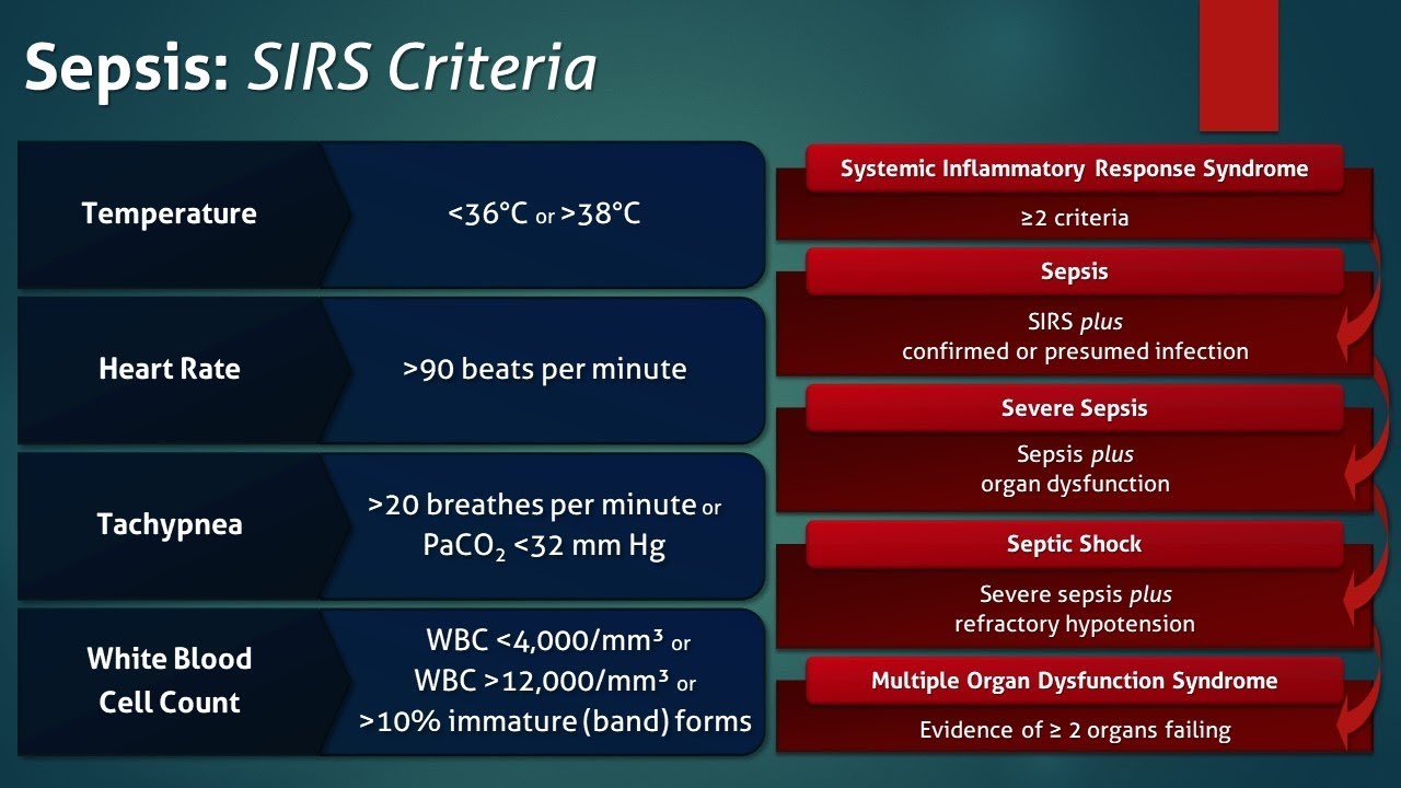
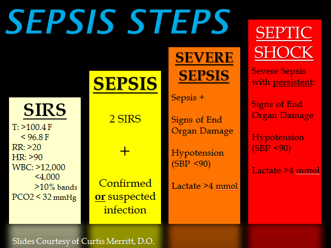
What the causes of SIRS can be:

The etiology of systemic inflammatory response syndrome (SIRS) is broad and includes infectious and noninfectious conditions, surgical procedures, trauma, medications, and therapies.

The following is partial list of the infectious causes of SIRS:

  • Bacterial sepsis
  • Burn wound infections
  • Candidiasis
  • Cellulitis
  • Cholecystitis
  • Community-acquired pneumonia
  • Diabetic foot infection
  • Erysipelas
  • Infective endocarditis
  • Influenza
  • Intra-abdominal infections (eg, diverticulitis, appendicitis)
  • Gas gangrene
  • Meningitis
  • Nosocomial pneumonia
  • Pseudomembranous colitis
  • Pyelonephritis
  • Septic arthritis
  • Toxic shock syndrome
  • Urinary tract infections (male and female)
  • *The following is a partial list of the noninfectious causes of SIRS:
  • Acute mesenteric ischemia
  • Adrenal insufficiency
  • Autoimmune disorders
  • Burns
  • Chemical aspiration
  • Cirrhosis
  • Cutaneous vasculitis
  • Dehydration
  • Drug reaction
  • Electrical injuries
  • Erythema multiforme
  • Hemorrhagic shock
  • Hematologic malignancy
  • Intestinal perforation
  • Medication side effect (eg, from theophylline)
  • Myocardial infarction
  • Pancreatitis
  • Seizure
  • Substance abuse – Stimulants such as cocaine and amphetamines
  • Surgical procedures
  • Toxic epidermal necrolysis
  • Transfusion reactions
  • Upper gastrointestinal bleeding
  • VasculitisThe treatment is don’t get it since it is hard to get rid of especially for people over 65 and in hospitals.  There is no one Rx for it.  If you’re unfortunate enough to be diagnosed with SIRS the sooner you get diagnosed with it including being in stage one as opposed to three the higher the odds the turn out will be for you.  Again the key is prevention; don’t get it. There is no one antidote to this SIRS

PREVENTION IS THE KEY ANTIDOTE!   So stay healthy and out of  hospitals!

QUOTE FOR THE WEEKEND:

“Sepsis is a clinical syndrome that complicates severe infection and is characterized by the systemic inflammatory response syndrome (SIRS), immune dysregulation, microcirculatory derangements, and end-organ dysfunction. In this syndrome, tissues remote from the original insult display the cardinal signs of inflammation, including vasodilation, increased microvascular permeability, and leukocyte accumulation.

Although inflammation is an essential host response, the onset and progression of sepsis center upon a “dysregulation” of the normal response, usually with an increase in both proinflammatory and antiinflammatory mediators, initiating a chain of events that leads to widespread tissue injury.”

Uptodate.com (https://www.uptodate.com/contents/systemic-inflammatory-response-syndrome-sirs-and-sepsis-in-children-definitions-epidemiology-clinical-manifestations-and-diagnosis)

Part I Sepsis & SIRS (systemic inflammatory response syndrome)-Not new in hospitals.

SIRS was first described by Dr. William R. Nelson, of the University of Toronto, in a presentation to the Nordic Micro Circulation meeting in Geilo, Norway-February 1983.  In 1992, the American College of Chest Physicians (ACCP) and the Society of Critical Care Medicine (SCCM) introduced definitions for systemic inflammatory response syndrome (SIRS), sepsis, severe sepsis, septic shock, and multiple organ dysfunction syndrome (MODS), they are interrelated with each other in SIRS.  The idea behind defining SIRS was to define a clinical response to a nonspecific insult of either infectious or noninfectious origin. SIRS is defined as 2 or more of the following variables:

  • Fever of more than 38°C (100.4°F) or less than 36°C (96.8°F)
  • Heart rate of more than 90 beats per minute
  • Respiratory rate of more than 20 breaths per minute or arterial carbon dioxide tension (PaCO 2) of less than 32 mm Hg, which is normally in our body at 35-45 mm Hg whereas the oxygen= PaO2 in our body greater than 80mm Hg for the norm.
  • Abnormal white blood cell count (>12,000/µL or < 4,000/µL or >10% immature [band] forms)

SIRS is nonspecific and can be caused by ischemia, inflammation, trauma, infection, or several insults combined. Thus, SIRS is not always related to infection but commonly is.  SIRS is an inflammatory state affecting the whole body, frequently a response of the immune system to infection, but not always.  It is frequently related to sepsis, a condition in which individuals meet criteria for SIRS and have a known infection.

It is the body’s response to an infectious or noninfectious insult to it. Although the definition of SIRS refers to it as an “inflammatory” response, it actually has pro- and anti-inflammatory components.  SIRS describes the host response to a critical illness of infectious or noninfectious cause, such as burns, trauma, and pancreatitis. More specific definitions are as follows: Sepsis is SIRS resulting from a presumed or known site of infection. Severe sepsis is sepsis with an acute associated multiple organ failure.

What causes sepsis?

Bacterial infections are the most common cause of sepsis. Sepsis can also be caused by fungal, parasitic, or viral infections. The source of the infection can be any of a number of places throughout the body. Common sites and types of infection that can lead to sepsis include:

  • The abdomen—An inflammation of the appendix (appendicitis), bowel problems, infection of the abdominal cavity (peritonitis), and gallbladder or liver infections
  • The central nervous system—Inflammation or infections of the brain or the spinal cord
  • The lungs—Infections such as pneumonia
  • The skin—Bacteria can enter skin through wounds or skin inflammations, or through the openings made with intravenous (IV) catheters (tubes inserted into the body to administer or drain fluids). Conditions such as cellulitis (inflammation of the skin’s connective tissue) can cause sepsis.
  • The urinary tract (kidneys or bladder)—Urinary tract infections are especially likely if the patient has a urinary catheter to drain urine

Who is at risk for sepsis?

Sepsis can strike anyone, but those at particular risk include:

  • People with weakened immune systems
  • Patients who are in the hospital
  • People with pre-existing infections or medical conditions
  • People with severe injuries, such as large burns or bullet wounds
  • People with a genetic tendency for sepsis
  • The very old or very young

What are the symptoms of sepsis?

Because of the many sites on the body from which sepsis can originate, there is a wide variety of symptoms. The most prominent are:

  • Decreased urine output
  • Fast heart rate
  • Fever
  • Or the opposite Hypothermia (very low body temperature)
  • Shaking
  • Chills
  • Warm skin or a skin rash
  • Confusion or delirium
  • Hyperventilation (rapid breathing)

How is sepsis diagnosed?

A person may have sepsis if he or she has:

  • A high or low white blood cell count
  • A low platelet count
  • Acidosis (too much acid in the blood); in the hospital what is checked is lactic acid blood level.
  • A blood culture that is positive for bacteria
  • Abnormal kidney or liver function

How is sepsis treated?

The most important intervention in sepsis is quick diagnosis and prompt treatment. Patients diagnosed with severe sepsis are usually placed in the intensive care unit (ICU) of the hospital for special treatment. The doctor will first try to identify the source and the type of infection, and then administer antibiotics to treat the infection. (Note: antibiotics are ineffective against infections caused by viruses; if anything what is used is antiviral medications.)

The doctor also administers IV fluids to prevent blood pressure from dropping too low. In some cases, vasopressor medications (which constrict blood vessels) are needed to achieve an adequate blood pressure. Some patients are given new drug therapies, such as activated protein C (APC). And finally, if organ failures occur, appropriate supportive care is provided (for example, dialysis for kidney failure, mechanical ventilation for respiratory failure, etc.).  Commonly what is used when initially sepsis is diagnosed is Vancomycin with other antibiotics like Imipenum, Cefepime, and others depending on what the blood culture shows as the microorganism if SIRS is caused by a bacterial infection (many times it is).

QUOTE FOR FRIDAY:

“As yet, there is no complete cure for SMA. However, the discovery of the genetic cause of SMA has led to the development of several treatment options that affect the genes involved in SMA — a gene replacement therapy called Zolgensma, and two drugs, called nusinersen (Spinraza) and risdiplam (Evyrsdi).”

Boston Children’s Hospital (https://www.childrenshospital.org/conditions/spinal-muscular-atrophy-sma)

QUOTE FOR THURSDAY:

“There is a 1 in 4 chance the child has SMA, 1 in 4 chance the child inherited 2 healthy copies of the SMN1 gene and a 1 in 2 chance the child is a carrier.  While there’s no cure for SMA, treatment can help. Home modifications, medications, assistive devices, physical and occupational therapy, and feeding and breathing assistance are all things that can make living with SMA easier.”.

healthline (https://www.healthline.com/health/spinal-muscular-atrophy/spinal-muscular-atrophy-facts-stats#types-and-symptoms)


Part III Awareness of Spinal Muscular Atrophy (SMA)-Causes and diagnosing!


Causes of muscle atrophy

Unused muscles can waste away if you are not active. Even after it begins, this type of atrophy can often be reversed with exercise and improved nutrition.

Muscle atrophy can also happen if you are bedridden or unable to move certain body parts due to a medical condition. Astronauts, for example, can also experience some muscle atrophy after a few days of weightlessness.

Other causes for muscle atrophy include:

  • lack of physical activity for an extended period of time
  • aging
  • alcohol-associated myopathy, a pain and weakness in muscles due to excessive drinking over long periods of time
  • burns
  • injuries, such as a torn rotator cuff or broken bones
  • malnutrition
  • spinal cord or peripheral nerve injuries
  • stroke
  • long-term corticosteroid therapy

Diseases can cause muscles to waste away or can make movement difficult, leading to muscle atrophy. These include:

  • amyotrophic lateral sclerosis (ALS), also known as Lou Gehrig’s disease, affects nerve cells that control voluntary muscle movement
  • dermatomyositis, causes muscle weakness and skin rash
  • Guillain-Barré syndrome, an autoimmune disease that leads to nerve inflammation and muscle weakness
  • multiple sclerosis, an autoimmune disease in which the body destroys the protective coverings of nerves
  • muscular dystrophy, an inherited disease that causes muscle weakness
  • neuropathy, damage to a nerve or nerve group, resulting in loss of sensation or function
  • osteoarthritis, causes reduced motion in the joints
  • polio, a viral disease affecting muscle tissue that can lead to paralysis
  • polymyositis, an inflammatory disease
  • rheumatoid arthritis, a chronic inflammatory disease that affects the joints
  • spinal muscular atrophy, a hereditary disease causing arm and leg muscles to waste away

Causes of Spinal Muscular Atrophy:

It is caused by a loss of specialized nerve cells, called motor neurons that control muscle movement.  In this disease we have an insufficient amount of SMN protein, which leads to permanent loss of motor neurons (Destruction).  The weakness tends to be more severe in the muscles that are close to the center of the body (proximal) compared to muscles away from the body’s center (distal). The muscle weakness usually worsens with age.

SMA is an auto-somal recessive disease. This means that (most of the time) both parents must carry the genetic mutation for a child to have the condition.

The gene affected in SMA is the “survival of motor neuron” gene (SMN1 and SMN2). In 95 percent of SMA cases, both copies of the SMN1 gene are missing. All people with SMA have a number of copies of the SMN2 gene. But the SMN2 gene produces only a small amount of functional SMN protein; the more copies of the SMN2 gene a child has, the milder the disease.

If someone in your family has SMA, your chance of being an SMA carrier significantly increases. When both parents are carriers, there is a 1 in 4 (25 percent) chance with each pregnancy that they will have a child with SMA.

The SMA foundation states that the disease generally manifests early in life and is the leading genetic cause of death in infants and toddlers. … One in 40 to one in 50 people (approximately 6 million Americans) are carriers of the SMA gene.

Spinal muscular atrophy (SMA) affects 1 per 8,000 to 10,000 people worldwide. Spinal muscular atrophy type I is the most common type, accounting for about half of all cases. Types II and III are the next most common and types 0 and IV are rare.

Mutations in the SMN1 gene cause all types of spinal muscular atrophy described above. The number of copies of the SMN2 gene modifies the severity of the condition and helps determine which type develops.

The SMN1 and SMN2 genes both provide instructions for making a protein called the survival motor neuron (SMN) protein. Normally, most functional SMN protein is produced from the SMN1 gene, with a small amount produced from the SMN2 gene. Several different versions of the SMN protein are produced from the SMN2 gene, but only one version is functional; the other versions are smaller and quickly broken down. The SMN protein is one of a group of proteins called the SMN complex, which is important for the maintenance of motor neurons.  Motor neurons transmit signals from the brain and spinal cord that tell skeletal muscles to tense (contract), which allows the body to move.

Most people with spinal muscular atrophy are missing a piece of the SMN1 gene, which impairs SMN protein production. A shortage of SMN protein leads to motor neuron death, and as a result, signals are not transmitted between the brain and muscles. Muscles cannot contract without receiving signals from the brain, so many skeletal muscles become weak and waste away, leading to the signs and symptoms of spinal muscular atrophy.

Typically, people have two copies of the SMN1 gene and one to two copies of the SMN2 gene in each cell. However, the number of copies of the SMN2 gene varies, with some people having up to eight copies. In people with spinal muscular atrophy, having multiple copies of the SMN2 gene is usually associated with less severe features of the condition that develop later in life. The SMN protein produced by the SMN2 genes can help make up for the protein deficiency caused by SMN1 gene mutations. People with spinal muscular atrophy type 0 usually have one copy of the SMN2 gene in each cell, while those with type I generally have one or two copies, those with type II usually have three copies, those with type III have three or four copies, and those with type IV have four or more copies. Other factors, many unknown, also contribute to the variable severity of spinal muscular atrophy.

Spinal muscular atrophy is inherited in an autosomal recessive pattern, which means both copies of the SMN1 gene in each cell have mutations. In most cases, the parents of an individual with an autosomal recessive condition each carry one copy of the mutated gene, but they typically do not show signs and symptoms of the condition. In rare cases, a person with spinal muscular atrophy inherits an SMN1 gene mutation from one parent and acquires a new mutation in the other copy of the gene that occurs during the formation of reproductive cells (eggs or sperm) or in early embryonic development. In these cases, only one parent is a carrier of the SMN1 gene mutation.

Individuals who have more than the usual two copies of the SMN2 gene usually do not inherit the extra copies from a parent. They typically arise during a random error when making new copies of DNA (replication) in an egg or sperm cell or just after fertilization.

Remember SMA is an autosomal recessive disease and this means that (most of the time) both parents must carry the genetic mutation for a child to have the condition.

The gene affected in SMA is the “survival of motor neuron” gene (SMN1 and SMN2). In 95 percent of SMA cases, both copies of the SMN1 gene are missing. All people with SMA have a number of copies of the SMN2 gene. But the SMN2 gene produces only a small amount of functional SMN protein; the more copies of the SMN2 gene a child has, the milder the disease.

If someone in your family has SMA, your chance of being an SMA carrier significantly increases. Remember when both parents are carriers, there is a 1 in 4 (25 percent) chance with each pregnancy that they will have a child with SMA.

Diagnosing: 

To find the cause of symptoms, your healthcare provider will perform a physical exam and get a medical history. Your physician may also order one or more of these tests to diagnose SMA:

  • Blood test: An enzyme and protein blood test can check for high levels of creatine kinase. Deteriorating muscles release this enzyme into the bloodstream.
  • Nerve conduction test: An electromyogram (EMG) measures the electrical activity of nerves muscles and nerves.
  • Muscle biopsy: Rarely, a physician may perform a muscle biopsy. This procedure involves removing a small amount of muscle tissue and sending it to a lab for examination. A biopsy can show atrophy, or loss of muscle.
  • Genetic test: This blood test identifies problems with the SMN1 gene. As a diagnostic tool, a genetic test is 95% effective at finding the altered SMN1 gene. Some states test for SMA as part of routine newborn screenings.

DNA testing is the preferred testing.

Genetic testing is a type of medical test that identifies changes in chromosomes, genes, or proteins. The results of a genetic test can confirm or rule out a suspected genetic condition or help determine a person’s chance of developing or passing on a genetic disorder. More than 1,000 genetic tests are currently in use, and more are being developed.

Several methods can be used for genetic testing:

  • Molecular genetic tests (or gene tests) study single genes or short lengths of DNA to identify variations or mutations that lead to a genetic disorder.
  • Chromosomal genetic tests analyze whole chromosomes or long lengths of DNA to see if there are large genetic changes, such as an extra copy of a chromosome, that cause a genetic condition.
  • Biochemical genetic tests study the amount or activity level of proteins; abnormalities in either can indicate changes to the DNA that result in a genetic disorder.

Genetic testing is voluntary. Because testing has benefits as well as limitations and risks, the decision about whether to be tested is a personal and complex one. A geneticist or genetic counselor can help by providing information about the pros and cons of the test and discussing the social and emotional aspects of testing.

Newborn Testing: To test if the routine newborn screening dried blood spots can be used to test if missing 2 copies of SMN1 gene, a status indicating spinal muscular atrophy.

QUOTE FOR WEDNESDAY:

“Those who might have gotten Skeletal Muscular Atrophy (SMA) are within the approximately 10,000 to 25,000 children and adults are living with SMA in the United States. It’s a rare disease that affects one out of 6,000 to 10,000 children.

A person with SMA inherits two copies of a missing or faulty (mutated) survival motor neuron 1 (SMN1) gene. One faulty gene comes from the mother and the other comes from the father. An adult can have a single copy of the defective gene that causes SMA and not know it.

About six million Americans (1 in 50) carry the mutated SMN1 gene. These carriers have one healthy SMN1 gene and one missing or defective SMN1 gene. Carriers don’t develop SMA. There’s a 1 in 4 chance that two carriers will have a child with SMA.”

Cleveland Clinic (https://my.clevelandclinic.org/health/diseases/14505-spinal-muscular-atrophy-sma)


QUOTE FOR TUESDAY:

What we need to know about spinal muscular atrophy (SMA):

  • Spinal muscular atrophy (SMA) is a disorder affecting the motor neurons—nerve cells that control voluntary muscle movement. These cells are located in the spinal cord.
  • Because the muscles cannot respond to signals from the nerves, they atrophy — weaken and shrink — from inactivity.
  • One in every 6,000 babies is born with SMA. It is one of the most prevalent genetic disorders affecting young children and a major cause of death in infancy.
  • SMA can strike children at any age. SMA in infancy and early childhood is associated with worse outcomes, while patients who develop symptoms later in childhood or in adolescence usually have a more positive prognosis.
  • SMA does not affect sensory nerves or intellect, but it has been observed that many patients with SMA are highly intelligent.

Part I Awareness of Spinal Muscular Atrophy (SMA)-What SMA is, staging of SMA & the types of MS atrophy there are?

What is Spinal Muscular Atrophy?

Spinal muscular atrophy is a genetic disorder characterized by weakness and wasting in muscles used for movement (skeletal muscles). It is caused by a loss of specialized nerve cells, called motor neurons that control muscle movement.  In this disease we have an insufficient amount of SMN protein, which leads to permanent loss of motor neurons (Destruction).  The weakness tends to be more severe in the muscles that are close to the center of the body (proximal) compared to muscles away from the body’s center (distal). The muscle weakness usually worsens with age. There are many types of spinal muscular atrophy that are caused by changes in the same genes. The types differ in age of onset and severity of muscle weakness; however, there is overlap between the types. Other forms of spinal muscular atrophy and related motor neuron diseases, such as spinal muscular atrophy with progressive myoclonic epilepsy, spinal muscular atrophy with lower extremity predominance, X-linked infantile spinal muscular atrophy, and spinal muscular atrophy with respiratory distress type 1 are caused by mutations in other genes.

Spinal muscular atrophy type 0 is evident before birth and is the rarest and most severe form of the condition. Affected infants move less in the womb, and as a result they are often born with joint deformities (contractures). They have extremely weak muscle tone (hypotonia) at birth. Their respiratory muscles are very weak and they often do not survive past infancy due to respiratory failure. Some infants with spinal muscular atrophy type 0 also have heart defects that are present from birth (congenital).

STAGING OF SPINAL MUSCULAR ATROPHY:

Spinal muscular atrophy type I (also called Werdnig-Hoffmann disease) is the most common form of the condition. It is a severe form of the disorder with muscle weakness evident at birth or within the first few months of life. Most affected children cannot control their head movements or sit unassisted. Children with this type may have swallowing problems that can lead to difficulty feeding and poor growth. They can also have breathing problems due to weakness of respiratory muscles and an abnormally bell-shaped chest that prevents the lungs from fully expanding. Most children with spinal muscular atrophy type I do not survive past early childhood due to respiratory failure.

Spinal muscular atrophy type II (also called Dubowitz disease) is characterized by muscle weakness that develops in children between ages 6 and 12 months. Children with this type can sit without support, although they may need help getting to a seated position. However, as the muscle weakness worsens later in childhood, affected individuals may need support to sit. Individuals with spinal muscular atrophy type II cannot stand or walk unaided. They often have involuntary trembling (tremors) in their fingers, a spine that curves side-to-side , and respiratory muscle weakness that can be life-threatening. The life span of individuals with spinal muscular atrophy type II varies, but many people with this condition live into their twenties or thirties.

Spinal muscular atrophy type III (also called Kugelberg-Welander disease) typically causes muscle weakness after early childhood. Individuals with this condition can stand and walk unaided, but over time, walking and climbing stairs may become increasingly difficult. Many affected individuals require wheelchair assistance later in life. People with spinal muscular atrophy type III typically have a normal life expectancy.

Spinal muscular atrophy type IV is rare and often begins in early adulthood. Affected individuals usually experience mild to moderate muscle weakness, tremors, and mild breathing problems. People with spinal muscular atrophy type IV have a normal life expectancy.

JOHN HOPKINS MEDICINE states these types of SMA:

Type Age at onset Symptoms, rate of progression, and life expectancy
Becker adolescence to early adulthood Symptoms are almost identical to Duchenne, but less severe; progresses more slowly than Duchenne; survival into middle age. As with Duchenne, disease is almost always limited to males.
Congenital birth Symptoms include general muscle weakness and possible joint deformities; disease progresses slowly; shortened life span.
Duchenne 2 to 6 years Symptoms include general muscle weakness and wasting; affects pelvis, upper arms, and upper legs; eventually involves all voluntary muscles; survival beyond 20s is rare. Seen in boys only. Very rarely can affect woman, who have much milder symptoms and a better prognosis.
Distal 40 to 60 years Symptoms include weakness and wasting of muscles of the hands, forearms, and lower legs; progression is slow; rarely leads to total incapacity.
Emery-Dreifuss childhood to early teens Symptoms include weakness and wasting of shoulder, upper arm, and shin muscles; joint deformities are common; progression is slow; sudden death may occur from cardiac problems.
Facioscapulohumeral childhood to early adults Symptoms include facial muscle weakness and weakness with some wasting of shoulders and upper arms; progression is slow with periods of rapid deterioration; life span may be many decades after onset.
Limb-Girdle late childhood to middle age Symptoms include weakness and wasting, affecting shoulder girdle and pelvic girdle first; progression is slow; death is usually due to cardiopulmonary complications.
Myotonic 20 to 40 years Symptoms include weakness of all muscle groups accompanied by delayed relaxation of muscles after contraction; affects face, feet, hands, and neck first; progression is slow, sometimes spanning 50 to 60 years.
Oculopharyngeal 40 to 70 years Symptoms affect muscles of eyelids and throat causing weakening of throat muscles, which, in time, causes inability to swallow and emaciation from lack of food; progression is slow.