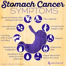
Part I What is Amyloidosis?


Amyloidosis (am-uh-loi-DO-sis) is a rare disease that occurs when a substance called amyloid builds up in your organs. Amyloid is an abnormal protein that is usually produced in your bone marrow and can be deposited in any tissue or organ.
The cause of primary amyloidosis is unknown. The condition is related to abnormal and excess production of antibodies by a type of immune cell called plasma cells. Clumps of abnormal proteins build up in certain organs. This reduces their ability to work correctly.
Symptoms depend on the organs affected. Amyloidosis frequently affects the heart, kidneys, liver, spleen, nervous system and digestive tract. Severe amyloidosis can lead to life-threatening organ failure. This disease can affect the tongue, intestines, skeletal and smooth muscles, nerves, skin, ligaments, heart, liver, spleen, and kidneys. Skin changes, such as thickening or easy bruising, and purplish patches around the eyes, difficulty swallowing,
Symptoms include: enlarged tongue, difficulty swallowing, feeling full quickly when eating, and significant weight loss, diarrhea possibly with blood or constipation, abnormal heart rhythm, fatigue, numbness of hands or feet, shortness of breath, hoarseness or changing voice, swelling of your ankles and legs and joint pain.
Amyloid is produced in your bone marrow and can be deposited in any tissue or organ affecting the tissue and organs like stated previously. The specific cause of this condition depends on the type of amyloidosis present.
Unfortunately, there are many different types of amyloidosis. Here are the types:
– Immunoglobulin light chain (AL) amyloidosis. This is the most common type, can affect your heart, kidneys, skin, nerves and liver. It was previously known as primary amyloidosis. It occurs when your bone marrow produces abnormal antibodies that can’t be broken down. The antibodies are deposited in your tissues as amyloid, interfering with normal function. Most people diagnosed with AL amyloidosis, the most common type, are age 50 or older, although earlier onset can occur. This is hard in preventing this disease from occurring. Nearly 70 percent of people with AL amyloidosis are men.
– AA amyloidosis, mostly affects your kidneys but occasionally your digestive tract, liver or heart. It was previously known as secondary amyloidosis. It occurs along with chronic infectious or inflammatory diseases, such as rheumatoid arthritis or inflammatory bowel disease. One way to prevent this is through good health to prevent chronic infections, possibly rheumatoid arthritis or inflammatory bowel disease.
– Hereditary (familial) amyloidosis, is an inherited disorder that often affects the liver, nerves, heart and kidneys. One type is caused by a certain amyloid (transthyretin amyloid) that can affect the nervous system or the heart. This is hereditary and unlikely to prevent but see your MD regularly to be checked for it every 6 months or yearly. See what your MD suggests if you know this is in your family. African-Americans have a greater risk of this type than do Caucasians. It is thought to be a significant cause of heart failure in African-American men.
– Dialysis-related amyloidosis, develops when proteins in blood are deposited in joints and tendons which end up causing pain, stiffness and fluid in the joints, as well as carpal tunnel syndrome. This type generally affects people on long-term dialysis. Dialysis can’t always remove large proteins from the blood. If you’re on dialysis, abnormal proteins can build up in your blood and eventually be deposited in tissue. The good news regarding this type of amyloidosis is this condition is less common with modern dialysis techniques.
IN THE END WHAT THIS CONDITION LEAVES AS AN FINAL RESULT TO INTEFERE WITH NORMAL FUNCTION OF TISSUES/ORGANS IN THE HUMAN BODY:
1.) Amyloid can harm the kidneys’ filtering system=our toileting system of the human body which takes toxics out of the bloodstream dumping them into the urinary bladder that our body voids out of the body through urinating. If this organ, the kidneys are affected, this ends up causing protein to leak from your blood into your urine. The kidneys’ ability to remove waste products from your body is lowered, which may eventually lead to kidney failure (if 100% failure occurs now hemodialysis starts causing the amyloidosis to be at risk for just getting worse, see above dialysis-related amyloidosis).
2.) Amyloid can harm the nervous system, which affects our electrical system of the human body. You may experience pain, numbness or tingling of the fingers or numbness, lack of feeling or a burning sensation in your toes or the soles of your feet. If amyloid affects the nerves that control your bowel function, you may experience periods of alternating constipation and diarrhea. Sometimes amyloidosis affects nerves that control blood pressure, and you may experience dizziness or near fainting when standing too quickly, as a result of a drop in your blood pressure due to orthostatic hypotension occurring secondary to the nerves affecting the B/P.
3.) Amyloid if it affects the engine of the human body it will cause reduction of that organ’s function being the heart. It causes your heart’s ability to fill with blood between heartbeats. Less blood is filled up in each chamber of the heart at the normal level an this causes the heart to pump out less cardiac output with each beat, especially the left ventricle of the heart that pumps out oxygenated blood throughout the body. This causes less oxygenated blood to our tissues and like plumbing when the pipes (in this case the arteries with veins) cause back up in the running of blood which go back to the lungs first (does not take long) all the way down to the feet (this takes time). This in the end can make you experience shortness of breath due to fluid build up in the lungs. If amyloidosis affects your heart’s electrical system, your heart rhythm may be disturbed. This can put you into arrhythmias the longer you don’t take care of the situation the worse the rhythm will get.
You may go to your general practitioner first depending on your insurance coverage but if it is questionable or your GP thinks it amyloidosis you should be referred to a doctor who specializes in blood disorders (hematologist). Be prepared with questions already written out at home when you see your doctor so you don’t walk out of the office knocking your head saying “I forgot to ask him…”. That could be from signs and symptoms, causes, risk factors, tests, treatments/medications, etc…

QUOTE FOR FRIDAY:

“Hemochromatosis can be difficult to diagnose. Early symptoms such as stiff joints and fatigue may be due to conditions other than hemochromatosis.

Many people with the disease don’t have any symptoms other than high levels of iron in their blood. Hemochromatosis may be identified because of irregular blood test results after testing is done for other reasons.  There are two key tests to detect iron overload.

The goal is to remove some of your blood so that your iron levels return to normal.”.

MAYO Clinic


Part II Stomach Cancer – Signs/Symptoms and how it’s diagnosed.

Symptoms

Early on, stomach cancer may cause:

  • Indigestion
  • Feeling bloated after you eat a meal
  • Heartburn
  • Slight nausea
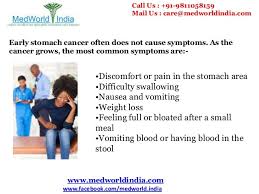
  • Loss of appetite                                                                                                As stomach tumors grow, you may have more serious symptoms, such as:
    • Stomach pain
    • Blood in your stool
    • Vomiting
    • Weight loss for no reason
    • Trouble swallowing
    • Yellowish eyes or skin
    • Swelling in your stomach
    • Constipation or diarrhea
    • Weakness or feeling tired
    • Heartburn

Just having indigestion or heartburn after a meal doesn’t mean you have cancer. But if you feel these symptoms a lot, talk to your doctor. He can see if you have other risk factors and test you to look for any problems.

Stomach cancers are usually found when a person goes to the doctor because of signs or symptoms they are having. The doctor will take a history and examine the patient. If stomach cancer is suspected, tests will be needed to confirm the diagnosis.

Medical history and physical exam

When taking your medical history, the doctor will ask you questions about your symptoms (eating problems, pain, bloating, etc.) and possible risk factors to see if they might suggest stomach cancer or another cause. The physical exam gives your doctor information about your general health, possible signs of stomach cancer, and other health problems. In particular, the doctor will feel your abdomen for any abnormal changes.

If your doctor thinks you might have stomach cancer or another type of stomach problem, he or she will refer you to a gastroenterologist, a doctor who specializes in diseases of the digestive tract, who will examine you and do further testing.

Upper endoscopy

Upper endoscopy (also called esophagogastroduodenoscopy or EGD) is the main test used to find stomach cancer. It may be used when someone has certain risk factors or when signs and symptoms suggest this disease may be present.

During this test, the doctor passes an endoscope, which is a thin, flexible, lighted tube with a small video camera on the end, down your throat. This lets the doctor see the lining of your esophagus, stomach, and first part of the small intestine. If abnormal areas are seen, biopsies (tissue samples) can be taken using instruments passed through the endoscope. The tissue samples are sent to a lab, where they are looked at under a microscope to see if cancer is present.

When seen through an endoscope, stomach cancer can look like an ulcer, a mushroom-shaped or protruding mass, or diffuse, flat, thickened areas of mucosa known as linitis plastica. Unfortunately, the stomach cancers in hereditary diffuse gastric cancer syndrome often cannot be seen during endoscopy.

Endoscopy can also be used as part of a special imaging test known as endoscopic ultrasound, which is described below.

This test is usually done after you are given medication to make you sleepy (sedation). If sedation is used, you will need someone to take you home (not just a cab).

Endoscopic ultrasound

Ultrasound uses sound waves to produce images of organs such as the stomach. During a standard ultrasound, a wand-shaped probe called a transducer is placed on the skin. It gives off sound waves and detects the echoes as they bounce off internal organs. The pattern of echoes is processed by a computer to produce a black and white image on a screen.

In endoscopic ultrasound (EUS), a small transducer is placed on the tip of an endoscope. While you are sedated, the endoscope is passed down the throat and into the stomach. This lets the transducer rest directly on the wall of the stomach where the cancer is. It lets the doctor look at the layers of the stomach wall, as well as the nearby lymph nodes and other structures just outside the stomach. The picture quality is better than a standard ultrasound because of the shorter distance the sound waves have to travel.

EUS is most useful in seeing how far a cancer may have spread into the wall of the stomach, to nearby tissues, and to nearby lymph nodes. It can also be used to help guide a needle into a suspicious area to get a tissue sample (EUS-guided needle biopsy).

Biopsy

Your doctor may suspect cancer if an abnormal-looking area is seen on endoscopy or an imaging test, but the only way to tell for sure if it is really cancer is by doing a biopsy. During a biopsy, the doctor removes a sample of the abnormal area.

Biopsies to check for stomach cancer are most often obtained during upper endoscopy. If the doctor sees any abnormal areas in the stomach lining during the endoscopy, instruments can be passed down the endoscope to biopsy them.

Some stomach cancers are deep within the stomach wall, which can make them hard to biopsy with standard endoscopy. If the doctor suspects cancer might be deeper in the stomach wall, endoscopic ultrasound can be used to guide a thin, hollow needle into the wall of the stomach to get a biopsy sample.

Biopsies may also be taken from areas of possible cancer spread, such as nearby lymph nodes or suspicious areas in other parts of the body.

Testing biopsy samples

Biopsy samples are sent to a lab to be looked at under a microscope. The samples are checked to see if they contain cancer, and if they do, what kind it is (for example, adenocarcinoma, carcinoid, gastrointestinal stromal tumor, or lymphoma).

If a sample contains adenocarcinoma cells, it may be tested to see if it has too much of a growth-promoting protein called HER2/neu (often just shortened to HER2). The HER2/neu gene instructs the cells to make this protein. Tumors with increased levels of HER2/neu are called HER2-positive.

Stomach cancers that are HER2-positive can be treated with drugs that target the HER2/neu protein, such as trastuzumab (Herceptin®).

The biopsy sample may be tested in 2 different ways:

  • Immunohistochemistry (IHC): In this test, special antibodies that stick to the HER2/neu protein are applied to the sample, which cause cells to change color if many copies are present. This color change can be seen under a microscope. The test results are reported as 0, 1+, 2+, or 3+.
  • Fluorescent in situ hybridization (FISH): This test uses fluorescent pieces of DNA that specifically stick to copies of the HER2/neu gene in cells, which can then be counted under a special microscope.

Often the IHC test is used first.

  • If the results are 0 or 1+, the cancer is HER2-negative. People with HER2-negative tumors are not treated with drugs (like trastuzumab) that target HER2.
  • If the test comes back 3+, the cancer is HER2-positive. Patients with HER2-positive tumors may be treated with drugs like trastuzumab.
  • When the result is 2+, the HER2 status of the tumor is not clear. This often leads to testing the tumor with FISH.

Imaging tests

Imaging tests use x-rays, magnetic fields, sound waves, or radioactive substances to create pictures of the inside of your body. Imaging tests may be done for a number of reasons, including:

  • To help find out whether a suspicious area might be cancerous
  • To learn how far cancer may have spread
  • To help determine if treatment has been effective

Upper gastrointestinal (GI) series

This is an x-ray test to look at the inner lining of the esophagus, stomach, and first part of the small intestine. This test is used less often than endoscopy to look for stomach cancer or other stomach problems, as it may miss some abnormal areas and does not allow the doctor to take biopsy samples. But it is less invasive than endoscopy, and it might be useful in some situations.

For this test, the patient drinks a white chalky solution containing a substance called barium. The barium coats the lining of the esophagus, stomach, and small intestine. Several x-ray pictures are then taken. Because x-rays can’t pass through the coating of barium, this will outline any abnormalities of the lining of these organs.

A double-contrast technique may be used to look for early stomach cancer. With this technique, after the barium solution is swallowed, a thin tube is passed into the stomach and air is pumped in. This makes the barium coating very thin, so even small abnormalities will show up.

Computed tomography (CT or CAT) scan

The CT scan is an x-ray test that produces detailed cross-sectional images of your body. Instead of taking one picture, like a standard x-ray, a CT scanner takes many pictures as it rotates around you. A computer then combines these pictures into images of slices of the part of your body being studied.

Before the test, you may be asked to drink 1 or 2 pints of a contrast solution and/or receive an intravenous (IV) line through which a contrast dye is injected. This helps better outline structures in your body.

The IV contrast can cause some flushing (redness and warm feeling). Some people are allergic and get hives, or rarely have more serious reactions like trouble breathing and low blood pressure. Be sure to tell the doctor if you have any allergies or have ever had a reaction to any contrast material used for x-rays.

A CT scanner has been described as a large donut, with a narrow table that slides in and out of the middle opening. You will need to lie still on the table while the scan is being done. CT scans take longer than regular x-rays, and you might feel a bit confined by the ring while the pictures are being taken.

CT scans show the stomach fairly clearly and often can confirm the location of the cancer. CT scans can also show the organs near the stomach, such as the liver, as well as lymph nodes and distant organs where cancer might have spread. The CT scan can help determine the extent (stage) of the cancer and whether surgery may be a good treatment option.

CT-guided needle biopsy: CT scans can also be used to guide a biopsy needle into a suspected area of cancer spread. The patient remains on the CT scanning table while a doctor moves a biopsy needle through the skin toward the mass. CT scans are repeated until the needle is within the mass. A fine-needle biopsy sample (tiny fragment of tissue) or a core-needle biopsy sample (a thin cylinder of tissue) is then removed and looked at under a microscope.

Magnetic resonance imaging (MRI) scan

MRI scans use radio waves and strong magnets instead of x-rays. The energy from the radio waves is absorbed by the body and then released in a pattern formed by the type of body tissue and by certain diseases. A computer translates the pattern into a very detailed image of parts of the body. A contrast material might be injected just as with CT scans, but this is used less often.

Most doctors prefer to use CT scans to look at the stomach. But an MRI may sometimes provide more information. MRIs are often used to look at the brain and spinal cord.

MRI scans take longer than CT scans, often up to an hour. You may have to lie inside a narrow tube, which is confining and can upset people with a fear of enclosed spaces. Special, open MRI machines can help with this if needed, although the images may not be as sharp in some cases. The MRI machine makes loud buzzing noises that you may find disturbing. Some places provide headphones to block this noise out.

Positron emission tomography (PET) scan

In this test, radioactive substance (usually a type of sugar related to glucose, known as FDG) is injected into a vein. (The amount of radioactivity used is very low and will pass out of the body over the next day or so.) Because cancer cells are growing faster than normal cells, they use sugar much faster, so they take up the radioactive material. After about an hour, you are moved onto a table in the PET scanner. You lie on the table for about 30 minutes while a special camera creates a picture of areas of radioactivity in the body.

PET is sometimes useful if your doctor thinks the cancer might have spread but doesn’t know where. The picture is not finely detailed like a CT or MRI scan, but it provides helpful information about the whole body. Although PET scans can be useful for finding areas of cancer spread, they aren’t always helpful in certain kinds of stomach cancer because these types don’t take up glucose very much.

Some machines can do both a PET and CT scan at the same time (PET/CT scan). This lets the doctor compare areas of higher radioactivity on the PET with the more detailed appearance of that area on the CT. PET/CT may be more helpful than PET alone for stomach cancer. This can help show if the cancer has spread beyond the stomach to other parts of the body, in which case surgery might not be a good treatment.

Chest x-ray

This test can help find out if the cancer has spread to the lungs. It might also determine if there are any serious lung or heart diseases present. This test is not needed if a CT scan of the chest has been done.

Other tests

Laparoscopy

If this procedure is done, it is usually only after stomach cancer has already been found. Although CT or MRI scans can create detailed pictures of the inside of the body, they can miss some tumors, especially if they are very small. Doctors might do a laparoscopy before any other surgery to help confirm a stomach cancer is still only in the stomach and can be removed completely with surgery. It may also be done before chemotherapy and/or radiation if these are planned before surgery.

This procedure is done in an operating room with the patient under general anesthesia (in a deep sleep). A laparoscope (a thin, flexible tube) is inserted through a small surgical opening in the patient’s side. The laparoscope has a small video camera on its end, which sends pictures of the inside of the abdomen to a TV screen. Doctors can look closely at the surfaces of the organs and nearby lymph nodes, or even take small samples of tissue. If it doesn’t look like the cancer has spread, sometimes the doctor will “wash” the abdomen with saline (salt water). The fluid (called peritoneal washings) is then removed and checked to see if it contains cancer cells. If it does, the cancer has spread, even if the spread couldn’t be seen.

Sometimes laparoscopy is combined with ultrasound to give a better picture of the cancer.

Lab tests

When looking for signs of stomach cancer, a doctor may order a blood test called a complete blood count (CBC) to look for anemia (which could be caused by the cancer bleeding into the stomach). A fecal occult blood test may be done to look for blood in stool (feces) that isn’t visible to the naked eye.

The doctor might recommend other tests if cancer is found, especially if you are going to have surgery. For instance, blood tests will be done to make sure your liver and kidney functions are normal and that your blood clots normally. If surgery is planned or you are going to get medicines that can affect the heart, you may also have an electrocardiogram (EKG) and echocardiogram (an ultrasound of the heart) to make sure your heart is functioning well.


Part III Part Treatment to Stomach Cancer:

Many treatments can fight stomach cancer. The one you and your doctor choose will depend on how long you’ve had the disease or how much it has spread in your body, called the stage of your cancer.

Surgery. Your doctor might remove part of your stomach or other tissues nearby that have cancer cells. Surgery gets rid of the tumor and stops cancer from spreading to other parts of your body. If your disease is in a more advanced stage, your doctor might need to remove all of your stomach=Gastrectomy or in some other cases the Surgeon may only have to remove part of the stomach=Partial Gastrectomy.

Some tumors can keep food from moving in and out of your stomach. In that case, you might have surgery to put in a stent, a device that keeps the pathways open.

Chemotherapy. Drugs kill your cancer cells or keep them from growing. You can take them as pills or through an IV at a clinic. Chemo usually takes several weeks. The drugs can cause side effects, but your doctor can help you find ways to feel better during treatment.

Radiation. High-energy waves or particles can kill cancer cells and shrink tumors. Your doctor may use an X-ray or other machine to beam radiation at the spot where your tumor is.

Chemoradiation. Your doctor might use this mix of chemotherapy and radiation to shrink your tumor before surgery.

Targeted drugs. These newer drugs are different because they fight only cancer cells. Other treatments, like chemo and radiation, can kill healthy cells along with diseased ones. As a result, targeted therapies have fewer side effects than these other treatments.

How Can I Prevent Stomach Cancer?

Treat stomach infections. If you have ulcers from an H. pylori infection, get treatment. Antibiotics can kill the bacteria, and other drugs will heal the sores in the lining of your stomach to cut your risk of cancer.

Eat healthy. Get more fresh fruits and vegetables on your plate every day. They’re high in fiber and in some vitamins that can lower your cancer risk. Avoid very salty, pickled, cured, or smoked foods like hot dogs, processed lunch meats, or smoked cheeses. Keep your weight at a healthy level, too. Being overweight or obese can also raise your risk of the disease.

QUOTE FOR THE DAY:


“After a cancer diagnosis, staging provides important information about the extent of cancer in the body and anticipated response to treatment.”


American Cancer Society


Go to striveforgoodhealth.com and learn more on the types of treatments given to patients with stom

QUOTE FOR THURSDAY:

“Hemochromatosis is a disorder in which the body can build up too much iron in the skin, heart, liver, pancreas, pituitary gland, and joints. Hemochromatosis is a disorder in which the body can build up too much iron in the skin, heart, liver, pancreas, pituitary gland, and joints. Too much iron is toxic to the body and over time the high levels of iron can damage tissues and organs and lead to serious problems with these organs mentioned above.”

Centers for Disease Control and Prevention CDC (https://www.cdc.gov/genomics/disease/hemochromatosis.htm)


QUOTE FOR WEDNESDAY:

“Scoliosis, a common, sometimes hereditary spinal condition, is a curvature of the spine that affects 2% to 3% of the population in the United States, or an estimated 6 million to 9 million people.  “If your child has scoliosis, don’t be afraid of it,” Vitale says. “The key is being aware of it and getting the diagnosis early so you can begin treatment. When treated early, there’s a lot of hope for stopping curve progression and allowing kids to live a perfectly active, normal life.”.

(https://www.cuimc.columbia.edu/news/8-facts-know-about-scoliosis)

Columbia University Irving Medical Center Dr. Michael G. Vitale                  (https://www.cuimc.columbia.edu/news/8-facts-know-about-scoliosis)

QUOTE FOR TUESDAY:

“On an airplane, an oxygen mask descends in front of you. What do you do? As we all know, the first rule is to put on your own oxygen mask before you assist anyone else. Only when we first help ourselves can we effectively help others. Caring for yourself is one of the most important—and one of the most often forgotten—things you can do as a caregiver. When your needs are taken care of, the person you care for will benefit, too.”.

Family Caregiver Alliance

Health and Safety for Caregivers

Safety Tips for Health Givers in Hospitals and Long Term Care Facilities through All American Homecare:

1 – Medical facilities such as hospitals and long-term care facilities will have people that maintain the grounds and buildings. However, as an at-home caregiver, you’ll be visiting patients’ homes that are in a variety of conditions. Be sure to watch your step while approaching the home, making sure that you don’t trip on uneven concrete and keep an eye out for rotting wood.

Once inside, do not remove your shoes, or change into a pair of shoes that you only wear indoors (it is important to keep your safety in mind while also respecting the wishes or culture of your patient). Your shoes will protect you from slipping or stubbing a toe.

Also be mindful of slippery hard surfaces, open cupboards, and other things that can cause injury when you are engrossed in helping your patient.

2 –When visiting patients, keep your guard up and be aware of your surroundings. Unlike the controlled environments of hospitals and other facilities, the homes of patients can be unpredictable.

Trust your instincts and never go into a situation that makes you feel unsafe. Be sure to call your supervisor to figure out how to proceed in these situations. Also learn to recognize warning signs and if you feel threatened, immediately leave and inform your supervisor of the situation.

3 – It’s vital that you use safe lifting practices each time you transfer a patient. This will help reduce incidents of back injury and muscle strains. Here’s a refresher on proper lifting techniques:

  • Keep your head and neck in proper alignment with your spine
  • Maintain the natural curve of your spine; bend with your hips and knees, not your spine
  • Avoid twisting your body when carrying the person
  • Always keep the patient being moved close to your body
  • Keep your feet shoulder-width apart to maintain your balance
  • Use the muscles in your legs to lift and/or pull

4 – There are plenty of tools for home healthcare workers to aid them in their daily tasks. These can include lifting and transfer aides. If you are helping a patient change a light bulb, don’t stand on a chair to do it; instead, use a step stool.

5 – Your uniform (often scrubs or a pair of slacks and a polo) ensures that you are properly dressed for the job. If you don’t have a company uniform, ensure that your clothing allows you to move freely. Also, wear close-toed shoes with good traction and support.

6 – Household spills and incontinence accidents increase the risk of falls for both the caregiver and the patient. Clutter can also lead to tripping and falling. Clean up these and other safety hazards immediately.

7 – Many of your patients will have pets. Not only can animals cause you to become distracted or interfere with your work, even the friendliest pet can turn on you. Your best bet is to avoid the animals in the home and focus on your patient.

8 – One of the biggest risks to home healthcare workers is going past their limits. Make sure that you know your limits. If a patient is too heavy for you to transfer on your own, ask for help. Also, pace yourself and build flexibility into your schedule. Injury-inducing shortcuts just aren’t worth it!

Taking time to stretch throughout the day can help reduce muscle fatigue and strains. When it’s time to take a break, take it. Keep yourself hydrated throughout the day and eat a meal and snacks. Being dehydrated and not eating can lead to feelings of tiredness and making dangerous mistakes.

9 – Here are a few other basic safety protocols you need to follow:

  • Confirm with your clients by phone before you visit.
  • Have detailed directions to your client’s home or use a GPS device.
  • Keep your vehicle in good working condition and fill up your gas tank.
  • Don’t text or talk on the phone while driving.
  • Keep your car doors locked and your windows closed.
  • Lock your bag and other possessions in the trunk of your car.
  • Most importantly, make sure that someone knows where you are at all times.

By following these safety guidelines, you’ll be able to provide better care for your patients and have a satisfying career that you enjoy each day!

QUOTE FOR MONDAY:

“Sickle cell disease (SCD) is a group of conditions that affect the red blood cells. Normal red blood cells are round and flexible. A person with sickle cell disease has red blood cells that are hard, sticky, and crescent-shaped like crescent moons or sickles. People with sickle cell disease often experience intense pain and anemia because of their abnormal red blood cells.

SCD is one of the most common hereditary disorders in the United States, affecting 1 in 1,900 infants. Of the 66,000 infants born in Minnesota annually, about 25 infants are born with SCD and more than 1,000 infants are born with a sickle cell trait (SCT).

Currently, the only known cure for SCD is a blood or marrow transplant (BMT). A blood or marrow transplant is a procedure that takes healthy stem cells from a donor and puts them into someone else”

Department of Health in Minnesota (https://www.health.state.mn.us/diseases/sicklecell/about.html)


QUOTE FOR THE WEEKEND:

“Mpox, formerly called monkeypox, is a rare disease similar to smallpox caused by a virus. It’s found mostly in areas of Africa, but has been seen in other regions of the world. It causes flu-like symptoms such as fever and chills, and a rash that can take weeks to clear. There’s no proven treatment for mpox, but it usually goes away on its own.

For decades, mpox was mostly seen in Africa. But it’s occasionally found in other countries, including the United States.

In the summer of 2021, a case of mpox was found in a U.S. resident who had traveled from Nigeria to the United States. Then, 2022 brought outbreaks to regions outside of Africa, including Europe, the Americas and Australia.”

Cleveland Clinic (https://my.clevelandclinic.org/health/diseases/22371-monkeypox)

What is monkey pox?

Monkeypox was first discovered in 1958 when two outbreaks of a pox-like disease occurred in colonies of monkeys kept for research, hence the name ‘monkeypox.’ The first human case of monkeypox was recorded in 1970 in the Democratic Republic of the Congo (DRC) during a period of intensified effort to eliminate smallpox. Since then, monkeypox has been reported in people in several other central and western African countries: Cameroon, Central African Republic, Cote d’Ivoire, Democratic Republic of the Congo, Gabon, Liberia, Nigeria, Republic of the Congo, and Sierra Leone. The majority of infections are in Democratic Republic of the Congo.

There are two distinct genetic clades of the monkeypox virus – the Central African (Congo Basin) clade and the West African clade. The Congo Basin clade has historically caused more severe disease and was thought to be more transmissible. The geographical division between the two clades has so far been in Cameroon – the only country where both virus clades have been found.

Monkeypox is a viral zoonosis (a virus transmitted to humans from animals) with symptoms very similar to those seen in the past in smallpox patients, although it is clinically less severe. Monkeypox virus is an enveloped double-stranded DNA virus that belongs to the Orthopoxvirus genus of the Poxviridae family.   With the eradication of smallpox in 1980 and subsequent cessation of smallpox vaccination, monkeypox has emerged as the most important orthopoxvirus for public health. Monkeypox primarily occurs in Central and West Africa, often in proximity to tropical rainforests and has been increasingly appearing in urban areas. Animal hosts include a range of rodents and non-human primates.

Various animal species have been identified as susceptible to monkeypox virus.. This includes rope squirrels, tree squirrels, Gambian pouched rats, dormice, non-human primates and other species. Uncertainty remains on the natural history of monkeypox virus and further studies are needed to identify the exact reservoir(s) and how virus circulation is maintained in nature.

Monkeypox cases in people have occurred outside of Africa linked to international travel or imported animals, including cases in the United States, as well as Israel, Singapore, and the United Kingdom.

The natural reservoir of monkeypox remains unknown. However, African rodents and non-human primates (like monkeys) may harbor the virus and infect people.

Symptoms of Monkey Pox:

People with monkeypox get a rash that may be located on or near the genitals (penis, testicles, labia, and vagina) or anus (butthole) and could be on other areas like the hands, feet, chest, face, or mouth.

  • The rash will go through several stages, including scabs, before healing.
  • The rash can initially look like pimples or blisters and may be painful or itchy.

Other symptoms of monkeypox can include:

  • Fever
  • Chills
  • Swollen lymph nodes
  • Exhaustion
  • Muscle aches and backache
  • Headache
  • Respiratory symptoms (e.g. sore throat, nasal congestion, or cough)

Monkeypox symptoms usually start within 3 weeks of exposure to the virus. If someone has flu-like symptoms, they will usually develop a rash 1-4 days later.

A person with monkeypox can spread it to others from the time symptoms start until the rash has fully healed and a fresh layer of skin has formed. Some people have been found to have infection but no symptoms. To date, however, there is no evidence that monkeypox spreads from people with no symptoms. CDC will continue to monitor for new or changing information about transmission.

Monkeypox can more accurately be described as “sexually transmissible.” In other words, sex is just one of the ways that monkeypox can be spread. In the past, monkeypox outbreaks have been linked to direct exposure to infected animals and animal products, with limited person-to-person spread. In the current monkeypox outbreak, the virus is spreading primarily through close personal contact. This may include contact with infectious lesions or respiratory secretions via close, sustained skin-to-skin contact that occurs during sex. However, any close, sustained skin-to-skin contact with someone who has monkeypox can spread the virus. The contact does not have to be exclusively intimate or sexual.

Monkeypox can spread from person to person through direct contact with the infectious rash, scabs, or body fluids. It also can be spread by respiratory secretions during prolonged, face-to-face contact, or during intimate physical contact, such as kissing, cuddling, or sex.

A person with monkeypox can spread it to others from the time symptoms start until the rash has fully healed and a fresh layer of skin has formed. Some people have been found to have infection but no symptoms. To date, however, there is no evidence that monkeypox spreads from people with no symptoms. CDC will continue to monitor for new or changing information about transmission.

When to get tested:

  • Currently, testing is only recommended if you have a rash consistent with monkeypox.
  • If you think you have monkeypox or have had close personal contact with someone who has monkeypox, consider taking precautions and visit a healthcare provider to help you decide if you need to be tested for monkeypox.

How its diagnosed:

  • You will likely need to fill out paperwork before you get tested.
  • To get a specimen to test, the healthcare provider will use a swab to rub vigorously across lesions of your rash. They will take swabs from more than one lesion.
  • This swabbing may be uncomfortable but is necessary to get enough material to detect the monkeypox virus from the specimens.
  • The specimens will be tested in a lab to see if the monkeypox virus is detected.
  • Results are usually available within a few days.
  • While you are waiting for your results, take precautions to avoid getting or spreading monkeypox virus to others.

The CDC states there is no treatment specifically for monkeypox.   But because the viruses that cause monkeypox and smallpox are similar, antiviral drugs developed to protect against smallpox may be used to treat monkeypox effectively.”  At at this time there’s currently not an approved antiviral treatment for monkeypox.  Monkeypox is usually a self-limited disease with symptoms lasting from two weeks to four weeks.

Most people with monkeypox get better on their own without treatment. Following diagnosis, your healthcare provider will monitor your condition and try to relieve your symptoms like preventing dehydration and give you antibiotics to treat secondary bacterial infections if they develop.

The CDC states “Two vaccines may be used for the prevention of monkeypox disease:

  • JYNNEOS vaccine is approved for the prevention of monkeypox and smallpox. During the current outbreak, JYNNEOS is the main vaccine being used in the United States.
  • ACAM2000 vaccine is approved for immunization against smallpox and made available for use against monkeypox under an Expanded Access Investigational New Drug (EA-IND) protocol.
  • People may be vaccinated after exposure to monkeypox virus to help prevent monkeypox disease (i.e., post-exposure prophylaxis).
  • No data are currently available on the clinical efficacy or effectiveness of JYNNEOS or ACAM2000 for monkeypox disease.”

The CDC states “As of November 29, 2022 The total U.S. cases are 29, 325 and the total deaths in the U.S. 15.”