QUOTE FOR MONDAY:

“Too much cortisol can cause some of the main symptoms of Cushing syndrome — a fatty hump between the shoulders, a rounded face, and pink or purple stretch marks on the skin. Cushing syndrome also can cause high blood pressure or bone loss. Sometimes, it can cause type 2 diabetes.  The people most commonly affected by Cushing syndrome are children, teenagers and adults, mostly those ages 25 to 50. People who take cortisol medication (for example, to treat asthma and rheumatoid arthritis) are especially vulnerable. Some 70% of people with Cushing syndrome are women or people assigned female at birth (AFAB) and 30% are men or people assigned male at birth (AMAB).”

Cleveland Clinic (https://my.clevelandclinic.org/health/diseases/5497-cushing-syndrome)

PART 2 What is Cushings Syndrome regarding complications, diagnosis and how its treated.

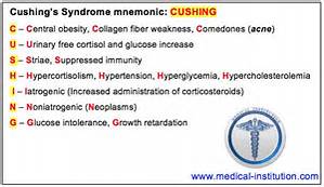
Complications of Cushings Syndrome include:

Diabetes (High or Low blood glucose levels)

Enlargement of pituitary tumor and other complications from the tumor growth

Fractures due to osteoporosis which are common in older people

High blood pressure which could be life threatening

Kidney stones from the increase in cortisol and other chemicals filtered through the kidneys

Serious infections which could lead to further secondary infections

Although diagnosis can be challenging, it is important being correctly diagnosed with Cushing’s disease is the first step toward regaining control of your health because an accurate diagnosis helps your doctor determine the best course of action. Cushing’s disease may be challenging to diagnose because of several factors:

Cushing’s disease does not cause the same symptoms in everyone

Cushing’s disease may cause the same symptoms as other medical conditions that are more common

The tumor that causes Cushing’s disease can be too small to be found on MRI scans

The tumor can become “inactive” so that it releases less cortisol at certain times

Inactive tumors can fool tests for Cushing’s disease that measure the levels of hormones in your urine, blood, or saliva

The process and tests used to diagnose Cushing’s disease

Ways to take control of your health once you are diagnosed

Cushing’s disease can be mistaken for other conditions

Doctors don’t always think to look for Cushing’s disease because it is rare and causes symptoms that can make them think you have a different condition. Because these other conditions are more common, your doctor will probably rule them out before testing you for Cushing’s disease. These conditions include:

-Pregnancy

-Depression or other psychiatric disorders

-Alcoholism

-Weight problems (obesity) caused by improper diet and/or exercise

-Poorly controlled diabetes mellitus

-Polycystic ovarian syndrome (PCOS)

How is Cushing’s syndrome diagnosed?

The process of diagnosing Cushing’s disease—There are generally 3 phases in the diagnostic process and you will likely see more than one doctor, including an endocrinologist.

Phase 1: Confirm clinical suspicion-Your doctor will to a physical exam.

The doctor will measure if you have the symptoms that are known to be caused by hypercortisolism.
Your doctor may look for:

Purplish streaks on your body (called striae)

Weight gain

Fatty tissue around your mid-section

Thinning of your arms and legs

Fatty tissue that creates a hump on your back

Redness or roundness in your face

Bruises on your body

Thinning of your skin

Excessive facial/hair growth (hirtusism) if you’re female

Acne

Weakness

Medical conditions that are unusual for a patient’s age

— High blood pressure (called hypertension)

— Diabetes

— High cholesterol

— Osteoporosis

— Infection

— Heart disease

__Evaluation

What you can expect is your doctor will do a physical examine your entire body to look for specific physical signs of Cushing’s disease and may do tests to better understand your symptoms, what we call diagnostic tooling.

Phase II: Confirm hypercortisolism
Medical tests will be performed to measure the level of cortisol in the body. These tests should only be done when a form of Cushing’s syndrome(including Cushing’s disease) is highly suspected.

-24 hour urinary free cortisol (UFC) –

Measures the level of cortisol in your urine over a 24-hour period. If the levels are too high, then you may have Cushing’s disease or Cushing’s syndrome. This test is often used because it only measures the type of cortisol that causes Cushing’s disease, called “circulating cortisol,” and may be more accurate than other tests that measure cortisol levels

Because of the difficulty in obtaining 24-hour urine collections in many outpatients, some physicians use a l-mg overnight dexamethasone suppression test. For this test, the patient takes l mg of dexamethasone orally at 11 p.m., and the plasma cortisol level is measured at 8 a.m. the following day (normal value: 5 μg per dL or less [140 nmol per L]). The reported sensitivity of this test is 98 percent; the reported specificity is 80 percent.4

Obesity, chronic illness, chronic alcoholism and depression can cause false-positive results (pseudo-Cushing’s syndrome) on the 1-mg dexamethasone suppression test and mildly elevated free cortisol values on the 24-hour urine collection.

If the result of the dexamethasone suppression test is abnormal or the 24-hour urinary free cortisol level is mildly elevated, a confirmatory test for Cushing’s syndrome is needed. The 24-hour urine collection for urinary free cortisol excretion can be used to confirm the result of the l-mg dexamethasone suppression test. Normal findings on both tests provide strong evidence against the presence of Cushing’s syndrome.4 However, when Cushing’s syndrome is still strongly suspected based on the clinical findings, negative tests should be repeated; the tests should also be performed again in three to six months.

Phase III: Determine if Cushing’s disease is the cause of hypercortisolism

-Adrenocorticotropic Hormone (ACTH) test-

Measures if the amount of ACTH in your blood is higher than normal.

If your ACTH levels are high or normal, then you may have a tumor that is producing ACTH.

ACTH-producing tumors are most often found on the pituitary (Cushing’s disease).

ACTH-producing tumors may be in other areas of the body (called ectopic Cushing’s syndrome) If your ACTH levels are low, you may have Cushing’s syndrome due to a different cause or another condition. Additional tests will be done to confirm whether or not you have Cushing’s syndrome

Treatment of cushings syndrome is by castigation of the under lying cause.

Treatments for Cushing’s syndrome are contrived to pass your body’s cortisol production to normal. By indurate, or even distinctly lowering cortisol levels, you’ll feel evident improvements in your signs and symptoms. Left untreated, however, Cushing’s syndrome can finally induce to death. The treatment choice depend on the cause. For example:

-If a tumour in an adrenal gland is the reason, an operation to withdraw it will cure the condition.

– For adrenal hyperplasia, both adrenal glands may require to be withdraw. You will then require to take lifelong replacement therapy of several adrenal hormones.

-Other tumours in the body that produce ‘ectopic’ ACTH may be able to be removed, depending on the kind of tumour, where it is, etc.

-Medication to block the production or consequence of cortisol may be an choice.

QUOTE FOR THE WEEKEND:

“Cushing syndrome happens when the body has too much of the hormone cortisol for a long time. This can result from the body making too much cortisol, or from taking medicines called glucocorticoids, which affect the body the same way as cortisol.  Too much cortisol can cause some of the main symptoms of Cushing syndrome — a fatty hump between the shoulders, a rounded face, and pink or purple stretch marks on the skin. Cushing syndrome also can cause high blood pressure or bone loss. Sometimes, it can cause type 2 diabetes.”

MAYO CLINIC (https://www.mayoclinic.org/diseases-conditions/cushing-syndrome/symptoms-causes/syc-20351310)

Part I What is Cushings Syndrome?

                                                 cushingssyndrome

                                                   cushings


Cushing’s syndrome describes the signs and symptoms associated with prolonged exposure to inappropriately high levels of the hormone cortisol. This can be caused by taking glucocorticoid drugs, or diseases that result in excess cortisol, adrenocorticotropic hormone (ACTH), or CRH levels.

Cushing’s syndrome appears when the body’s tissues are display to immoderate levels of cortisol for long periods of time.

There are two types of the disease and they are known as exogenous and endogenous. Exogenous Cushing syndrome is caused by something outside of the body, like when hormones are given to a patient during a RX for another condition. Endogenous is caused by natural causing problems within the body. Endogenous is likely to be hereditary and not caused by an outside force like a steroid complex.

 Causes of Cushing Syndrome:

The most common cause of Cushing’s syndrome is exogenous administration of glucocorticoids prescribed by a health care practitioner to treat other diseases (called iatrogenic Cushing’s syndrome). This can be an effect of corticosteroid treatment of a variety of disorders such as asthma and rheumatoid arthritis, or in immunosuppression after an organ transplant.

Administration of synthetic ACTH(adrenocorticotropichormone) is also possible, but ACTH is less often prescribed due to cost and lesser utility. Although rare, Cushing’s syndrome can also be due to the use of medroxyprogesterone In this form of Cushing’s, the adrenal glands atrophy due to lack of stimulation by ACTH, since glucocorticoids downregulate production of ACTH. Cushing syndrome in childhood usually results from use of glucocorticoid medication.

Endogenous Cushing’s syndrome results from some derangement of the body’s own system of secreting cortisol. Normally, ACTH is released from the pituitary gland when necessary to stimulate the release of cortisol from the adrenal glands.

In pituitary Cushing’s, a benign pituitary adenoma secretes ACTH. This is also known as Cushing’s disease and is responsible for 70% of endogenous Cushing’s syndrome.

In adrenal Cushing’s, excess cortisol is produced by adrenal gland tumors, hyperplastic adrenal glands, or adrenal glands with nodular adrenal hyperplasia.

Tumors outside the normal pituitary-adrenal system can produce ACTH (occasionally with CRH) that affects the adrenal glands. This etiology is called ectopic or paraneoplastic Cushing’s disease and is seen in diseases like small cell lung cancer.

Finally, rare cases of CRH-secreting tumors (without ACTH secretion) have been reported, which stimulates pituitary ACTH production.

Pseudo-Cushing’s syndrome

 Elevated levels of total cortisol can also be due to estrogen found in oral contraceptive pills that contain a mixture of estrogen and progesterone, leading to Pseudo-Cushing’s syndrome. Estrogen can cause an increase of cortisol-binding globulin and thereby cause the total cortisol level to be elevated. However, the total free cortisol, which is the active hormone in the body, as measured by a 24 hour urine collection for urinary free cortisol, is normal.

Epidemiology

 Iatrogenic Cushing’s syndrome (caused by treatment with corticosteroids) is the most common form of Cushing’s syndrome.

Symptoms of Cushings Syndrome:

 Symptom of cushings syndrome include fat deposits close to the face neck and trunk; weariness; muscular weakness; salt and water retention; acne; leisurely bruising; menstlruall irregularities; and signs (in women) of virilisation, such as increase of the voice, commute in body, shape, loss of scalp hair, and extend in facial and body hair. Complications of cushings syndrome include advanced blood pressure, The symptoms and signs of cushings syndrome induced by a chronic redundant of corticosteroid hormones in the blood. The redundant may be acquired by a tumour of the outer part (cortex) of the adrenal gland, or may be referable to over inspiration of the adrenal glands by a tumour the pituitary gland.

Many children and teenagers with Cushing’s syndrome will exhibit various symptoms of the following:

-extreme weight gain

-growth retardation

-missed periods in teenage girls

-excess hair growth

-acne

-reddish-blue streaks on the skin

-high blood pressure

-tiredness and weakness

-either very early or late puberty

Adults with the disease may also have symptoms of intense weight gain, redundant hair growth, high blood pressure, and skin difficulties. In addition, they may show:

-muscle and bone weakness

-moodiness, irritability, or depression

-sleep disturbances

-high blood sugar

-menstrual disorders in women and diminished fertility in men

Complications of this disease include:

 -Diabetes (High or Low blood glucose levels)

-Enlargement of pituitary tumor and other complications from the tumor growth

-Fractures due to osteoporosis which are common in older people

-High blood pressure which could be life threatening

-Kidney stones from the increase in cortisol and other chemicals filtered through the kidneys

-Serious infections which could lead to further secondary infections


Tune in Monday for Part II on Cushing’s Disease in how it is treated.


QUOTE FOR FRIDAY:

“Gastrointestinal stromal tumor, GIST, is a relatively uncommon type of cancer that occurs in the gastrointestinal tract. GIST’s belong to a class of diseases called sarcomas. Experts estimate that 4,500 to 6,000 GIST’s are diagnosed each year in the United States. GIST’s can develop anywhere in the GI tract but occur most often in the stomach (approximately 60%).”

GIST Cancer Awareness Foundation (gistawareness.org)

Gastrointestinal stromal tumo – GIST.


Cancer starts when cells in the body begin to grow out of control. Cells in nearly any part of the body can become cancer, and then can spread to other areas of the body.

Gastrointestinal stromal tumors (GISTs) are not common, and the exact number of people diagnosed with these tumors each year is not known. Until the late 1990s, not much was known about these tumors (and doctors didn’t have good ways of identifying them with lab tests), so many of them ended up being classified as other kinds of cancers.

Current estimates for the total number of GIST cases diagnosed each year in the United States range from about 4,000 to about 6,000.

A gastrointestinal stromal tumor (GIST) is a type of cancer that begins in the digestive system. GISTs happen most often in the stomach and small intestine.

A GIST is a growth of cells that’s thought to form from a special type of nerve cells. These special nerve cells are in the walls of the digestive organs. They play a part in the process that moves food through the body.

The GI tract processes food for energy and rids the body of solid waste. After food is chewed and swallowed, it goes through the esophagus, a tube that carries food down the throat and chest to the stomach. The esophagus joins the stomach just beneath the diaphragm (the thin band of muscle below the lungs).

The stomach is a sac-like organ that helps the digestive process by mixing the food with gastric juices. The food and gastric juices are then emptied into the small intestine. The small intestine, which is about 20 feet long, continues breaking down the food and absorbs most of the nutrients into the bloodstream.

The small intestine joins the large intestine, the first part of which is the colon, a muscular tube about 5 feet long. The colon absorbs water and mineral nutrients from the remaining food matter. The waste left after this process (stool) goes into the rectum, where it is stored until it passes out of the body through the anus.

Gastrointestinal stromal tumors (GISTs) are uncommon cancers that start in special cells in the wall of the gastrointestinal (GI) tract, also known as the digestive tract. To understand GISTs, it helps to know something about the structure and function of the GI tract.

Gastrointestinal stromal tumors (GISTs) start in very early forms of special cells in the wall of the GI tract called the interstitial cells of Cajal (ICCs). ICCs are sometimes called the “pacemakers” of the GI tract because they signal the muscles in the GI tract to contract to move food and liquid along.

More than half of GISTs start in the stomach. Most of the others start in the small intestine, but GISTs can start anywhere along the GI tract. A small number of GISTs start outside the GI tract in nearby areas such as the omentum (an apron-like layer of fatty tissue that hangs over the organs in the abdomen) or the peritoneum (the thin lining over the organs and walls inside the abdomen).

Some GISTs seem to be much more likely than others to grow into other areas or spread to other parts of the body. Doctors look at certain factors to help tell whether a GIST is likely to grow and spread quickly, such as:

  • The size of the tumor
  • Where it’s located in the GI tract
  • How fast the tumor cells are dividing (its mitotic rate, described in Tests for Gastrointestinal Stromal Tumors)

Small GISTs may cause no symptoms, and they may grow so slowly that they don’t cause problems at first. As a GIST grows, it can cause signs and symptoms. They might include:

  • Abdominal pain
  • A growth you can feel in your abdomen
  • Fatigue
  • Nausea
  • Vomiting
  • Cramping pain in the abdomen after eating
  • Not feeling hungry when you would expect to
  • Feeling full if you eat only a small amount of food
  • Dark-colored stools caused by bleeding in the digestive system

GISTs can happen in people at any age, but they are most common in adults and very rare in children. The cause of most GISTs isn’t known. A small number are caused by genes passed from parents to children.

GISTs are not the same as other, more common types of GI tract cancers that develop from other types of cells.

Cancers can occur anywhere in the GI tract − from the esophagus to the anus. Most cancers that start in the GI tract, including most esophagus cancers, stomach cancers, and colon and rectum cancers, start in the gland cells that line almost all of the GI tract. The cancers that develop in these cells are called adenocarcinomas.

Cancers can also start in squamous cells, which are flat cells that line some parts of the GI tract, like the upper part of the esophagus and the end of the anus. Cancers starting in these cells are called squamous cell carcinomas.

The GI tract also has neuroendocrine cells. These cells have some features in common with nerve cells but also have other features in common with hormone-producing (endocrine) cells. Cancers that develop from these cells are called neuroendocrine tumors (NETs). These cancers are rare in the GI tract. Carcinoid tumors are an example of a neuroendocrine tumor found in the GI tract.

Other rare types of cancer in the GI tract include different types of soft tissue sarcomas, such as:

  • Leiomyosarcomas: cancers of smooth muscle cells
  • Angiosarcomas: cancers of blood vessel cells
  • Malignant peripheral nerve sheath tumors (MPNSTs): cancers of cells that support and protect nerves

GISTs are different from these other types of GI tract cancers. They start in different types of cells, need different types of treatment, and have a different prognosis (outlook). This is why doctors need to figure out whether a person with a tumor in the GI tract has a GIST, some other type of cancer, or a non-cancerous condition.

How it is diagnosed:

First you M.D. will do a physical exam.

They may also do the following tests:

  • Upper endoscopy.
  • Computed tomography (CT) scan.
  • Positron emission tomography (PET).
  • Biopsy to obtain tissue for a pathologist to examine under a microscope.

Treatment:

The most common treatment is surgery to remove the tumors.

Survival:

Survival rates vary depending on tumor size and activity at the time of diagnosis, your overall health and the tumor’s response to treatment. Overall, data from the U.S. National Cancer Institute (NCI) show that 85% of people with GIST were alive five years after diagnosis.

QUOTE FOR THURSDAY:

The entire digestive tract helps with our immune system, but some scientists and doctors think the appendix may be a place for our body to store certain healthy types of gut bacteria that otherwise could be altered or changed during an intestinal illness or with overuse of antibiotics.

Appendicitis is an inflammation of the appendix. The appendix is a finger-shaped pouch that sticks out from the colon on the lower right side of the belly, also called the abdomen.

Appendicitis causes pain in the lower right part of the belly. However, in most people, pain begins around the belly button and then moves. As inflammation worsens, appendicitis pain typically increases and eventually becomes serious.

Although anyone can develop appendicitis, most often it happens in people between the ages of 10 and 30. Treatment of appendicitis is usually antibiotics and, in most instances, surgery to remove the appendix.”

MAYO CLINIC (https://www.mayoclinic.org/diseases-conditions/appendicitis/symptoms-causes/syc-20369543)

The appendix of the human body & Appendicitis.

The appendix is a small pouch attached to the large intestine. The appendix is a small, finger-shaped pouch of intestinal tissue located between the small intestine (cecum) and large intestine (colon). The appendix is a small finger-shaped tube that branches off the first part of the large intestine.

Appendicitis (means append=appendix and itis=inflammation.  Appendicitis causes pain in your lower right abdomen. However, in most people, pain begins around the navel and then moves. As inflammation worsens, appendicitis pain typically increases and eventually becomes severe.

Although anyone can develop appendicitis, most often it occurs in people between the ages of 10 and 30. Standard treatment is surgical removal of the appendix.

Signs and symptoms of appendicitis may include:

  • Sudden pain that begins on the right side of the lower abdomen
  • Sudden pain that begins around your navel and often shifts to your lower right abdomen
  • Pain that worsens if you cough, walk or make other jarring movements
  • Nausea and vomiting
  • Loss of appetite
  • Low-grade fever that may worsen as the illness progresses
  • Constipation or diarrhea
  • Abdominal bloating
  • Flatulence

The site of your pain may vary, depending on your age and the position of your appendix. When you’re pregnant, the pain may seem to come from your upper abdomen because your appendix is higher during pregnancy.

Strongly suggestive of appendicitis is pushing down on the R lower quadrant and upon letting go the pain is severe compared to when pushing down.  It is called “rebound effect”.

Diagnostic Tests for confirming appendicitis:

  • Blood test. This allows your doctor to check for a high white blood cell count, which may indicate an infection.
  • Urine test. Your doctor may want you to have a urinalysis to make sure that a urinary tract infection or a kidney stone isn’t causing your pain.
  • Rectal exam
  • Imaging tests. Your doctor may also recommend an abdominal X-ray, an abdominal ultrasound, computerized tomography (CT) scan or magnetic resonance imaging (MRI) to help confirm appendicitis or find other causes for your pain.

Treatment:

Appendicitis treatment usually involves surgery to remove the inflamed appendix. Before surgery you may be given a dose of antibiotics to treat infection.  There are times though antibiotics may only be used and the MD see’s if the appendicitis is resolved; it depends on the MD and the severity of the appendicitis.

Surgery to remove the appendix (appendectomy)

Appendectomy can be performed as open surgery using one abdominal incision about 2 to 4 inches (5 to 10 centimeters) long (laparotomy). Or the surgery can be done through a few small abdominal incisions (laparoscopic surgery). During a laparoscopic appendectomy, the surgeon inserts special surgical tools and a video camera into your abdomen to remove your appendix.

In general, laparoscopic surgery allows you to recover faster and heal with less pain and scarring. It may be better for older adults and people with obesity.

But laparoscopic surgery isn’t appropriate for everyone. If your appendix has ruptured and infection has spread beyond the appendix or you have an abscess, you may need an open appendectomy, which allows your surgeon to clean the abdominal cavity.

Expect to spend one or two days in the hospital after your appendectomy.

Draining an abscess before appendix surgery

If your appendix has burst and an abscess has formed around it, the abscess may be drained by placing a tube through your skin into the abscess. Appendectomy can be performed several weeks later after controlling the infection.


QUOTE FOR WEDNESDAY:

“A widowmaker heart attack happens when you have a blockage in the biggest artery in your heart. That means blood can’t move through your left anterior descending (LAD) artery, which provides 50% of your heart muscle’s blood supply. Immediate treatment is crucial for a chance at survival.

A widowmaker heart attack is a type of heart attack in which you have a full blockage in your heart’s biggest artery. This artery, the left anterior descending (LAD) artery, sends oxygen-rich blood to your heart’s left ventricle. This lower chamber pumps oxygen-rich blood to your aorta, which sends it to your body.

Healthcare providers call a heart attack a “myocardial infarction,” which means you don’t have enough blood going to your heart muscle (myocardium). Your heart muscle can die without enough blood flow.

A heart attack is a medical emergency. Call 911 or your local emergency number if you think you’re having a heart attack.”

Cleveland Clinic (https://my.clevelandclinic.org/health/diseases/24507-widowmaker-heart-attack)


What is the Black Widow?

When it comes to the heart, time is critical. Blood carries oxygen to the body. When blood flow is blocked, cells can die. The more time that passes before treatment, the bigger the risk of death.

A complete blockage in the main artery that feeds the front and side walls of the heart and the septum, a muscular boundary between the left and right chambers.

“That’s a big deal” ,says Dr. Todd Caulfield, the Providence interventional cardiologist who operates on patient’s like this. “People call this the widow maker.”, he states.  Nationwide about 300,000 people suffer cardiac arrest outside a hospital every year, according to the federal Centers for Disease Control and Prevention. Only 8 percent survive.

The heart pumps blood to the body and brain. The heart also needs blood so it pumps blood to itself, too. The heart’s blood supply is essentially three main arteries: one that goes to the front; the other that goes to the back; and the final artery that connects to the side of the heart. When one of those arteries is blocked, the heart doesn’t get enough blood and oxygen, causing a heart attack. This is simply due to the block that doesn’t allow oxygen to the heart tissue that that artery feeds oxygenated blood.  This causes ischemia (lack of oxygen perfusion to tissue).  It causes pain first (chest pain=angina) and if the block continues than it will lead to infarction meaning death of the tissue not receiving enough (partial block) or no oxygen (a complete block).  The exact same thing happens in the brain, which causes a stroke; or the foot causing first pain leading to blackish skin of the foot tissue=necrotic tissue which ends up being amputated (like in some patients with  diabetes or peripheral vascular disease).  So lack of oxygen to our tissue parts anywhere can cause death to the tissue and depending on the tissue area cause death (like with the heart, due to the heart being the engine of the human body).

Interventional Cardiologist Randy Goodroe says patients are getting treatment quicker for heart attacks, more now than ever before. So the once grim reality of suffering from “the widowmaker” – a heart attack that results in immediate death – has become somewhat of a misnomer.

It’s called a “widowmaker”, Dr. Goodroe added, because it would typically kill you if the artery doesn’t get opened fast enough.

There are two different kinds of heart attacks. One is called an ST-elevation myocardial infarction, or STEMI, where the artery is totally blocked (i.e. the widowmaker), and we are on the clock to get the artery opened as soon as possible. There’s also a non-ST-elevation myocardial infarction (NSTEMI) where an artery is partially or temporarily blocked, and is still urgent to get the artery cleared, but not emergent.

Common Risk Factors:

Age is definitely a risk factor. We start seeing coronary disease in people who are between 50 and 60-years-old, and obviously, the older the person, the older the arteries, the higher the risk. Diabetes is another one of the greatest risk factors. Smoking is the strongest modifiable risk factor, meaning it’s the worst thing people can do to themselves to accelerate the disease process. High blood pressure and family history are also risk factors for heart attacks.

How to detect it if in the ER with symptoms & than repair it:

If a patient enters the ER with heart attack symptoms, quickly performed is an electrocardiogram (EKG) including cardiac enzymes to and echocardiogram or stress test to detect a totally obstructed artery. The artery is like a pipe or a tube., so we put a tiny wire through the blockageThose patients are very quickly taken to the cardiac catheterization lab where an angiogram is done to detect if there is and how bad the blockage is, if present.  Than with an angioplasty (balloons) at the end of the angiogram catheter break up the blockage and stents inserted in the area of blockage to get the blood flowing to the heart again.  The balloon is taken out and a stent is slid over that also has a balloon on it and once the stent goes up, it props open the blockage.  Blockage is repaired.