QUOTE FOR TUESDAY:

“Gastroesophageal reflux disease, or GERD, happens when acid from your stomach backs up into your esophagus, the tube connecting your stomach to your mouth.

This digestive disorder affects the ring of muscle between your esophagus and your stomach. This ring is called the lower esophageal sphincter (LES). If you have GERD, you may get heartburn or acid indigestion. Doctors think that some people may have it because of a condition called hiatal hernia. In most cases, you can ease your GERD symptoms through diet and lifestyle changes. But some people may need medication or surgery.”

Web M.D. (WebMD.com/GERD: Symptoms, Causes, Treatments, Remedies for Relief)

What is GERD or ACID REFLUX? Learn the symptoms, complications, risk factors, & treatments.

What is acid reflux?

A gastrointestinal (GI) disease that is caused by a valve located between the esophagus and stomach, which normally is a strong muscular ring of tissue. This valve normally opens and closes completely preventing food backing up (called reflux) in the esophagus allowing the contents to reach the throat to the mouth. With acid reflux, what happens is this valve gets over expanded frequently to the point the valve gets overstretched and no longer fits over the opening between the esophagus and the stomach with a tight fit. Due to loss of the elasticity it now is allowing leakage from the stomach with both food and fluids going up the esophagus to the throat to the mouth due to the action of reflux, during the digestion process. What happens during digestion the stomach contents with digestive fluids from the stomach (which are acidic) are refluxed, that are NOT normally in the esophagus. In conclusion, reflux sends stomach contents that’s returning (an acidic environment) upward in the esophagus which is now in an environment not use to being exposed to the digestive fluids=acidity, which are needed to perform the digestion process of our foods/fluids that are in the stomach only. This leakage of the valve is the cause of this problem occurring=GERD or GASTROINTESTINAL REFLUX.

When you don’t have GERD food and fluids, the nutrients we swallow goes down the esophagus to the valve where it opens letting the contents into the stomach.  Than digestion takes place in about 1-2 hours after eating with no feeling of heartburn or foods or fluids we ate don’t come back up in our throat to the mouth. Normally the valve is tight enough in preventing reflux=no leakage going back up to the mouth with food and acid during digestion (the primary purpose of its function).  This means during digestion the food gets into the stomach which is broken down into smaller particles by the digestive acid fluids allowing the nutrients to pass into the bloodstream with the waste products staying in the stomach to reach the rectum.  So when stomach digestion is complete it passes all the waste products onto the smaller intestines to the larger intestines to the rectum to be evacuated, without leakage or reflux.  With GERD this contents does go backward sending it to the mouth again.

The signs and symptoms (s/s) of GERD or acid reflux:

1-Heartburn is the classic GERD symptom. It’s best described as a burning sensation in the chest and/or discomfort in the upper belly or abdomen accompanied by a feeling of fullness.
2- Regurgitation is the involuntary return of partially digested food from the stomach into the mouth. This uncomfortable symptom is commonly caused by GERD, since the esophageal sphincter (valve) is damaged to such a severe degree that the stomach juices (acidotic) can freely reflux to the level of the throat or mouth.
3-Pain present behind the sternum (chest pain) to the upper mid abdomen (where the stomach is). If severe call your M.D. or doctor to have evaluated (especially if in the chest).
4-Chronic cough to hoarseness                                                                       5-Recurrent pneumonias                                                                         6-Bloating 7-Nausea 8-Vomiting (yellow/green)
9-Lump in the throat 10-Difficulty swallowing                                                   11-Chronic sore throat 12-Laryngitis 13-Post nasal drip
14-Ear Aches 15-Tooth decay or gingivitis (inflammation of the gums) or bad breath this is due to the acid fluids with the foods and fluids regurgitated back to the mouth from the stomach.

Complications that are caused by GERD when left untreated:

1-Narrowing of the esophagus called esophageal stricture. This is due to damage to cells in the lower esophageal from acid exposure that leads to scarring of the tissue. The scar tissue narrows the food pathway causing difficulty to swallow called dysphagia.
2-Esophagitis – inflammation of the esophagus. This constant backwash of acid can irritate the lining of your esophagus. Over time, the inflammation can cause complications such as bleeding or breathing problems leading into the next problem. 3-Esophageal Ulcers – Due to frequent exposure to acidic fluids and foods to the esophagus the mucosa gets irritated so bad it will even erode the mucosa causing skin ulceration. The esophagus environment is not use to the stomach’s = acidic. Take the outer skin of the body, if exposed long enough to acidic chemicals the skin will burn. Same principle for the esophagus constantly exposed to the environment of the stomach’s content of acidic fluids every time digestion takes place.
4-Precancerous changes to the esophagus (Barrett’s esophagus). In Barrett’s esophagus, the color and composition of the tissue lining the lower esophagus change. These changes are associated with risk of esophageal cancer. The risk of cancer is low. Cancer is rare but can happen (adenocarcinoma of the esophagus).

Risk factors:

Conditions that increase the risk of GERD would
include: Obesity, Pregnancy, Smoking, Dry Mouth, Diabetes, Asthma, Connective Tissue Disorders like scleroderma, delayed stomach empting, Zollinger-Ellison syndrome (ZES) (This is a rare disorder characterized by one or more tumors in the pancreas, duodenum, or both. The tumors cause the stomach to make too much acid, leading to peptic ulcers in the duodenum. The tumors are sometimes cancerous and spread to other areas of the body.).

Treatment:

The key to treatment is prevention but if already diagnosed GERD than it would be maintenance. There is no one answer but start with being checked by your physician if you have any symptoms indicative of this diagnosis. Start with a getting a very good diagnostic tool ordered by your doctor called an Upper GI series (endoscopy) and when it’s done it will tell the M.D. a lot in what’s going on.

Then there is medications as a remedy, that can be useful, they are classified as proton pump inhibitors to H2 Inhibitors with more.

Another great key to the treatment is your LIFESTYLE=Diet (not eating acid foods, not eating fast), activity/exercise, your height compared to your weight (BMI or simply what you weigh) and lastly if you practice healthy vs. unhealthy habits.

If you would like to learn more about this come back tomorrow to my web page when I go further on the topic GERD (part 2) regarding the diet for the disease.

QUOTE FOR MONDAY:

“Idiopathic pulmonary fibrosis (IPF) is considered a rare, sporadic disease. According to the National Institutes of Health (NIH), about 100,000 people in the United States have IPF. Approximately 30,000 to 40,000 new cases are diagnosed each year. Worldwide, IPF affects 13 to 20 out of every 100,000 people.

IPF can be difficult to diagnose, mainly because there are few if any symptoms in its earliest stages. Additionally, symptoms of IPF — such as a dry, hacking cough, shortness of breath, and chest discomfort — mimic other conditions.

However, with IPF, breathing becomes so difficult that even being at rest is straining on the body. Other common symptoms include extreme fatigue and clubbing, where the fingertips and nails are enlarged and rounded. People with IPF may also experience unintended weight loss or aching muscles and joints.”

Healthline (www.healthline.com/IPF: Statistics, Facts, and You)

Part II Pulmonary Fibrosis Awareness – The symptoms, complications, how its diagnosed and the treatments for it.

Symptoms

Signs and symptoms of pulmonary fibrosis may include:

  • Shortness of breath (dyspnea)
  • A dry cough
  • Fatigue
  • Unexplained weight loss
  • Aching muscles and joints
  • Widening and rounding of the tips of the fingers or toes (clubbing)

The course of pulmonary fibrosis — and the severity of symptoms — can vary considerably from person to person. Some people become ill very quickly with severe disease. Others have moderate symptoms that worsen more slowly, over months or years.

Some people may experience a rapid worsening of their symptoms (acute exacerbation), such as severe shortness of breath, that may last for several days to weeks. People who have acute exacerbations may be placed on a mechanical ventilator. Doctors may also prescribe antibiotics, corticosteroid medications or other medications to treat an acute exacerbation.

Complications

Complications of pulmonary fibrosis may include:

  • High blood pressure in your lungs (pulmonary hypertension). Unlike systemic high blood pressure, this condition affects only the arteries in your lungs. It begins when the smallest arteries and capillaries are compressed by scar tissue, causing increased resistance to blood flow in your lungs.This in turn raises pressure within the pulmonary arteries and the lower right heart chamber (right ventricle). Some forms of pulmonary hypertension are serious illnesses that become progressively worse and are sometimes fatal.
  • Right-sided heart failure (cor pulmonale). This serious condition occurs when your heart’s lower right chamber (ventricle) has to pump harder than usual to move blood through partially blocked pulmonary arteries.
  • Respiratory failure. This is often the last stage of chronic lung disease. It occurs when blood oxygen levels fall dangerously low.
  • Lung cancer. Long-standing pulmonary fibrosis also increases your risk of developing lung cancer.
  • Lung complications. As pulmonary fibrosis progresses, it may lead to complications such as blood clots in the lungs, a collapsed lung or lung infections.

Diagnosis

To diagnose your condition, your doctor may review your medical and family history, discuss your signs and symptoms, review any exposure you’ve had to dusts, gases and chemicals, and conduct a physical exam. During the physical exam, your doctor will use a stethoscope to listen carefully to your lungs while you breathe. He or she may also suggest one or more of the following tests.

Imaging tests

  • Chest X-ray. A chest X-ray shows images of your chest. This may show the scar tissue typical of pulmonary fibrosis, and it may be useful for monitoring the course of the illness and treatment. However, sometimes the chest X-ray may be normal, and further tests may be required to explain your shortness of breath.
  • Computerized tomography (CT) scan. CT scanners use a computer to combine X-ray images taken from many different angles to produce cross-sectional images of internal structures in the body. A high-resolution CT scan can be particularly helpful in determining the extent of lung damage caused by pulmonary fibrosis. Also, some kinds of fibrosis have characteristic patterns.
  • Echocardiogram. An echocardiogram uses sound waves to visualize the heart. It can produce still images of your heart’s structures, as well as videos that show how your heart is functioning. This test can evaluate the amount of pressure occurring in the right side of your heart.

Lung function tests

  • Pulmonary function testing. Several types of pulmonary function tests may be conducted. In a test called spirometry, you exhale quickly and forcefully through a tube connected to a machine. The machine measures how much air your lungs can hold and how quickly you can move air in and out of your lungs. Other tests may be conducted to measure your lung volumes and diffusing capacity.
  • Pulse oximetry. This simple test uses a small device placed on one of your fingers to measure the oxygen saturation in your blood. Oximetry can serve as a way to monitor the course of the disease.
  • Exercise stress test. An exercise test on a treadmill or stationary bike may be used to monitor your lung function when you’re active.
  • Arterial blood gas test. In this test, your doctor tests a sample of your blood, usually taken from an artery in your wrist. The oxygen and carbon dioxide levels in the sample are then measured.

Tissue sample (biopsy)

If other tests haven’t diagnosed the condition, doctors may need to remove a small amount of lung tissue (biopsy). The biopsy is then examined in a laboratory to diagnose pulmonary fibrosis or rule out other conditions. The tissue sample may be obtained in one of these ways:

  • Bronchoscopy. In this procedure, your doctor removes very small tissue samples — generally no larger than the head of a pin — using a small, flexible tube (bronchoscope) that’s passed through your mouth or nose into your lungs. The tissue samples are sometimes too small for an accurate diagnosis. The biopsy may also be used to rule out other conditions.The risks of bronchoscopy are generally minor and might include a temporary sore throat or discomfort in your nose from the passage of the bronchoscope. However, serious complications can include bleeding or a deflated lung.During bronchoscopy, your doctor may conduct an additional procedure called bronchoalveolar lavage. In this procedure, your doctor injects salt water through a bronchoscope into a section of your lung, and then immediately suctions it out. The solution that’s withdrawn contains cells from your air sacs.Although bronchoalveolar lavage samples a larger area of the lung than other procedures do, it may not provide enough information to diagnose pulmonary fibrosis. It might also be used to rule out other conditions.
  • Surgical biopsy. Although a surgical biopsy is more invasive and has potential complications, it may be the only way to obtain a large enough tissue sample to make an accurate diagnosis. This procedure may be done as a minimally invasive surgery, called video-assisted thoracoscopic surgery (VATS), or as an open surgery (thoracotomy).During VATS, your surgeon inserts surgical instruments and a small camera through two or three small incisions between your ribs. The camera allows your surgeon to view your lungs on a video monitor while removing tissue samples from your lungs. This procedure is performed after you’ve been given a general anesthetic, so you’ll be asleep during the procedure.During open surgery (thoracotomy), a surgeon removes a lung sample through an incision in the chest between your ribs. The procedure takes place after you’ve been given a general anesthetic.

Blood tests

Doctors may also order blood tests to evaluate your liver and kidney function, and to test for and rule out other conditions.

Treatments

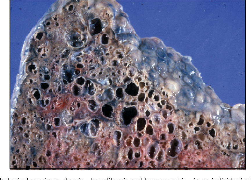
The lung scarring that occurs in pulmonary fibrosis can’t be reversed, and no current treatment has proved effective in stopping progression of the disease. Some treatments may improve symptoms temporarily or slow the disease’s progression. Others may help improve quality of life. Doctors will evaluate the severity of your condition to determine the most appropriate treatment for your condition.

Medications

Your doctor may recommend newer medications, including pirfenidone (Esbriet) and nintedanib (Ofev). These medications may help slow the progression of idiopathic pulmonary fibrosis. Both medications have been approved by the Food and Drug Administration (FDA). Additional medications and new formulations of these medications are being developed but have not yet been FDA approved.

Nintedanib can cause side effects such as diarrhea and nausea. Side effects of pirfenidone include rash, nausea and diarrhea.

Researchers continue to study medications to treat pulmonary fibrosis.

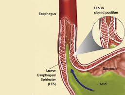
Doctors may recommend anti-acid medications to treat gastroesophageal reflux disease (GERD), a digestive condition that commonly occurs in people with idiopathic pulmonary fibrosis.

Oxygen therapy

Using oxygen can’t stop lung damage, but it can:

  • Make breathing and exercise easier
  • Prevent or lessen complications from low blood oxygen levels
  • Reduce blood pressure in the right side of your heart
  • Improve your sleep and sense of well-being

You may receive oxygen when you sleep or exercise, although some people may use it all the time. Some people carry a canister of oxygen, making them more mobile.

Pulmonary rehabilitation

Pulmonary rehabilitation can help you manage your symptoms and improve your daily functioning. Pulmonary rehabilitation programs focus on:

  • Physical exercise to improve your endurance
  • Breathing techniques that may improve lung efficiency
  • Nutritional counseling
  • Counseling and support
  • Education about your condition

Lung transplant

Lung transplantation may be an option for people with pulmonary fibrosis. Having a lung transplant can improve your quality of life and allow you to live a longer life. However, a lung transplant can involve complications such as rejection and infection. Your doctor may discuss with you if a lung transplant may be appropriate for your condition.

QUOTE FOR THE WEEKEND:

“Pulmonary fibrosis is a lung disease that occurs when lung tissue becomes damaged and scarred. This thickened, stiff tissue makes it harder for the lungs to work properly. Pulmonary fibrosis worsens over time. Some people can stay stable for a long time, but the condition gets worse faster in others. As it gets worse, people become more and more short of breath.”

MAYO CLINIC (Pulmonary fibrosis – Symptoms and causes – Mayo Clinic)

Part I Pulmonary Fibrosis Awareness Month-Risk Factors and Causes!


Pulmonary fibrosis is a lung disease that occurs when lung tissue becomes damaged and scarred. This thickened, stiff tissue makes it more difficult for your lungs to work properly. As pulmonary fibrosis worsens, you become progressively more short of breath.

The scarring associated with pulmonary fibrosis can be caused by a multitude of factors. But in most cases, doctors can’t pinpoint what’s causing the problem. When a cause can’t be found, the condition is termed idiopathic pulmonary fibrosis.

The lung damage caused by pulmonary fibrosis can’t be repaired, but medications and therapies can sometimes help ease symptoms and improve quality of life. For some people, a lung transplant might be appropriate.

Risk factors

Factors that make you more susceptible to pulmonary fibrosis include:

  • Age. Although pulmonary fibrosis has been diagnosed in children and infants, the disorder is much more likely to affect middle-aged and older adults.
  • Sex. Idiopathic pulmonary fibrosis is more likely to affect men than women.
  • Smoking. Far more smokers and former smokers develop pulmonary fibrosis than do people who have never smoked. Pulmonary fibrosis can occur in patients with emphysema.
  • Certain occupations. You have an increased risk of developing pulmonary fibrosis if you work in mining, farming or construction or if you’re exposed to pollutants known to damage your lungs.
  • Cancer treatments. Having radiation treatments to your chest or using certain chemotherapy drugs can increase your risk of pulmonary fibrosis.
  • Genetic factors. Some types of pulmonary fibrosis run in families, and genetic factors may be a component.

Causes

Pulmonary fibrosis scars and thickens the tissue around and between the air sacs (alveoli) in your lungs. This makes it more difficult for oxygen to pass into your bloodstream. The damage can be caused by many different factors — including long-term exposure to certain toxins, certain medical conditions, radiation therapy and some medications.

Occupational and environmental factors

Long-term exposure to a number of toxins and pollutants can damage your lungs. These include:

  • Silica dust
  • Asbestos fibers
  • Hard metal dusts
  • Coal dust
  • Grain dust
  • Bird and animal droppings

Radiation treatments

Some people who receive radiation therapy for lung or breast cancer show signs of lung damage months or sometimes years after the initial treatment. The severity of the damage may depend on:

  • How much of the lung was exposed to radiation
  • The total amount of radiation administered
  • Whether chemotherapy also was used
  • The presence of underlying lung disease

Medications

Many drugs can damage your lungs, especially medications such as:

  • Chemotherapy drugs. Drugs designed to kill cancer cells, such as methotrexate (Trexall, Otrexup, others) and cyclophosphamide, can also damage lung tissue.
  • Heart medications. Some drugs used to treat irregular heartbeats, such as amiodarone (Cordarone, Nexterone, Pacerone), may harm lung tissue.
  • Some antibiotics. Antibiotics such as nitrofurantoin (Macrobid, Macrodantin, others) or ethambutol can cause lung damage.
  • Anti-inflammatory drugs. Certain anti-inflammatory drugs such as rituximab (Rituxan) or sulfasalazine (Azulfidine) can cause lung damage.

Medical conditions

Lung damage can also result from a number of conditions, including:

  • Dermatomyositis
  • Polymyositis
  • Mixed connective tissue disease
  • Systemic lupus erythematosus
  • Rheumatoid arthritis
  • Sarcoidosis
  • Scleroderma
  • Pneumonia

Many substances and conditions can lead to pulmonary fibrosis. Even so, in most cases, the cause is never found. Pulmonary fibrosis with no known cause is called idiopathic pulmonary fibrosis.

Researchers have several theories about what might trigger idiopathic pulmonary fibrosis, including viruses and exposure to tobacco smoke. Also, some forms of idiopathic pulmonary fibrosis run in families, and heredity may play a role in idiopathic pulmonary fibrosis.

Many people with idiopathic pulmonary fibrosis may also have gastroesophageal reflux disease (GERD) — a condition that occurs when acid from your stomach flows back into your esophagus. Ongoing research is evaluating if GERD may be a risk factor for idiopathic pulmonary fibrosis, or if GERD may lead to a more rapid progression of the condition. However, more research is needed to determine the association between idiopathic pulmonary fibrosis and GERD.

QUOTE FOR FRIDAY:

“The New Year is often approached with new goals and mindsets, many of which focus on health, including reducing alcohol consumption. The Dry January campaign is one approach to help those who are sober curious or simply interested in cutting back in the new year.

Dry January is a month-long journey to stop drinking. It initially started as a campaign in the UK in 2013 under the organization Alcohol Change UK and quickly caught on worldwide as a growing trend. Since this campaign launched, nearly a quarter of all Americans have participated in Dry January each year.

The World Health Organization warns that no level of alcohol consumption is safe for health, and therefore, abstaining from alcohol is the best way to prevent alcohol-related diseases that can affect your heart, kidneys, brain, and liver. Alcohol consumption is a leading preventable cause of death in the U.S., according to a 2022 study published in JAMA Network Open, and it seems that alcohol consumption in the United States is not stopping or slowing down anytime soon.

According to a 2023 Gallup survey:

62% of Americans consume alcohol
69% of these adults say they last had a drink within the past week. This includes 32% whose most recent drink was in the last 24 hours.
20% of adults in this survey say they drink more than they should
Beer and liquor sales have increased and especially are on the rise during the holiday season in December.
Younger generations drink less, & older generations continue to drink more.”

Alcohol Help (alcohol.com/Dry January 2025: The Ultimate Guide)


Try the dry free alcohol challenge for one month & learn how its good for the body.

Harvard Health Publishing states the following:

“If you’d like to cut down your alcohol consumption or start the new year with a clean slate, join in the Dry January challenge by choosing not to drink beer, wine, or spirits for one month. Dry January began in 2012 as a public health initiative from Alcohol Change UK, a British charity. Now millions take part in this health challenge every year.

While past observational studies suggested a link between drinking a moderate amount of alcohol and health benefits for some people, more recent research has questioned whether any amount of alcohol improves health outcomes. And heavier drinking or long-term drinking can increase physical and mental problems, especially among older adults. Heart and liver damage, a higher cancer risk, a weakened immune system, memory issues, and mood disorders are common issues.

Yet, cutting out alcohol for even a month can make a noticeable difference in your health. Regular drinkers who abstained from alcohol for 30 days slept better, had more energy, and lost weight, according to a study in BMJ Open. They also lowered their blood pressure and cholesterol levels and reduced cancer-related proteins in their blood.

Tips for a successful Dry January

A month may seem like a long time, but most people can be successful. Still, you may need assistance to stay dry in January. Here are some tips:

  • Find a substitute non-alcoholic drink. For social situations, or when you crave a cocktail after a long day, reach for alcohol-free beverages like sparkling water, soda, or mocktails (non-alcoholic cocktails.)
    Non-alcoholic beer or wine also is an option, but some brands still contain up to 0.5% alcohol by volume, so check the label. “Sugar is often added to these beverages to improve the taste, so try to choose ones that are low in sugar,” says Dawn Sugarman, a research psychologist at Harvard-affiliated McLean Hospital in the division of alcohol, drugs, and addiction.
  • Avoid temptations. Keep alcohol out of your house. When you are invited to someone’s home, bring your non-alcoholic drinks with you.
  • Create a support group. Let friends and family know about your intentions and encourage them to keep you accountable. Better yet, enlist someone to do the challenge with you.
  • Use the Try Dry app. This free app from the UK helps you track your drinking, set personal goals, and offers motivational information like calories and money saved from not drinking. It’s aimed at cutting back on or cutting out alcohol, depending on your choices.
  • Don’t give up. If you slip up, don’t feel guilty. Just begin again the next day.

Check your feelings

Sugarman recommends people also use Dry January to reflect on their drinking habits. It’s common for people to lose their alcohol cravings and realize drinking need not occupy such an ample space in their lives. If this is you, consider continuing for another 30 days, or just embrace your new attitude toward drinking where it’s an occasional indulgence.

If you struggle during the month, or give up after a week or so, you may need extra help cutting back. Talk to your doctor about getting the help you need.

The Rethinking Drinking site created by the National Institute on Alcohol Abuse and Alcoholism (NIAAA) is also an excellent resource. For the record, NIAAA recommends limiting alcohol to two daily drinks or less for men and no more than one drink a day for women.

Be aware of problems that might crop up

Dry January can reveal signs of potential alcohol problems, including symptoms of alcohol withdrawal ranging from mild to serious, depending on how much you usually drink.

  • Mild symptoms include anxiety, shaky hands, headache, nausea, vomiting, sweating, and insomnia.
  • Severe symptoms often kick in within two or three days after you stop drinking. They can include hallucinations, delirium, racing heart rate, and fever.

“If you suffer alcohol withdrawal symptoms at any time, you should seek immediate medical help,” says Sugarman.”

QUOTE FOR THURSDAY:

“An estimated 4.22 million U.S. adults had glaucoma in 2022, and 1.49 million had vision-affecting glaucoma, with “substantial variation” in prevalence by region and demographics, according to a research study published in JAMA Ophthalmology

“Up-to-date estimates of how many people have glaucoma in a certain area are vital for addressing the problem,” said Joshua Ehrlich, MD, MPH, lead author of the study and associate professor of ophthalmology and visual sciences at Michigan Medicine. “This kind of information is important for formulating evidence-based policy and public health solutions.”

To estimate the prevalence of glaucoma and related vison loss on a state and county level, Ehrich and colleagues conducted a meta-analysis of data from the National Health and Nutrition Examination Survey, Medicare and IBM MarketScan claims, population-based studies of eye disease and population estimates from the U.S. Census Bureau.”

Glaucoma Research Foundation (Glaucoma Study Shows Higher Prevalence Than Previous Estimates – Glaucoma Research Foundation Glaucoma.org)

Part III Glaucoma National Awareness: The types of treatments of Glaucoma!

If you are diagnosed with glaucoma, it is important to set a regular schedule of examinations with your eye doctor to monitor your condition and make sure that your prescribed treatment is effectively maintaining a safe eye pressure.

Treatments

The Treatment of Glaucoma: The damage caused by glaucoma can’t be reversed. But treatment and regular checkups can help slow or prevent vision loss, especially in you catch the disease in its early stage. The goal of glaucoma treatment is to lower pressure in your eye (intraocular pressure). Depending on your situation, your options may include eyedrops, laser treatment or surgery. 

Remember The damage caused by glaucoma can’t be reversed. But treatment and regular checkups can help slow or prevent vision loss, especially in you catch the disease in its early stage. The goal of glaucoma treatment is to lower pressure in your eye (intraocular pressure). Depending on your situation, your options may include eyedrops, laser treatment or surgery.

Eyedrops

Glaucoma treatment often starts with prescription eyedrops. These can help decrease eye pressure by improving how fluid drains from your eye or by decreasing the amount of fluid your eye makes. Depending on how low your eye pressure needs to be, more than one of the eyedrops below may need to be prescribed.

Prescription eyedrop medications include:

  • Prostaglandins. These increase the outflow of the fluid in your eye (aqueous humor), thereby reducing your eye pressure. Medicines in this category include latanoprost (Xalatan), travoprost (Travatan Z), tafluprost (Zioptan), bimatoprost (Lumigan) and latanoprostene bunod (Vyzulta). Possible side effects include mild reddening and stinging of the eyes, darkening of the iris, darkening of the pigment of the eyelashes or eyelid skin, and blurred vision. This class of drug is prescribed for once-a-day use.
  • Beta blockers. These reduce the production of fluid in your eye, thereby lowering the pressure in your eye (intraocular pressure). Examples include timolol (Betimol, Istalol, Timoptic) and betaxolol (Betoptic). Possible side effects include difficulty breathing, slowed heart rate, lower blood pressure, impotence and fatigue. This class of drug can be prescribed for once- or twice-daily use depending on your condition.
  • Alpha-adrenergic agonists. These reduce the production of aqueous humor and increase outflow of the fluid in your eye. Examples include apraclonidine (Iopidine) and brimonidine (Alphagan P, Qoliana). Possible side effects include an irregular heart rate, high blood pressure, fatigue, red, itchy or swollen eyes, and dry mouth. This class of drug is usually prescribed for twice-daily use but sometimes can be prescribed for use three times a day.
  • Carbonic anhydrase inhibitors. These medicines reduce the production of fluid in your eye. Examples include dorzolamide (Trusopt) and brinzolamide (Azopt). Possible side effects include a metallic taste, frequent urination, and tingling in the fingers and toes. This class of drug is usually prescribed for twice-daily use but sometimes can be prescribed for use three times a day.
  • Rho kinase inhibitor. This medicine lowers eye pressure by suppressing the rho kinase enzymes responsible for fluid increase. It is available as netarsudil (Rhopressa) and is prescribed for once-a-day use. Possible side effects include eye redness, eye discomfort and deposits forming on the cornea.
  • Miotic or cholinergic agents. These increase the outflow of fluid from your eye. An example is pilocarpine (Isopto Carpine). Side effects include headache, eye ache, smaller pupils, possible blurred or dim vision, and nearsightedness. This class of medicine is usually prescribed to be used up to four times a day. Because of potential side effects and the need for frequent daily use, these medications are not prescribed very often anymore.

Because some of the eyedrop medicine is absorbed into your bloodstream, you may experience some side effects unrelated to your eyes. To minimize this absorption, close your eyes for one to two minutes after putting the drops in. You may also press lightly at the corner of your eyes near your nose to close the tear duct for one or two minutes. Wipe off any unused drops from your eyelid.

If you have been prescribed multiple eyedrops or you need to use artificial tears, space them out so that you are waiting at least five minutes in between types of drops.

Oral medications

If eyedrops alone don’t bring your eye pressure down to the desired level, your doctor may also prescribe an oral medication, usually a carbonic anhydrase inhibitor. Possible side effects include frequent urination, tingling in the fingers and toes, depression, stomach upset, and kidney stones.

Surgery and other therapies

Other treatment options include laser therapy and various surgical procedures. The following techniques are intended to improve the drainage of fluid within the eye, thereby lowering pressure:

  • Laser therapy. Laser trabeculoplasty (truh-BEK-u-low-plas-tee) is an option if you have open-angle glaucoma. It’s done in your doctor’s office. Your doctor uses a small laser beam to open clogged channels in the trabecular meshwork. It may take a few weeks before the full effect of this procedure becomes apparent.
  • Filtering surgery. With a surgical procedure called a trabeculectomy (truh-bek-u-LEK-tuh-me), your surgeon creates an opening in the white of the eye (sclera) and removes part of the trabecular meshwork.
  • Drainage tubes. In this procedure, your eye surgeon inserts a small tube shunt in your eye to drain away excess fluid to lower your eye pressure.
  • Minimally invasive glaucoma surgery (MIGS). Your doctor may suggest a MIGS procedure to lower your eye pressure. These procedures generally require less immediate postoperative care and have less risk than trabeculectomy or installing a drainage device. They are often combined with cataract surgery. There are a number of MIGS techniques available, and your doctor will discuss which procedure may be right for you.

After your procedure, you’ll need to see your doctor for follow-up exams. And you may eventually need to undergo additional procedures if your eye pressure begins to rise again or other changes occur in your eye.