QUOTE FOR MONDAY:

“Sports help children develop physical skills, get exercise, make friends, have fun, learn teamwork, learn to play fair, and improve self-esteem.  It is important to remember that the attitudes and behavior taught to children in sports carry over to adult life. Parents should take an active role in helping their child develop good sportsmanship. To help your child get the most out of sports, you need to be actively involved.  Although this involvement takes time and creates challenges for work schedules, it allows you to become more knowledgeable about the coaching, team values, behaviors, and attitudes. Your child’s behavior and attitude reflect a combination of the coaching and your discussions about good sportsmanship and fair play.”

American Academy of Child and Adolescent Pschiatry (https://www.aacap.org/AACAP/Families_and_Youth/Facts_for_Families/FFF-Guide/Children-And-Sports-061.aspx)

Why youth sports are important!

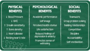
Regular physical activity benefits health in many ways, including helping build and maintain healthy bones, muscles, and joints; helping control weight and reduce fat; and preventing or delaying the development of high blood pressure (GAO, 2012). Exercise is one of the least expensive ways to stay healthy, with one study finding that exercise can prevent chronic diseases as effectively as medication (British Journal of Medicine, 2013). A comprehensive study and analysis of existing research found that leisure-time physical activity is associated with reduced risk of 13 different types of cancer, including breast, colon, liver and myeloid leukemia (National Institutes of Health, 2016).

Sports participation is a significant predictor of young adults’ participation in sports and physical fitness activities. Adolescents who play sports are eight times as likely to be active at age 24 as adolescents who do not play sports (Sports Participation as Predictors of Participation in Sports and Physical Fitness Activities in Young Adulthood, Perkins, 2004). Three in four (77%) of adults aged 30+ who play sports today played sports as school-aged children. Only 3% of adults who play sports currently did not play when they were young (Robert Wood Johnson Foundation/Harvard University/NPR, 2015).

Obesity reduction. In a 2014 study published in the American Journal of Preventive Medicine, researchers analyzed obesity prevention strategies and their ability to reduce obesity by the year 2032. They found afterschool physical activity programs would reduce obesity the most, 1.8% among children ages 6 to 12. That’s twice the projected impact as any ban on child-directed fast-food advertising. An earlier study of college students found that “motives for sport participation are more desirable than those for exercise and may facilitate improved adherence to physical activity recommendations” (Kilpatrick, Journal of American College Health, 2005).

But the obesity epidemic continues. In 2018, a study published by the American Academy of Pediatrics showed overweight and obesity rates increased in all age groups among children ages 2 to 19 (Prevalence of Obesity and Severe Obesity in U.S. Children, 1999-2016). The rates generally increased with age, with 41.5% of teens being obese by 16 to 19 years old. Of particular concern were continued racial and ethnic disparities. White and Asian children showed significantly lower rates of obesity than Hispanic and African-American children. Researchers also found a sharp increase in obesity from 2015 to 2016 compared to the previous cycle among children ages 2 to 5, especially boys. Girls 16 to 19 years old had a notable jump in overweight rates, from 36% in 2013-14 to 48% in 2015-16. Youth who have disabilities are 4.5 times less active and have obesity rates that are 38% higher than other youth (Physical Literacy in the United States: A Model, Strategic Plan, and Call to Action, 2015).

Not enough children are active. A 2018 study from Nationwide Children’s Hospital showed that just 5% of youth ages 5 to 18 reported meeting the federally recommended amount of exercise — 60 minutes per day. The study, which examined 7,822 children over three years, also found that 50 percent were insufficiently active and 5 percent reported no physical activity. Researchers recommended that pediatricians should treat exercise like a vital sign, similar to height and weight, and engage patients in conversations about how to be more physically active. A 2020 study found that 60% of American children had inadequate levels of cardiorespiratory fitness, putting them at increased risk of chronic diseases at younger ages (American Heart Association, 2020). The Aspen Institute’s Healthy Sport Index is a tool that can help parents find the best sport for their child based on health benefits and risks. The resource includes original research that found boys generate more vigorous physical activity at high school practices than girls (North Carolina State University, 2018).

EDUCATIONAL BENEFITS

From the Sport for All, Play for Life report. click to enlarge

From the Sport for All, Play for Life report. click to enlarge

Organized sports activity helps children develop and improve cognitive skills, according to a study that tracked kids from kindergarten through fourth grade (Piche, 2014). Physical activity in general is associated with improved academic achievement, including grades and standardized test scores. Further, such activity can affect attitudes and academic behavior, including enhanced concentration, attention, and improved classroom behavior (GAO, 2012).

High school athletes are more likely than non-athletes to attend college and get degrees, and team captains and most valuable players achieve in school at even higher rates (U.S. Dept. of Education, 2005). Also, high school athletes are more likely to expect to graduate from a four-year college (73% girls, 59% boys) compared to non-athletes (67% girls, 53% boys), according to data collected for the Healthy Sport Index (Women’s Sports Foundation, 2018). A higher percentage of high school athletes also receive A/A- grades than non-athletes (Women’s Sports Foundation, 2018).

The benefits extend to the workplace. A survey of 400 female corporate executives found 94% played a sport and that 61% say sports contributed to their career success (EY Women Athletes Business Network/espnW, 2014).

PSYCHOSOCIAL BENEFITS

A correlation has been found between regular exercise and mental health among students in general as they move into the teenage years. Among students who exercised six to seven days a week, 25.1% felt sad for two weeks or more in the past 12 months, compared to 35.7% of students who reported exercising on zero to one day (Women’s Sports Foundation, 2004). Of students who exercised six to seven days, 15% reported suicidal ideation, and 6.4% reported a suicide attempt in the past year, compared to 24.6% and 10.3% of students who exercised zero to one day, respectively (Journal of American Academy of Child & Adolescent Psychiatry, 2015). A 2019 study found that children who reported no exercise were twice as likely to have mental health problems, particularly related to anxiety and depression, compared with those who met the recommendation of an hour a day, and a 2020 study suggested that the more physical activity teenagers participated in, the less likely they were to report depression as 18-year-olds (The New York Times, 2020).

Physical activity, and sports in particular, can positively affect aspects of personal development among young people, such as self-esteem, goal-setting, and leadership. However, evidence indicates that the quality of coaching is a key factor in maximizing positive effects (GAO, 2012).

Both male and female high school athletes are less likely to smoke cigarettes and suffer from loneliness and low self-esteem, when compared to non-athlete peers, according to research used for the Healthy Sport Index (Women’s Sports Foundation, 2018). High school athletes, though, are more likely to binge drink alcohol, with youth in contact sports  (football, lacrosse, wrestling) showing the riskiest off-field behavior).


QUOTE FOR THE WEEKEND:

“National Burn Awareness Week is a window of opportunity for organizations to mobilize burn, fire, and life safety educators to unite in sharing a common burn awareness and prevention message in our communities.  Avoid a dangerous balancing act.  Never carry a child or be right next to a child when carrying or sitting with a hot food plate or hot drink in your hand.”

American Burn Association (https://ameriburn.org/education/burn-reconstruction/)

QUOTE FOR FRIDAY:

“Nurses working in preanesthesia and postanesthesia care, ambulatory surgery, and pain management are dedicated to caring for patients and their families. They are passionate about what they do as nurses. Every year, the American Society for Perianesthesia Nurses (ASPAN) recognizes and honors perianesthesia nurses across the country during this week.”

American Board of Peri-Anesthesia Nursing Certification ABPANC  (https://www.cpancapa.org/nurse-leaders/perianesthesia-nurse-awareness-week/)

QUOTE FOR THURSDAY:

“Most cardiac arrests occur when a diseased heart’s electrical system malfunctions. This malfunction causes an abnormal heart rhythm such as ventricular tachycardia or vent.”ricular fibrillation. Some cardiac arrests are also caused by extreme slowing of the heart’s rhythm (bradycardia).  There are other rhythms that can also cause cardiac arrest.”

American Heart Association (https://www.heart.org/en/health-topics/cardiac-arrest/causes-of-cardiac-arrest)

Certain cardiac rhythms can lead to a cardiac arrest & how!

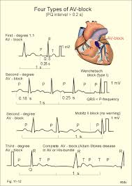
HeartBlocks1

The rhythms above are heart blocks (HB) that occur in the bottom of the upper chambers which can occur in some people. There is 1st degree HB where you can live a completely normal life with but 2nd and 3rd degree HB needs treatment (usually a pacemaker) by cardiologist surgeon.  After treatment with 2nd and 3rd degree HB you can live a completely normal life with follow up with your cardiologist and yearly pacemaker checks.

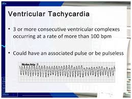
In this rhythm below the Ventricular Tachycardia is with a point on the top but than flips upside down (commonly called Torsedes Pointes).  This is commonly due to Magnesium Level low and IV Magnesium in the hospital is given 1 to 2 gm.
ventrhy4

This  rhythm above with a pulse=also a rhythm pulsating in different areas of the heart in the ventricles only causing the rhythm not to look identical throughout the tele strip above = Polymorphic V- Tac- meaning the stimulus in the ventricles to make the heart beat is coming from different areas of the ventricles for each beat.  Each jagged tooth is a beat that makes up the whole strip shown above for Ventricular Tachycardia.

Than when the atriums aren’t working as the natural pacemaker that took over for the sinus node but now they don’t work so now the ventricles take over and the rhythms of all ventricle rhythms are with NO p waves since the atriums are not working so no p wave is involved but we have QRS waves but their wide in measurement because the rhythm starts in the ventricles. The rhythms are PVC (Premature Ventricular Contractions), Idioventricular Rhythm, Ventricular tachycardia (Monomorphic and Polymorphic-rhythm getting more irregular. When regular and monomorphic=looking identical with every ventricular beat or contraction as opposed to polymorphic=not looking identical each contraction but each one is a ventricular contraction), Torsades De Pointes Ventricular Tachycardia (the rhythm starts upright but turns upside down but each contraction without a p wave and a wide contraction meaning a ventricular contraction), and Ventricular Fibrillation, to asystole.

Here’s what they look like:

 Accelerated Idioventricular Rhythm

Accelerated idioventricular rhythm occurs when three or more ventricular escape beats appear in a sequence. Heart rate will be 50-100 bpm. The QRS complex will be wide (0.12 sec. or more).

A regular QRS measures less than 0.12 which is with all atriums rhythms.

 Asystole

Asystole is the state of no cardiac electrical activity and no cardiac output. Immediate action is required.

Idioventricular Rhythm

Idioventricular rhythm is a slow rhythm of under 50 bpm. It indicates that then ventricules are producing escape beats.
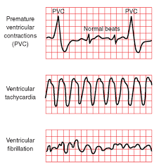
Premature Ventricular Complex (above 1st strip)

Premature ventricular complexes (PVCs) occur when a ventricular site generates an impulse. This happens before the next regular sinus beat. Look for a wide QRS complex, equal or greater than 0.12 sec. The QRS complex shape can be bizarre. The P wave will be absent.

Premature Ventricular Complex – Bigeminy a QRS after every 2 regular beats

Premature Ventricular Complex – Trigeminy a QRS after every 3 regular beats

Premature Ventricular Complex – Quadrigeminy a QRS after every 4 regular beats

 Ventricular Fibrillation (in above strip-3rd one)

Ventricular fibrillation originates in the ventricules and it chaotic. No normal EKG waves are present. No heart rate can be observed. Ventricular fibrillation is an emergency condition requiring immediate action.

Ventricular Tachycardia  (in above strip-2nd one)

A sequence of three PVCs in a row is ventricular tachycardia. The rate will be 120-200 bpm. Ventricular Tachycardia has two variations, monomorphic and polymorphic. These variations are discussed separately.

Ventricular Tachycardia Monomorphic

Monomorphic ventricular tachycardia occurs when the electrical impulse originates in one of the ventricules. The QRS complex is wide. Rate is above 100 bpm.  Each V tac beat looks identical like in the strip above.

Ventricular Tachycardia Polymorphic

Polymorphic ventricular tachycardia has QRS complexes that very in shape and size. If a polymorphic ventricular tachycardia has a long QT Interval, it could be Torsade de Pointes.  The strip shows the pulses are not identical=polymorphic since the pulse beats are coming from all different areas of the ventricles.

Torsade de Pointes  (the rhythm strip at the top under Heart Blocks)

Torsade de Pointes is a special form of ventricular tachycardia. The QRS complexes vary in shape and amplitude and appear to wind around the baseline.  This is an example or polymorphic ventricular tachycardia.

Ventricular ending line needs to be treated stat to be switched back to atrial rhythm since the heart is missing ½ of the conduction it’s to normally receive from the atriums and if not reversed the heart will go into failure to heart attack or to asystole flat line and go into a cardiac arrest.

With PVCs=Premature Ventricle Contractions asymptomatic we just closely monitor the pt and telemetry the pt is on. Now a pt with PVCs and symtomatic usually meds with 0xygen (sometimes 02 alone resolves it but other times with meds) but if it gets worse into V Tachycardia the treatment is below.

Idioventricular Rhythm (IVR)is usually with a slow brady pulse and needs meds.   Accelerated IVR (AIVR) is usually hemodynamically tolerated and self-limited; thus, it rarely requires treatment.

Occasionally, patients may not tolerate AIVR due to (1) loss of atrial-ventricular synchrony, (2) relative rapid ventricular rate, or (3) ventricular tachycardia or ventricular fibrillation degenerated from AIVR (extremely rare). Under these situations, atropine can be used to increase the underlying sinus rate to inhibit AIVR.

Other treatments for AIVR, which include isoproterenol, verapamil, antiarrhythmic drugs such as lidocaine and amiodarone, and atrial overdriving pacing are only occasionally used today.

Patients with AIVR should be treated mainly for its underlying causes, such as digoxin toxicity, myocardial ischemia, and structure heart diseases. Beta-blockers are often used in patients with myocardial ischemia-reperfusion and cardiomyopathy

With Ventricular rhythms with fast pulse over 100 with symptomatic signs for the patient we may use as simple as valsalva pressure on the neck that medical staff only do but when pt is in asymptomatic (no symptoms) Ventricular Tachycardia (V-Tac) to even medications but when symptomatic if in V-Tac start cardioversion with a pulse if no pulse called pulseless V-Tac we use a defibrillator since there is no pulse there is no QRS to pace with in having the shock hit at the R wave, why? NO PULSE.

Treatment for Torsade de Pointes is Magnesium deficiency and Mag. Supplement given IV 2gms. Usually effective but if necessary the same as above as directed for it with a pulse or the other V Tac. (without a pulse)-See above.

Ventricular Fibrillation is when the ventricles are just quivering and the atriums in any ventricular rhythm doing nothing. The pt needs CPR and ASAP a defibrillator in hopes the shock will knock the rhythm back to a normal sinus or some form of a real rhythm.

Asystole which is a straight line, no pulse and this is CPR with epinephrine or Vasopressin 40 for only the replacement of the 1st or 2nd dose of Epinephrine 1mg. This is given 3-5 minutes (epinephrine). No defibrillation since no pulse. A rhythm may come back and if not the MD will call when CPR stops. Asystole is hard to resolve in most cases highier probability of resolution if in a hospital where close monitoring is done and its detected quicker.

The PURPOSE in treating any rhythm abnormal to the human heart is to reach the goal of a optimal or healthiest rhythm (a normal sinus rhythm , the best rhythm the heart can be in) and if not reaching an atrial rhythm.  We the medical field aim to reach a heart rhythm the patient can live with and hopefully reaching the best NSR-Normal Sinus Rhythm.  Normal sinus rhythm that is a rhythm starting from the upper right chamber extending to the left one and continues down on both sides to the bottom of the ventricles.  This rhythm is giving the most effective oxygen perfusion to the heart to allow it to do its function (pumping good oxygenated blood flow out of the left ventricle at the same time pumping highly carbon dioxide blood from the right side of the heart to the lungs to get more oxygen).   Doing this it allows the human body to get good amounts of oxygen to all our tissues=good overall oxygen perfusion to all tissues.  At the same time what happens is red blood cells from all tissues with mostly used up oxygen from the cell and more carbon dioxide in the cell are also being pumped by the heart to return to the right side  to the lungs to go through this whole process again in getting more oxygen in the RBCs which keeps us alive. A human without oxygen or low oxygen to their tissues or any tissue is going to reach cellular starvation which in turn causes starvation to the tissues (in general) or to a tissue (Ex. Diabetic the foot to lack of 02 to cyanotic purple tissue to necrotic black tissue=dead to amputated since the tissue is dead.).

Cardiac Arrest or Heart Attack are more likely to occur in  a irregular rhythm especially making the heart work to hard being RVR afib in the atriums that can lead easily to ventricular tachycardia to ventricular fibrillation and not treated immediately.

Cardiac Arrest is an electrical problem with the conduction of the heart whereas a Heart Attack is caused by a blockage of blood (Ex. coronary artery) to the heart that can lead to a bad rhythm due to lack of 0xygen that leads to worse rhythms as the heart gets more stressed out.

QUOTE FOR WEDNESDAY:

A stent can cause blood clotting, which may increase the risk of heart attack or stroke. The National Heart, Lung, and Blood Institute state that about 1 to 2 percent of people who have stented arteries develop a blood clot at the site of the stent. Doctors will usually prescribe one or more drugs to prevent clotting.”

MAYO CLINIC (https://www.mayoclinic.org/tests-procedures/coronary-angioplasty/about/pac-20384761)

Part II What are Cardiac Stents risks when getting one?

  stentstents part 2

Stents are used in cases of “restenosis”, which refers to the re-closing of arteries after balloon angioplasty. In carefully selected patients, the use of stents can dramatically reduce restenosis following balloon angioplasty or other catheter-based procedures. Stents are used frequently to hold open the arteries that have been damaged, torn, or dissected by balloon angioplasty or other catheter-based procedures. Like plumber or mechanic, get it.

Stents allow angioplasty to be done in patients with severe and long-segment obstruction of coronary arteries. As soon as the I initial part of the block is widened, a stent is place, which holds it open allowing further opening to proceed. Stents have also allowed angioplasty to be performed in patients with blocks of multiple vessels, and in multiple blocks in a single artery.

Risks of getting cardiac stents:

Risks include the standard risks of an interventional, catheter-based procedure, which should be specifically discussed with your doctor. Lesions treated with stents can “restenosis” (re- narrow with in weeks to months after the procedure) similar to restenosis associated with angioplasty. This is why patients after having a stent put in they are on a medication for example like Plavix for life to prevent this occurrence from happening. It is a anti platelet medication, meaning it doesn’t allow clotting to happen in the stent so blockage doesn’t reoccur through clotting.

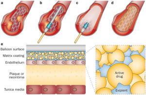
Many new technologies are being tested to reduce the problem of restenosis, meaning close up. These technologies include coating and coverings for the stent, new materials, and radiation. These new technologies are primarily experimental at this point and will reach soon to the market if not already. Technology allows the medical field to continuously expand and this will be replaced at one point but isn’t yet. Just give it time. At one point we had no CABG (coronary artery bypass) but now the stent in certain cases is replaced by angiograms to further expanding to angioplasties and stents furthering allowing the surgery to take place 1x hopefully if the patient is compliant in diet, exercise, and following the doctors orders with meds, activity and follow up visits (which are so vital).   A lot is up to the patient in caring for themselves to prevent having this take place again.

St John’s Hopkins Medicine states that the risks of getting a stent are the following:

Possible risks linked to angioplasty, stenting, atherectomy, and related procedures include:

  • Bleeding at the site where the catheter is put into the body (usually the groin, wrist, or arm)
  • Blood clot or damage to the blood vessel from the catheter
  • Blood clot within the treated blood vessel
  • Infection at the catheter insertion site
  • Abnormal heart rhythms
  • Heart attack
  • Stroke
  • Chest pain or discomfort
  • Rupture of the coronary artery or complete closing of the coronary artery, needing open-heart surgery
  • Allergic reaction to the contrast dye used
  • Kidney damage from the contrast dye

Follow up Instructions

Your doctor will recommend blood thinning medications following your stent procedure. These agents are usually given for one month post procedure along with aspirin and then continued indefinitely. Your doctor may also prescribe antibiotics for a period of time after the stent procedure, to be taken anytime you have a medical or dental procedure. Preventing infection.

MRI tests should not be done for at least eight weeks without your doctor’s approval. Metal detectors do not present a problem. Stents appear to be safe in the long-term; there are no long-term complications associated with a permanent stent.


QUOTE FOR TUESDAY:

“A stent is a tiny tube that can play a big role in treating your heart disease. It helps keep your arteries — the blood vessels that carry blood from your heart to other parts of your body, including the heart muscle itself — open.

Most stents are made out of wire mesh and are permanent. Some are made out of fabric. These are called stent grafts and are often used for larger arteries.

Others are made of a material that dissolves and that your body absorbs over time. They’re coated in medicine that slowly releases into your artery to prevent it from being blocked again.”

Web MD (https://www.webmd.com/heart-disease/what-is-stent)

.”

Part I What are Cardiac Stents?

stent

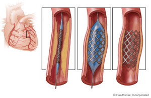
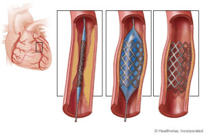
A stent is a wire mesh tube that is used to help hold open an artery. To simple understanding this concept think of a plumber or a mechanic. With a plumber sometimes they have to replace a certain area of a pipe that connects the water or like a mechanic replacing a certain area of piping (like the muffler piping connected infront of the muffler than can either can be replaced or just welded with piece of piping welded just to save money. Well a stent opens the artery that was clogged and its put in that place to reinforce that area of the artery to remain open to allow blood to get to that heart from that coronary artery and it will stay there life to keep the artery permanently patent to prevent the blockage from happening again with the synthetic mesh piece.

Description

Stents are used to hold open diseased coronary arteries (these arteries supply blood to the heart), as well as diseased arteries of the peripheral vascular system (PVS). Peripheral means away from the heart the PVS is the arteries that supply blood to the rest of the body (again away from the heart all the way down to the hands and feet).

There are variety of stents currently available.

For a surgeon to find out if you even need one, first usually a angiogram is performed and this is a catheter simply from the femerol artery or from your arm to the coronary arteries. If the MD sees you show a blockage 80% or more an angioplasty is performed which is a balloon at the end of this catheter that blows up and decompresses to give the effect like punching gloves. The balloon inflates and deflates over and over again till the blockage breaks open free and then a stent is put in that area to help keep it open permanently (patent).

Some stents have been compressed onto the outside of an angioplasty balloon catheter and delivered by inflating the balloon in the desired location. Other stents are “self- expanding” spring-loaded devices, which expand automatically upon deployment.

Stents remain in arteries permanently. The tissue lining the arteries actually grows over the metal mesh to cover the inner lumen of the stent.

Stent procedures have become very common like tonsillectomies were in childhood. Stents are sometimes used as an alternative to coronary artery bypass surgery, if the patient is a candidate. Stents are often used in combination with balloon angioplasty. One leads to the other depending on what the angioplasty displays for the surgeon on the T.V. in when they are doing the procedure and if the come up to a blockage high enough to perform the angioplasty followed with a stent it will be done.

Stents are used in cases of “restenosis”, which refers to the re-closing of arteries after balloon angioplasty. In carefully selected patients, the use of stents can dramatically reduce restenosis following balloon angioplasty or other catheter-based procedures. Stents are used frequently to hold open the arteries that have been damaged, torn, or dissected by balloon angioplasty or other catheter-based procedures. Like plumber or mechanic, get it.

Stents allow angioplasty to be done in patients with severe and long-segment obstruction of coronary arteries. As soon as the I initial part of the block is widened, a stent is place, which holds it open allowing further opening to proceed. Stents have also allowed angioplasty to be performed in patients with blocks of multiple vessels, and in multiple blocks in a single artery.

Angioplasty and stent placement are common procedures to open arteries in the heart that are clogged. These procedures are formally known as coronary angioplasty or percutaneous coronary intervention.

Angioplasty involves the use of a tiny balloon to widen the artery. A stent is a tiny wire-mesh tube that your doctor inserts into the artery. The stent stays in place to prevent the artery from closing. A cardiologist typically performs both procedures at the same time.

While angioplasty with stent placement addresses an individual blockage, it doesn’t fix the underlying cause of the blockage. To prevent further blockages and reduce your risk of other medical conditions, you may have to make certain lifestyle changes to prevent it happening again.  The procedure of the angioplasty to insert a stent if needed resolves the immediate acute problem that the underlining disease the pt had causing the blockage.  This could be due to high cholesterol, diabetes, CAD,  and atheroscerosis.