QUOTE FOR FRIDAY:

“Nurses working in preanesthesia and postanesthesia care, ambulatory surgery, and pain management are dedicated to caring for patients and their families. They are passionate about what they do as nurses. Every year, the American Society for Perianesthesia Nurses (ASPAN) recognizes and honors perianesthesia nurses across the country during this week.”

American Board of Peri-Anesthesia Nursing Certification ABPANC  (https://www.cpancapa.org/nurse-leaders/perianesthesia-nurse-awareness-week/)

QUOTE FOR THURSDAY:

“Most cardiac arrests occur when a diseased heart’s electrical system malfunctions. This malfunction causes an abnormal heart rhythm such as ventricular tachycardia or vent.”ricular fibrillation. Some cardiac arrests are also caused by extreme slowing of the heart’s rhythm (bradycardia).  There are other rhythms that can also cause cardiac arrest.”

American Heart Association (https://www.heart.org/en/health-topics/cardiac-arrest/causes-of-cardiac-arrest)

Certain cardiac rhythms can lead to a cardiac arrest & how!

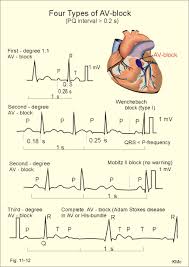
HeartBlocks1

The rhythms above are heart blocks (HB) that occur in the bottom of the upper chambers which can occur in some people. There is 1st degree HB where you can live a completely normal life with but 2nd and 3rd degree HB needs treatment (usually a pacemaker) by cardiologist surgeon.  After treatment with 2nd and 3rd degree HB you can live a completely normal life with follow up with your cardiologist and yearly pacemaker checks.

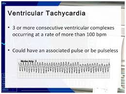
In this rhythm below the Ventricular Tachycardia is with a point on the top but than flips upside down (commonly called Torsedes Pointes).  This is commonly due to Magnesium Level low and IV Magnesium in the hospital is given 1 to 2 gm.

ventrhy4

This  rhythm above with a pulse=also a rhythm pulsating in different areas of the heart in the ventricles only causing the rhythm not to look identical throughout the tele strip above = Polymorphic V- Tac- meaning the stimulus in the ventricles to make the heart beat is coming from different areas of the ventricles for each beat.  Each jagged tooth is a beat that makes up the whole strip shown above for Ventricular Tachycardia.

Than when the atriums aren’t working as the natural pacemaker that took over for the sinus node but now they don’t work so now the ventricles take over and the rhythms of all ventricle rhythms are with NO p waves since the atriums are not working so no p wave is involved but we have QRS waves but their wide in measurement because the rhythm starts in the ventricles. The rhythms are PVC (Premature Ventricular Contractions), Idioventricular Rhythm, Ventricular tachycardia (Monomorphic and Polymorphic-rhythm getting more irregular. When regular and monomorphic=looking identical with every ventricular beat or contraction as opposed to polymorphic=not looking identical each contraction but each one is a ventricular contraction), Torsades De Pointes Ventricular Tachycardia (the rhythm starts upright but turns upside down but each contraction without a p wave and a wide contraction meaning a ventricular contraction), and Ventricular Fibrillation, to asystole.

Here’s what they look like:

 Accelerated Idioventricular Rhythm

Accelerated idioventricular rhythm occurs when three or more ventricular escape beats appear in a sequence. Heart rate will be 50-100 bpm. The QRS complex will be wide (0.12 sec. or more).

A regular QRS measures less than 0.12 which is with all atriums rhythms.

 Asystole

Asystole is the state of no cardiac electrical activity and no cardiac output. Immediate action is required.

Idioventricular Rhythm

Idioventricular rhythm is a slow rhythm of under 50 bpm. It indicates that then ventricules are producing escape beats.

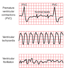
Premature Ventricular Complex (above 1st strip)

Premature ventricular complexes (PVCs) occur when a ventricular site generates an impulse. This happens before the next regular sinus beat. Look for a wide QRS complex, equal or greater than 0.12 sec. The QRS complex shape can be bizarre. The P wave will be absent.

Premature Ventricular Complex – Bigeminy a QRS after every 2 regular beats

Premature Ventricular Complex – Trigeminy a QRS after every 3 regular beats

Premature Ventricular Complex – Quadrigeminy a QRS after every 4 regular beats

 Ventricular Fibrillation (in above strip-3rd one)

Ventricular fibrillation originates in the ventricules and it chaotic. No normal EKG waves are present. No heart rate can be observed. Ventricular fibrillation is an emergency condition requiring immediate action.

Ventricular Tachycardia  (in above strip-2nd one)

A sequence of three PVCs in a row is ventricular tachycardia. The rate will be 120-200 bpm. Ventricular Tachycardia has two variations, monomorphic and polymorphic. These variations are discussed separately.

Ventricular Tachycardia Monomorphic

Monomorphic ventricular tachycardia occurs when the electrical impulse originates in one of the ventricules. The QRS complex is wide. Rate is above 100 bpm.  Each V tac beat looks identical like in the strip above.

Ventricular Tachycardia Polymorphic

Polymorphic ventricular tachycardia has QRS complexes that very in shape and size. If a polymorphic ventricular tachycardia has a long QT Interval, it could be Torsade de Pointes.  The strip shows the pulses are not identical=polymorphic since the pulse beats are coming from all different areas of the ventricles.

Torsade de Pointes  (the rhythm strip at the top under Heart Blocks)

Torsade de Pointes is a special form of ventricular tachycardia. The QRS complexes vary in shape and amplitude and appear to wind around the baseline.  This is an example or polymorphic ventricular tachycardia.

Ventricular ending line needs to be treated stat to be switched back to atrial rhythm since the heart is missing ½ of the conduction it’s to normally receive from the atriums and if not reversed the heart will go into failure to heart attack or to asystole flat line and go into a cardiac arrest.

With PVCs=Premature Ventricle Contractions asymptomatic we just closely monitor the pt and telemetry the pt is on. Now a pt with PVCs and symtomatic usually meds with 0xygen (sometimes 02 alone resolves it but other times with meds) but if it gets worse into V Tachycardia the treatment is below.

Idioventricular Rhythm (IVR)is usually with a slow brady pulse and needs meds.   Accelerated IVR (AIVR) is usually hemodynamically tolerated and self-limited; thus, it rarely requires treatment.

Occasionally, patients may not tolerate AIVR due to (1) loss of atrial-ventricular synchrony, (2) relative rapid ventricular rate, or (3) ventricular tachycardia or ventricular fibrillation degenerated from AIVR (extremely rare). Under these situations, atropine can be used to increase the underlying sinus rate to inhibit AIVR.

Other treatments for AIVR, which include isoproterenol, verapamil, antiarrhythmic drugs such as lidocaine and amiodarone, and atrial overdriving pacing are only occasionally used today.

Patients with AIVR should be treated mainly for its underlying causes, such as digoxin toxicity, myocardial ischemia, and structure heart diseases. Beta-blockers are often used in patients with myocardial ischemia-reperfusion and cardiomyopathy

With Ventricular rhythms with fast pulse over 100 with symptomatic signs for the patient we may use as simple as valsalva pressure on the neck that medical staff only do but when pt is in asymptomatic (no symptoms) Ventricular Tachycardia (V-Tac) to even medications but when symptomatic if in V-Tac start cardioversion with a pulse if no pulse called pulseless V-Tac we use a defibrillator since there is no pulse there is no QRS to pace with in having the shock hit at the R wave, why? NO PULSE.

Treatment for Torsade de Pointes is Magnesium deficiency and Mag. Supplement given IV 2gms. Usually effective but if necessary the same as above as directed for it with a pulse or the other V Tac. (without a pulse)-See above.

Ventricular Fibrillation is when the ventricles are just quivering and the atriums in any ventricular rhythm doing nothing. The pt needs CPR and ASAP a defibrillator in hopes the shock will knock the rhythm back to a normal sinus or some form of a real rhythm.

Asystole which is a straight line, no pulse and this is CPR with epinephrine or Vasopressin 40 for only the replacement of the 1st or 2nd dose of Epinephrine 1mg. This is given 3-5 minutes (epinephrine). No defibrillation since no pulse. A rhythm may come back and if not the MD will call when CPR stops. Asystole is hard to resolve in most cases highier probability of resolution if in a hospital where close monitoring is done and its detected quicker.

The PURPOSE in treating any rhythm abnormal to the human heart is to reach the goal of a optimal or healthiest rhythm (a normal sinus rhythm , the best rhythm the heart can be in) and if not reaching an atrial rhythm.  We the medical field aim to reach a heart rhythm the patient can live with and hopefully reaching the best NSR-Normal Sinus Rhythm.  Normal sinus rhythm that is a rhythm starting from the upper right chamber extending to the left one and continues down on both sides to the bottom of the ventricles.  This rhythm is giving the most effective oxygen perfusion to the heart to allow it to do its function (pumping good oxygenated blood flow out of the left ventricle at the same time pumping highly carbon dioxide blood from the right side of the heart to the lungs to get more oxygen).   Doing this it allows the human body to get good amounts of oxygen to all our tissues=good overall oxygen perfusion to all tissues.  At the same time what happens is red blood cells from all tissues with mostly used up oxygen from the cell and more carbon dioxide in the cell are also being pumped by the heart to return to the right side  to the lungs to go through this whole process again in getting more oxygen in the RBCs which keeps us alive. A human without oxygen or low oxygen to their tissues or any tissue is going to reach cellular starvation which in turn causes starvation to the tissues (in general) or to a tissue (Ex. Diabetic the foot to lack of 02 to cyanotic purple tissue to necrotic black tissue=dead to amputated since the tissue is dead.).

Cardiac Arrest or Heart Attack are more likely to occur in  a irregular rhythm especially making the heart work to hard being RVR afib in the atriums that can lead easily to ventricular tachycardia to ventricular fibrillation and not treated immediately.

Cardiac Arrest is an electrical problem with the conduction of the heart whereas a Heart Attack is caused by a blockage of blood (Ex. coronary artery) to the heart that can lead to a bad rhythm due to lack of 0xygen that leads to worse rhythms as the heart gets more stressed out.

QUOTE FOR WEDNESDAY:

A stent can cause blood clotting, which may increase the risk of heart attack or stroke. The National Heart, Lung, and Blood Institute state that about 1 to 2 percent of people who have stented arteries develop a blood clot at the site of the stent. Doctors will usually prescribe one or more drugs to prevent clotting.”

MAYO CLINIC (https://www.mayoclinic.org/tests-procedures/coronary-angioplasty/about/pac-20384761)

Part II What are Cardiac Stents risks when getting one?

  stentstents part 2

Stents are used in cases of “restenosis”, which refers to the re-closing of arteries after balloon angioplasty. In carefully selected patients, the use of stents can dramatically reduce restenosis following balloon angioplasty or other catheter-based procedures. Stents are used frequently to hold open the arteries that have been damaged, torn, or dissected by balloon angioplasty or other catheter-based procedures. Like plumber or mechanic, get it.

Stents allow angioplasty to be done in patients with severe and long-segment obstruction of coronary arteries. As soon as the I initial part of the block is widened, a stent is place, which holds it open allowing further opening to proceed. Stents have also allowed angioplasty to be performed in patients with blocks of multiple vessels, and in multiple blocks in a single artery.

Risks of getting cardiac stents:

Risks include the standard risks of an interventional, catheter-based procedure, which should be specifically discussed with your doctor. Lesions treated with stents can “restenosis” (re- narrow with in weeks to months after the procedure) similar to restenosis associated with angioplasty. This is why patients after having a stent put in they are on a medication for example like Plavix for life to prevent this occurrence from happening. It is a anti platelet medication, meaning it doesn’t allow clotting to happen in the stent so blockage doesn’t reoccur through clotting.

Many new technologies are being tested to reduce the problem of restenosis, meaning close up. These technologies include coating and coverings for the stent, new materials, and radiation. These new technologies are primarily experimental at this point and will reach soon to the market if not already. Technology allows the medical field to continuously expand and this will be replaced at one point but isn’t yet. Just give it time. At one point we had no CABG (coronary artery bypass) but now the stent in certain cases is replaced by angiograms to further expanding to angioplasties and stents furthering allowing the surgery to take place 1x hopefully if the patient is compliant in diet, exercise, and following the doctors orders with meds, activity and follow up visits (which are so vital).   A lot is up to the patient in caring for themselves to prevent having this take place again.

St John’s Hopkins Medicine states that the risks of getting a stent are the following:

Possible risks linked to angioplasty, stenting, atherectomy, and related procedures include:

  • Bleeding at the site where the catheter is put into the body (usually the groin, wrist, or arm)
  • Blood clot or damage to the blood vessel from the catheter
  • Blood clot within the treated blood vessel
  • Infection at the catheter insertion site
  • Abnormal heart rhythms
  • Heart attack
  • Stroke
  • Chest pain or discomfort
  • Rupture of the coronary artery or complete closing of the coronary artery, needing open-heart surgery
  • Allergic reaction to the contrast dye used
  • Kidney damage from the contrast dye

Follow up Instructions

Your doctor will recommend blood thinning medications following your stent procedure. These agents are usually given for one month post procedure along with aspirin and then continued indefinitely. Your doctor may also prescribe antibiotics for a period of time after the stent procedure, to be taken anytime you have a medical or dental procedure. Preventing infection.

MRI tests should not be done for at least eight weeks without your doctor’s approval. Metal detectors do not present a problem. Stents appear to be safe in the long-term; there are no long-term complications associated with a permanent stent.


QUOTE FOR TUESDAY:

“A stent is a tiny tube that can play a big role in treating your heart disease. It helps keep your arteries — the blood vessels that carry blood from your heart to other parts of your body, including the heart muscle itself — open.

Most stents are made out of wire mesh and are permanent. Some are made out of fabric. These are called stent grafts and are often used for larger arteries.

Others are made of a material that dissolves and that your body absorbs over time. They’re coated in medicine that slowly releases into your artery to prevent it from being blocked again.”

Web MD (https://www.webmd.com/heart-disease/what-is-stent)

.”

Part I What are Cardiac Stents?

stent

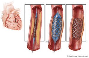
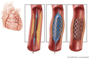
A stent is a wire mesh tube that is used to help hold open an artery. To simple understanding this concept think of a plumber or a mechanic. With a plumber sometimes they have to replace a certain area of a pipe that connects the water or like a mechanic replacing a certain area of piping (like the muffler piping connected infront of the muffler than can either can be replaced or just welded with piece of piping welded just to save money. Well a stent opens the artery that was clogged and its put in that place to reinforce that area of the artery to remain open to allow blood to get to that heart from that coronary artery and it will stay there life to keep the artery permanently patent to prevent the blockage from happening again with the synthetic mesh piece.

Description

Stents are used to hold open diseased coronary arteries (these arteries supply blood to the heart), as well as diseased arteries of the peripheral vascular system (PVS). Peripheral means away from the heart the PVS is the arteries that supply blood to the rest of the body (again away from the heart all the way down to the hands and feet).

There are variety of stents currently available.

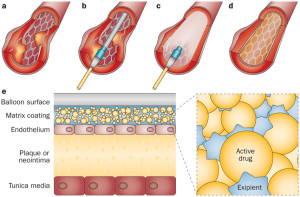
For a surgeon to find out if you even need one, first usually a angiogram is performed and this is a catheter simply from the femerol artery or from your arm to the coronary arteries. If the MD sees you show a blockage 80% or more an angioplasty is performed which is a balloon at the end of this catheter that blows up and decompresses to give the effect like punching gloves. The balloon inflates and deflates over and over again till the blockage breaks open free and then a stent is put in that area to help keep it open permanently (patent).

Some stents have been compressed onto the outside of an angioplasty balloon catheter and delivered by inflating the balloon in the desired location. Other stents are “self- expanding” spring-loaded devices, which expand automatically upon deployment.

Stents remain in arteries permanently. The tissue lining the arteries actually grows over the metal mesh to cover the inner lumen of the stent.

Stent procedures have become very common like tonsillectomies were in childhood. Stents are sometimes used as an alternative to coronary artery bypass surgery, if the patient is a candidate. Stents are often used in combination with balloon angioplasty. One leads to the other depending on what the angioplasty displays for the surgeon on the T.V. in when they are doing the procedure and if the come up to a blockage high enough to perform the angioplasty followed with a stent it will be done.

Stents are used in cases of “restenosis”, which refers to the re-closing of arteries after balloon angioplasty. In carefully selected patients, the use of stents can dramatically reduce restenosis following balloon angioplasty or other catheter-based procedures. Stents are used frequently to hold open the arteries that have been damaged, torn, or dissected by balloon angioplasty or other catheter-based procedures. Like plumber or mechanic, get it.

Stents allow angioplasty to be done in patients with severe and long-segment obstruction of coronary arteries. As soon as the I initial part of the block is widened, a stent is place, which holds it open allowing further opening to proceed. Stents have also allowed angioplasty to be performed in patients with blocks of multiple vessels, and in multiple blocks in a single artery.

Angioplasty and stent placement are common procedures to open arteries in the heart that are clogged. These procedures are formally known as coronary angioplasty or percutaneous coronary intervention.

Angioplasty involves the use of a tiny balloon to widen the artery. A stent is a tiny wire-mesh tube that your doctor inserts into the artery. The stent stays in place to prevent the artery from closing. A cardiologist typically performs both procedures at the same time.

While angioplasty with stent placement addresses an individual blockage, it doesn’t fix the underlying cause of the blockage. To prevent further blockages and reduce your risk of other medical conditions, you may have to make certain lifestyle changes to prevent it happening again.  The procedure of the angioplasty to insert a stent if needed resolves the immediate acute problem that the underlining disease the pt had causing the blockage.  This could be due to high cholesterol, diabetes, CAD,  and atheroscerosis.


QUOTE FOR MONDAY:

“Heart disease is the leading cause of death for men in the United States, killing 382,776 men in 2020—that’s about 1 in every 4 male deaths.  Heart disease is the leading cause of death for men of most racial and ethnic groups in the United States, including African Americans, American Indians or Alaska Natives, Hispanics, and whites. For Asian American or Pacific Islander men, heart disease is second only to cancer.”

Centers for Disease Control and Prevention (CDC)

QUOTE FOR THE WEEKEND:

“Fact: Cardiovascular disease is the No. 1 killer of women, causing 1 in 3 deaths each year. It’s a third of our mothers, sisters, friends, neighbors, coworkers and more. It’s a third of the women we can’t bear to live with it.

Fact: Cardiovascular disease impacts some women at higher rates than others, but the simple truth is that most cardiovascular diseases can still be prevented with education and healthy lifestyle changes.

Fact: Heart disease and stroke can affect a woman at any age, making it vital for all women to understand their personal risk factors and family history. Women can also experience unique life events that can impact their risk, including pregnancy and menopause. Furthermore, research shows that stress may impact health, making it important for women to understand the mind-body connection and how to focus on improving both their physical health and mental well-being.”

American Heart Association – Go Red for women (https://www.goredforwomen.org/en/about-heart-disease-in-women/facts)

How women differ from men in heart disease and why!

          Women and Heart DIsease+

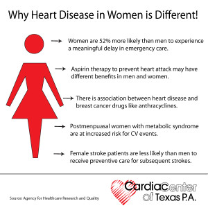
Many many women and their doctors don’t know that heart disease is the number one killer of women. Furthermore, the heart disease that is seen in women is often not quite the same as heart disease in men.

Let’s remember from Part I that Heart disease is an umbrella term that includes heart failure, coronary artery disease (CAD), arrhythmias, angina, and other heart-related infections, irregularities, and birth defects

These facts lead to two common (and sometimes tragic) misapprehensions held by many women and their doctors: That women don’t really get much heart disease, and when they do, it behaves pretty much like the heart disease that men get.

The truth is that not only is heart disease very common in women, but also, when women get heart disease it often acts quite differently than it does in men. Failing to understand these two fundamental truths leads to a lot of preventable deaths and disability in women with heart disease.

If you are a woman, you need to know the basics about heart disease – especially heart disease as it behaves in women.

When women have angina, they are more likely than men to experience “atypical” symptoms. Instead of chest pain, they are more likely to experience a hot or burning sensation, or even tenderness to touch, which may be located in the back, shoulders, arms or jaw – and often women have no chest discomfort at all. An alert doctor will think of angina whenever a patient describes any sort of fleeting, exertion-related discomfort located anywhere above the waist, and they really shouldn’t be thrown off by such “atypical” descriptions of symptoms. However, unless doctors are thinking specifically of the possibility of CAD, they are all too likely to write such symptoms off to mere musculoskeletal pain or gastrointestinal disturbances.

Women are more likely than men to have heart attack symptoms unrelated to chest pain, such as:

      • Neck, jaw, shoulder, upper back or abdominal discomfort.
      • Shortness of breath.
      • Right arm pain.
      • Nausea or vomiting.
      • Sweating.
      • Lightheadedness or dizziness.
      • Unusual fatigue.

Heart attacks (or myocardial infarctions)  also tend to behave differently in women.

Frequently, instead of the crushing chest pain that is considered typical for a heart attack, women may experience nausea, vomiting, indigestion, shortness of breath or extreme fatigue – but no chest pain. Unfortunately, these symptoms are also easy to attribute to something other than the heart. Furthermore, women (especially women with diabetes) are more likely than men to have “silent” heart attacks – that is, heart attacks without any acute symptoms at all, and which are diagnosed only at a later time, when subsequent cardiac symptoms occur.

The Diagnosis Of CAD in Women Can Be More Difficult.

Diagnostic tests that work quite well in men can be misleading in women. The most common problem is seen with stress testing – in women, the electrocardiogram (ECG) during exercise can often show changes suggesting CAD, whether CAD is present or not, making the study difficult to interpret. Many cardiologists routinely add an echocardiogram or a thallium study when doing a stress test in a woman, which greatly improves diagnostic accuracy.

In women with typical CAD, coronary angiography is every bit as useful as in men; it identifies the exact location of any plaques (i.e., blockages) within the coronary arteries, and guides therapeutic decisions. However, in women with atypical coronary artery disorders (to be discussed in the next section), coronary angiograms often appear misleadingly normal. Thus, in women angiography is often not the gold standard for diagnosis, as it is for most men.

CAD In Women Can Take Atypical Forms.

At least four atypical coronary artery disorders can occur in women, usually in younger (i.e., pre-menopausal) women. Each of these conditions can produce symptoms of angina with apparently “normal” coronary arteries (that is, coronary arteries that often appear normal on angiogram). The problem, obviously, is that if the physician trusts the results of the angiogram, he/she is likely to miss the real diagnosis.

DALLAS, February 19, 2013 — A new study show women’s heart disease awareness is increasing.  A study with the number of women aware that heart disease is the leading cause of death nearly is doubling in the last 15 years, but that this knowledge still lags in minorities and younger women, according to the American Heart Association (AHA).

Among the study’s major findings, researchers comparing women’s views about heart disease in 1997 and today found:

  • In 2012, 56 percent of women identified heart disease as the leading cause of death compared with 30 percent in 1997.
  • In 1997, women were more likely to cite cancer than heart disease as the leading killer (35 percent versus 30 percent); but in 2012, only 24 percent cited cancer.
  • In 2012, 36 percent of black women and 34 percent of Hispanic women identified heart disease as the top killer — awareness levels that white women had in 1997 (33 percent).
  • Women 25-34 years old had the lowest awareness rate of any age group at 44 percent.

Among the women surveyed in 2012, researchers found:

  • Racial and ethnic minorities reported higher levels of trust in their healthcare providers compared with whites, and were also more likely to act on the information provided—dispelling the myth that mistrust of providers contributes to disparities.
  • Compared with older women, younger women were more likely to report not discussing heart disease risk with their doctors (6 percent among those 25-34 versus 33 percent for those 65 and older).

Risk Factors for Heart Disease in Women – Those we can’t change = Nonmodifiable Factors:

Age and Family History, Gender, Ethnicity.

The risk of having heart disease increases with age and this is due to stiffening of heart muscles which makes the heart less efficient in pumping blood around the body. You can determine your heart age by using this tool, developed by the British Heart Foundation: https://www.bhf.org.uk/heart-health/risk-factors/check-your-heart-age.

Another risk factor you cannot change is if you have a history of heart disease among family members. This can double your risk, so if your mother, father, sister or brother has suffered from heart disease before the age of 60 you are at a greater risk of developing heart disease.

Modifiable Risk Factors – Those we can change are:

1-Smoking is the single largest preventable cause of death in Australia, and approximately 40% of women who smoke die due to heart disease, stroke or blood vessel disease. Smokers are 2-4 times more at risk of developing heart disease compared to non-smokers. In 2011/2012, over 1.3 million women in Australia smoked, and 89% of them did this on a daily basis. While these numbers are for women aged 15 and over, the largest group were in the 25-34 age group.

Passive smoking (exposure to the cigarette smoke of others) also causes an increase in the risk of developing heart disease, which increases further in people having high blood pressure or high cholesterol. Women who smoke and also take the contraceptive pill have a 10 times higher risk of having a heart attack.

2-Alcohol. Do you know that drinking too much alcohol increases the risk of heart disease? Excessive drinking causes more weight gain (due to increased calories!), increase in blood pressure and blood lipids. Over a long period of time it can weaken the heart muscle and cause abnormal heart rhythms. Try and not drink alcohol every day, limit it to two standard drinks at a time and aim for at least two alcohol free days a week and make sure you don’t increase the amount you drink on the other days. Periodically take a break from any alcohol for a week or more and you will notice many benefits including a better nights sleep.

3.High Blood Pressure or Hypertension. Your blood pressure is a measurement of how ‘hard’ your heart is working to push blood around your body, through the blood vessels. It can be a ‘silent’ killer and if you do not know your blood pressure then it is worth having it checked by your GP. Changing your lifestyle will reduce your blood pressure. A recent study suggests that keeping your blood pressure under 140/90 can increase your life expectancy by 5 years at the age of 50 years. You can assess your high blood pressure through your MD monthly or less expensive buy a b/p machine and check your b/p everyday especially if your on antihypertensive meds to make sure your b/p isn’t under 100/60 to prevent hypotension.

4.Diabetes. Do you have diabetes and if so, is it under control?

Diabetes doubles your risk of having heart disease. People who have uncontrolled diabetes are at risk of having heart disease at an earlier age. For pre-menopausal women, having diabetes cancels the protective effects of hormone present in women and significantly increases the risk of heart disease. Taking steps to find out what your blood sugar is and keeping it well-controlled is essential.

5.Obesity- Do you know your body fat content?  If you think that you are overweight then you put yourself at risk of having heart disease. Being overweight will increase your blood pressure and contribute to developing diabetes. In addition to that, women who carry weight around their middle (belly fat) as opposed to their hips are twice as likely to develop heart disease.

By taking the steps to reduce your weight, you can reduce your risk of heart disease. A great tool developed by National Heart Foundation of Australia calculates if you might be at risk: http://www.heartfoundation.org.au/healthy-eating/Pages/bmi-calculator.aspx

6- INACTIVE-Are you physically active every day? Recent research indicates that “sitting is the new smoking” and being physically inactive can double your risk of having heart disease. It is important to get some exercise every day, such as a 30 minute walk where you raise your heart rate. It also raises your serotonin levels (feel-good hormone) and can reduce depression

7- STRESS-We could almost ask – do you know anyone who is not stressed?! However, while everyday life is stressful, those people who are almost constantly stressed are at risk of adopting unhealthy behaviours in order to reduce their stress levels. Examples include increasing their alcohol intake or smoking in order to relax; or tending to eat more junk food because they are often short of time. All of these factors increase their risk of heart disease.

Women, stress and the risk of heart disease

Along with poor diet, lack of exercise and smoking, unmanaged stress may increase the risk for heart disease. Now medical experts are discovering that mental stress affects women in different, and in some cases, more devastating ways, especially if they already have coronary conditions. One study that

Heart disease is the leading cause of death for men and women in the United States. Every year, 1 in 4 deaths are caused by heart disease. The good news? Heart disease can often be prevented when people make healthy choices and manage their health conditions. Communities, health professionals, and families can work together to create opportunities for people to make healthier choices. Make a difference in your community: Spread the word about strategies for preventing heart disease and encourage people to live heart healthy lives