Archives

QUOTE FOR MONDAY:

“National Kidney Foundation Recognizes March as National Kidney Month and World Kidney Day as it Celebrates a Key Milestone

New York City, NY – (March 10, 2025) — The National Kidney Foundation (NKF) is recognizing a special milestone this March during National Kidney Month. NKF is celebrating 75 years of transforming lives through kidney health advocacy, research, and innovation. As part of the celebration, NKF is launching “75+You” this Thursday, as part of World Kidney Day on March 13th, to rally communities nationwide in the fight against kidney disease and highlight how, together, we can make an impact on millions of lives.

Did you know that one in ten people will have a kidney stone over the course of a lifetime? Recent studies have shown that kidney stone rates are on the rise across the country. Those in the know believe that some major misconceptions may be the culprit.”

National Kidney Foundation (75 Years of Transforming Kidney Health | National Kidney Foundation & Six Easy Ways to Prevent Kidney Stones | National Kidney Foundation)


Part I March is Kidney Month – The importance of kidneys and know its functions to understand kidney failure.

kidney 3

    kidney failure

The kidneys are important organs with many functions in the body, including producing hormones, absorbing minerals, and filtering blood and producing urine. While they are important and kidney failure can be fatal, a human only needs one healthy kidney to survive.

The kidneys are two bean-shaped organs that extract waste from blood, balance body fluids, form urine, and aid in other important functions of the body.

They reside against the back muscles in the upper abdominal cavity. They sit opposite each other on either side of the spine. The right kidney sits a little bit lower than the left to accommodate the liver.

When it comes to components of the urinary system, the kidneys are multi-functional powerhouses of activity, for if the kidneys aren’t working, meaning they don’t filter toxic wastes out of our blood stream (with other functions it does) than the waste products don’t get dumped into the urinary bladder from the renal tubes, called right and left ureters. In human anatomy, the ureters are tubes made of smooth muscle fibers that propel urine from the kidneys to the urinary bladder. If the kidneys are not working they are not filtering our blood (same principle as filtering beer to make it to perfection, the kidneys do it for our blood to be able to have the cells do their function to the optimal levels with keeping toxins out of the body in preventing many blood problems with more due to acidosis (toxin build up). In the adult, the ureters are usually 25–30 cm (10–12 in) long and ~3–4 mm in diameter.

The kidneys have multiple functions! Some of the core actions of a healthy kidney or kidneys of a human body include:

  • Waste excretion: There are many things your body doesn’t want inside of it. The kidneys filter out toxins, excess salts, and urea (a toxin), a nitrogen-based waste created by cell metabolism.
  • * Urea is an organic chemical compound and is essentially the waste produced by the body after metabolizing protein. Naturally the compound urea is produced when the liver breaks down protein or amino acids, and ammonia, the kidneys then transfer the urea from the blood to the urine, when they do filtering of the blood.  Urea is a byproduct of protein metabolism, the ending result. Extra nitrogen is expelled from the body through urea because it is extremely soluble (solid); it is a very efficient process. The average person excretes about 30 grams of urea a day, mostly through urine but a small amount is also secreted in perspiration. Synthetic versions of the chemical compound can be created in liquid or solid form and is often an ingredient found in fertilizers, animal food, and diuretics, just to name a few . Urea is what gives our urine the color yellow.         In the gastrointestinal tract, blood proteins are broken down into ammonia (could be due to high protein eating to drugs with actual conditions); and goes to the liver converting it to Urea. It is then released into the blood stream where the kidney’s take it up and eliminate it. Urea is then eliminated by the kidney’s, but not produced by it.  Urea is synthesized in the liver and transported through the blood to the kidneys for removal.
  •  A Healthy Kidney or Kidneys functions in the human body doing:
  • Water level balancing: As the kidneys are key in the chemical breakdown of urine, they react to changes in the body’s water level throughout the day. As water intake decreases, the kidneys adjust accordingly and leave water in the body instead of helping excrete it which aides in electrolyte balancing in the blood with keeping the body hydrated properly.
  • Blood pressure regulation: The kidneys need constant pressure to filter the blood. When it drops too low, the kidneys increase the pressure. One way is by producing a blood vessel-constricting protein (angiotensin) that also signals the body to retain sodium and water. Both the constriction and retention help restore normal blood pressure.
  • Red blood cell regulation: When the kidneys don’t get enough oxygen, they send out a distress call in the form of erythropoietin, a hormone that stimulates the bone marrow to produce more oxygen-carrying red blood cells.
  • Acid regulation: As cells metabolize, they produce acids. Foods we eat can either increase the acid in our body or neutralize it. If the body is to function properly, it needs to keep a healthy balance of these chemicals. The kidneys do that, too.Because of all of the vital functions the kidneys perform and the toxins they encounter, the kidneys are susceptible to various problems.
  • Acute kidney failure is a condition in which the kidneys suddenly lose their ability to function properly. This can occur for many reasons, including:

  • Infection
  • Blood-clotting disorders
  • Decreased blood flow caused by low blood pressure
  • Autoimmune kidney disorders
  • Urinary tract infections
  • Complications from pregnancy
  • Most people are born with two kidneys, but many people can live on just one. Kidney transplant surgeries with live donors are common medical procedures today. *
  • Chronic kidney failure – same as acute in that the kidney (s) loses its function. 
  • DehydrationDiseases and conditions that commonly cause chronic kidney disease include:
  • Type 1 or type 2 diabetes.
  • High blood pressure
  • Glomerulonephritis (gloe-mer-u-lo-nuh-FRY-tis), an inflammation of the kidney’s filtering units (glomeruli)
  • Interstitial nephritis, an inflammation of the kidney’s tubules and surrounding structures
  • Polycystic kidney disease
  • Prolonged obstruction of the urinary tract, from conditions such as enlarged prostate, kidney stones and some cancers
  • Vesicoureteral (ves-ih-koe-yoo-REE-ter-ul) reflux, a condition that causes urine to back up into your kidneys
  • Recurrent kidney infection, also called pyelonephritis (pie-uh-lo-nuh-FRY-tis)
  • A chronic condition caused the failure to happen called a secondary diagnosis.
  • Learn more about Acute  Chronic kidney failure this week coming up. 

QUOTE FOR THE WEEKEND:

“National Sleep Awareness Week is March 9 – 15 –You spend about one-third of your life sleeping, but it’s still something many struggle with. Researchers and experts also struggle with it because of the mysteries surrounding how and why we sleep and what happens to us while we do. Fortunately, advances in medical science are helping people find ways to get the sleep they need.

What is sleep?

Sleep is a normal body process that allows your body and brain to rest. At first glance, sleep is deceptively simple. For most people, it’s just a matter of getting comfortable, closing your eyes and drifting into slumber. But despite how simple it seems, sleep is one of the most complex and mysterious body processes known to science.”

Cleveland Clinic (Sleep: What It Is, Why It’s Important, Stages, REM & NREM)

World Sleep Week Awareness!

Tips to Making Better Sleep

May is Better Sleep Month! Take advantage of this time to create better sleep habits that lead to more restful sleep for a lifetime. We’ve created a list of tips on how to encourage better, healthier sleep at night, naturally. Challenge yourself to put them in practice for the rest of Better Sleep Month—and, hopefully, thereafter.

1.)  Create a Sleep Schedule

Try going to bed at the same time every night and waking up at the same time every morning, even on the weekends. Sticking to a sleep schedule will train your body to recognize your set bedtime and allow you to adapt to a proper waking and sleeping cycle. If you frequently change the time you wake up or go to bed, your body won’t be able to adjust to a regular sleep schedule. Make an effort to schedule times that leave room for about eight hours of sleep each night. To help you stay on track with your new May sleep schedule, mark your calendar with the times you wake up and go to bed each day. Using your sleep calendar to log which days you aren’t falling asleep at the proper time might help you identify the reasons why—for example, drinking too much coffee late in the afternoon or stress from a big day at work. Writing out your sleep schedule and any troubles will help you visualize the steps you can take to improve your sleeping patterns.

2.)  Stick to a Rigid Bedtime Routine

Daily activities impact your circadian rhythm and allow your body to anticipate upcoming events. Our bodies crave consistency, so developing a rigid bedtime routine will help signal to your body when it’s time to rest. Create a bedtime prep checklist to ensure you are preparing your body and brain for sleep the same way nightly. Good nighttime rituals include reading, meditating, brushing your teeth, closing the blinds, and getting under the covers. Your bedtime routine is personal to you, so take some time this month to think about the steps you’ll take to prepare yourself for quality sleep most comfortably.

3.)  Turn Off Electronics

Light cues from your environment can be detrimental to your sleep patterns and circadian rhythm. The light from electronics works as a stimulant in your brain, sending a confusing signal to wake up when it may actually be time for bed. Challenge yourself to keep electronics out of your bedroom this month, and record the nights you are successful on your sleep calendar for motivation. Put your phone and laptop in another room and click off your television before beginning your bedtime rituals. Try using a traditional alarm clock instead of your phone to avoid the temptation to check your texts or emails before bed. Your body and mind will thank you for the opportunity to relax and wind down in a dark space so you can easily fall and stay asleep.

Stop Hitting Snooze

Make May the month you stop hitting the snooze button in the morning. Although getting an extra five minutes of sleep sounds pleasant, it’s actually more harmful than beneficial for your body. Snoozing your alarm is detrimental to your natural sleeping patterns and damages the consistency your body has been working so hard to achieve. It can also make you feel more tired or groggy throughout the day. If you are an avid snoozer, try putting your alarm clock on the other side of the room this month to force yourself to get out of bed to turn it off.

Make Sleep a Priority in your life

Hold yourself accountable to making better sleep a priority today and thereafter. While it’s important to do this always, concentrating on healthy sleep habits for just one month could set you up for a lifetime of quality sleep. If you succeed at doing these sleeping changes, you might just have restful nights and productive days and reach the goal you have wanted for so long, good sleep.

QUOTE FOR FRIDAY:

“Key points

  • A TBI affects how the brain works.
  • TBI is a major cause of death and disability.
  • TBIs may be missed in older adults.
  • A TBI may lead to short- or long-term health problems.
  • A TBI during childhood may affect brain development.

There were over 69,000 TBI-related deaths in the United States in 2021.1 That’s about 190 TBI-related deaths every day.

TBIs affect the lives of people of all ages. Anyone can experience a TBI, but data suggest that some groups are at greater risk of dying from a TBI or experiencing long-term health problems after the injury .

Center for Disease Control and Prevention – CDC (Facts About TBI | Traumatic Brain Injury & Concussion )


Part III National Brain Injury Awareness Month!

What are the causes?

Common causes include falls, car or motorcycle crashes, vehicular accidents involving pedestrians, athletics, and assaults with or without a weapon.

Who is affected?

Approximately 1.5 to 2 million adults and children suffer a traumatic brain injury (TBI) each year in the United States. Most people who experience a head injury, about 1.1 million, will have a mild injury that does not require an admission to the hospital. Another 235,000 individuals will be hospitalized with a moderate to severe head injury, and approximately 50,000 will die.

How is a diagnosis made?

When a person is brought to the emergency room with a head injury, doctors will learn as much as possible about his or her symptoms and how the injury occurred. The person’s condition is assessed quickly to determine the extent of injury.

The Glasgow Coma Score (GCS) is a 15-point test used to grade a patient’s level of consciousness. Doctors assess the patient’s ability to 1) open his or her eyes, 2) ability to respond appropriately to orientation questions, (“What is your name? What is the date today?”), and 3) ability to follow commands (“Hold up two fingers, or give a thumbs up”). If unconscious or unable to follow commands, his or her response to painful stimulation is checked. A number is taken from each category and added together to get the total GCS score. The score ranges from 3 to 15 and helps doctors classify an injury as mild, moderate, or severe. Mild TBI has a score of 13-15. Moderate TBI has a score of 9-12, and severe TBI has a score of 8 and below.

Diagnostic imaging tests will be performed:

CT of TBI

Figure 3. CT scan shows a blood clot (hematoma) collecting under the bone (red arrows) and displacing brain (yellow arrow) to the other side of the skull.

  • Computed Tomography (CT) is a noninvasive X-ray that provides detailed images of anatomical structures within the brain. A CT scan of the head is taken at the time of injury to quickly identify fractures, bleeding in the brain, blood clots (hematomas) and the extent of injury (Fig. 3). CT scans are used throughout recovery to evaluate the evolution of the injury and to help guide decision-making about the patient’s care.
  • Magnetic Resonance Imaging (MRI) is a noninvasive test that uses a magnetic field and radiofrequency waves to give a detailed view of the soft tissues of the brain. A dye (contrast agent) may be injected into the patient’s bloodstream. MRI can detect subtle changes in the brain that cannot be seen on a CT scan.
  • Magnetic Resonance Spectroscopy (MRS) gives information about the metabolism of the brain. The numbers generated from this scan provide a general prognosis about the patient’s ability to recover from the injury.

What treatments are available?

Mild TBI usually requires rest and medication to relieve headache. Moderate to severe TBI require intensive care in a hospital. Bleeding and swelling in the brain can become an emergency that requires surgery. However, there are times when a patient does not require surgery and can be safely monitored by nurses and physicians in the neuroscience intensive care unit (NSICU).

The goals of treatment are to resuscitate and support the critically ill patient, minimize secondary brain injury and complications, and facilitate the patient’s transition to a recovery environment. Despite significant research, doctors only have measures to control brain swelling, but do not have a way to eliminate swelling from occurring.

Neurocritical care
Neurocritical care is the intensive care of patients who have suffered a life-threatening brain injury. Many patients with severe TBI are comatose or paralyzed; they also may have suffered injuries in other parts of the body. Their care is overseen by a neurointensivist, a specialty-trained physician who coordinates the patient’s complex neurological and medical care. Patients are monitored and awakened every hour for nursing assessments of their mental status or brain function.

Click to view larger image with labels

Figure 4. In the NSICU, the patient is connected to numerous machines, tubes, and monitors. The monitoring equipment provides information about body functions and helps guide care. Some equipment may take over certain functions, such as breathing, nutrition, and urination, until the patient’s body is able to do these things on its own.

Seeing a patient who has suffered a severe TBI can be shocking. It is possible that your loved one’s appearance will be altered because of facial injury and equipment that is used for monitoring. Numerous tubes, lines, and equipment may be used to closely monitor his or her heart rate, blood pressure, and other critical body functions. (Fig. 4)

ICP monitor

Figure 5. A brain oxygen and cerebral blood flow monitor is inserted into the brain tissue and secured to the skull with a bolt. A catheter is inserted into the ventricle of the brain to monitor intracranial pressure (ICP). If pressure is too high, the CSF fluid can be drained from the ventricles.

  • Intracranial pressure (ICP) monitor. A catheter is placed through a small hole in the skull and positioned inside the ventricle (fluid-filled area deep within the brain) to measure pressure inside the head (Fig. 5). The ICP monitor allows the NSICU team to intervene quickly if the pressure becomes too high. Typical intracranial pressure is less than 20 mmHg. However, there are times when a higher number is safe and acceptable.
  • Brain oxygen monitor (Licox). A catheter is placed through a small hole in the skull and positioned within the brain tissue. The Licox measures the oxygen level and temperature within the brain. Adjustments in the amount of oxygen given to the patient are often made to maximize the brain’s oxygen level. A cerebral blood flow monitor, called a Hemedex, is a newer monitor that is placed with the Licox and helps the NSICU team evaluate blood flow through the brain.
  • Ventilator. Some patients may require a ventilator, a machine that helps them breathe. The ventilator is connected to the patient by the endotracheal tube, or ET tube. The tube is placed into the patient’s mouth and down into the trachea, or windpipe. The tube allows the machine to push air into and out of the lungs, thereby helping the patient breathe.
  • Feeding tube. When patients are on a ventilator or have a decreased level of alertness, they may not be able to eat or get sufficient nutrition to meet their needs. A nasal-gastric feeding tube may be inserted through the patient’s nose and passed down the throat into the stomach. It delivers liquid nutrition as well as any medication that is required.
  • Seizures and EEG monitoring. A seizure is an abnormal electrical discharge from the brain. Approximately 24% of patients who suffer a TBI will have a seizure that is undetected unless they are monitored by an electroencephalogram (EEG). Seizures that are not visible to the human eye are referred to as non-convulsive seizures. Because these seizures are serious, all patients with a severe TBI are monitored with continuous EEG for 24 to 72 hours after injury.

Medication

  • Sedation and pain. After a head injury it may be necessary to keep the patient sedated with medications. These medications can be turned off quickly in order to awaken the patient and check their mental status. Because patients often have other injuries, pain medication is given to keep them comfortable.
  • Controlling intracranial pressure. Hypertonic saline is a medication used to control pressure within the brain. It works by drawing the extra water out of the brain cells into the blood vessels and allowing the kidneys to filter it out of the blood.
  • Preventing seizures. Patients who’ve had a moderate to severe traumatic brain injury are at higher risk of having seizures during the first week after their injury. Patients are given an anti-seizure medication (levetiracetam or phenytoin) to prevent seizures from occurring.
  • Preventing infection. Although every attempt is made to prevent infection, the risk is always present. Any device placed within the patient has the potential to introduce a microbe. If an infection is suspected, a test will be sent to a laboratory for analysis. If an infection is present, it will be treated with antibiotics.

Surgery

Surgery is sometimes necessary to repair skull fractures, repair bleeding vessels, or remove large blood clots (hematomas). It is also performed to relieve extremely high intracranial pressure.

  • Craniotomy involves cutting a hole in the skull to remove a bone flap so that the surgeon can access the brain. The surgeon then repairs the damage (e.g., skull fracture, bleeding vessel, remove large blood clots). The bone flap is replaced in its normal position and secured to the skull with plates and screws.

Figure 6. A large decompressive craniectomy is removed and the dura is opened to allow the brain to expand. Blood clots are removed and bleeding vessels are repaired. The bone flap is frozen and replaced about 6 weeks later.

  • Decompressive craniectomy involves removing a large section of bone so that the brain can swell and expand. This is typically performed when extremely high intracranial pressure becomes life threatening. At that time the patient is taken to the operating room where a large portion of the skull is removed to give the brain more room to swell (Fig. 6). A special biologic tissue is placed on top of the exposed brain and the skin is closed. The bone flap is stored in a freezer. One to 3 months after the swelling has resolved and the patient has stabilized from the injury, the bone flap is replaced in another surgery, called cranioplasty.

Other surgical procedures may be performed to aid in the patient’s recovery:

  • Tracheotomy involves making a small incision in the neck to insert the breathing tube directly into the windpipe. The ventilator will then be connected to this new location on the neck and the old tube is removed from the mouth.
  • Percutaneous Endoscopic Gastrostomy Tube (PEG) is a feeding tube inserted directly into the stomach through the abdominal wall. A small camera is placed down the patient’s throat into the stomach to aid with the procedure and to ensure correct placement of the PEG tube (see Surgical Procedures for Accelerated Recovery).

Clinical trials

Clinical trials are research studies in which new treatments—drugs, diagnostics, procedures, and other therapies—are tested in people to see if they are safe and effective. Research is always being conducted to improve the standard of medical care. Information about current clinical trials, including eligibility, protocol, and locations, are found on the Web. Studies can be sponsored by the National Institutes of Health (see clinicaltrials.gov) as well as private industry and pharmaceutical companies (see www.centerwatch.com).

Recovery & prevention

The recovery process varies depending on the severity of the injury, but typically progresses through stages: coma, confusion / amnesia, and recovery.

  • When a patient is in a coma, his or her eyes are closed and they show minimal reaction when spoken to or stimulated. Movements that may be seen at this time are basic reflexes or automatic responses to a stimulus. The brain wave activity in a comatose person is very different from that of a sleeping person.
  • When a patient begins to awaken, the first natural response is that of bodily protection. Patients at this stage will move away from any stimulus or tend to pull at items attached to them in an attempt to remove anything that is uncomfortable or irritating. His or her eyes may be open more often, but they may not be aware of their behavior or be able to interact in a meaningful way. It is common for a patient to respond to each stimulus (hearing, seeing, or touching) in the same way. Responses may include increased rate of breathing, moaning, moving, sweating, or a rise in blood pressure.
  • As the patient continues to wake up, their interactions may become more purposeful. They may look at a person and follow them around the room with their eyes, or follow simple commands such as “Hold up your thumb.” Patients tend to be confused and may have inappropriate or agitated behaviors.

Not all head injuries are the same. Patients recover at different rates and to varying degrees. It is difficult to determine at what point a patient will start understanding and interacting with their caregivers or family in a meaningful way. It is important to have patience; recovery from a brain injury can take weeks, months, or even years.

The Family’s Role
Many family members express feelings of helplessness when their loved one is in the NSICU. You are not alone. Please take care of yourself and use your energy wisely.

Visiting hours are limited in the NSICU. Too much stimulation can agitate the patient and raise his or her blood pressure. You can most effectively convey your concern by sitting quietly and holding your loved one’s hand. Be aware that the patient, though silent, may hear anything you say. Never speak as if the patient were not there.

As patients recover, they need help understanding what has happened to them during this “lost period of time.” Keep in mind that the recovery of consciousness is a gradual process – not just a matter of waking up. Progress is usually tracked in three areas: movement, thinking, and interacting. You can help by keeping a diary of their progress. Family photos may help with regaining memory.

Rehabilitation
Most patients are discharged from the hospital when their condition has stabilized and they no longer require intensive care. A social worker will work closely with the family as preparations are made for a return home or for transfer to a long-term care or rehabilitation center.

  • A long-term acute care (LTAC) facility is a place for patients who have stabilized from their initial injury but who still require a ventilator or frequent nursing care. Many patients are discharged to an LTAC to continue being weaned from the ventilator. Once off the ventilator, they can be moved to a rehabilitation or skilled nursing facility.
  • A rehabilitation facility is a place for patients who do not require a ventilator but who still require help with basic daily activities. Physical and occupational therapists work with patients to help them achieve their maximum potential for recovery. Rehab facilities are either Acute Inpatient Rehab that require patients to participate in 3 hours or more of rehab a day or a Skilled Nursing Facility (SNF) that provide 1-3 hours of rehab a day depending on what the patient can tolerate.

Recovering from a brain injury relies on the brain’s plasticity—the ability for undamaged areas of the brain to take over functions of the damaged areas. It also relies on regeneration and repair of nerve cells. And most importantly, on the patient’s hard work to relearn and compensate for lost abilities.

  • A physical therapist helps patients rebuild and maintain strength, balance, and coordination. They can work with the patient in any facility.
  • An occupational therapist helps patients to perform activities of daily living, such as dressing, feeding, bathing, toileting, and transferring themselves from one place to another. They also provide adaptive equipment if a patient has difficultly performing a task.
  • A speech therapist helps patients by monitoring their ability to safely swallow food and helping with communication and cognition.
  • A neuropsychologist helps patients relearn cognitive functions and develop compensation skills to cope with memory, thinking, and emotional needs.

Prevention

Tips to reduce the risk for a head injury:

  • Always wear your helmet when riding a bicycle, motorcycle, skateboard, or all-terrain vehicle.
  • Never drive under the influence of alcohol or drugs.
  • Always wear your seat belt and ensure that children are secured in the appropriate child safety seats.
  • Avoid falls in the home by keeping unsecured items off the floor, installing safety features such as non-slip mats in the bathtub, handrails on stairways, and keeping items off of stairs.
  • Avoid falls by exercising to increase strength, balance, and coordination.
  • Store firearms in a locked cabinet with bullets in a separate location.
  • Wear protective headgear while playing sports.

Reference for Part I and Part II: Mayfield Brain & Spine at 513-221-1100.

QUOTE FOR THURSDAY:

“There are two broad types of brain injuries penetrating and non-penetrating brain injuries:

  1. Penetrating TBI (also known as open TBI) happens when an object pierces the skull (e.g., a bullet, shrapnel, bone fragment, etc.) and enters the brain tissue. Penetrating TBI typically damages only part of the brain.
  2. Non-penetrating TBI (also known as closed head injury or blunt TBI) is caused by an external force strong enough to move the brain within the skull. Causes include falls, motor vehicle crashes, sports injuries, blast injury, or being struck by an object.”

National Institute of Neurological Disorders and Stroke (Traumatic Brain Injury (TBI) | National Institute of Neurological Disorders and Stroke)

Part II National Brain Injury Awareness Month!

Overview

Traumatic brain injury (TBI) is sudden damage to the brain caused by a blow or jolt to the head. Common causes include car or motorcycle crashes, falls, sports injuries, and assaults. Injuries can range from mild concussions to severe permanent brain damage. While treatment for mild TBI may include rest and medication, severe TBI may require intensive care and life-saving surgery. Those who survive a brain injury can face lasting effects in their physical and mental abilities as well as emotions and personality. Most people who suffer moderate to severe TBI will need rehabilitation to recover and relearn skills.

What is a traumatic brain injury?

TBI is an injury to the brain caused by a blow or jolt to the head from blunt or penetrating trauma. The injury that occurs at the moment of impact is known as the primary injury. Primary injuries can involve a specific lobe of the brain or can involve the entire brain. Sometimes the skull may be fractured, but not always. During the impact of an accident, the brain crashes back and forth inside the skull causing bruising, bleeding, and tearing of nerve fibers (Fig. 1). Immediately after the accident the person may be confused, not remember what happened, have blurry vision and dizziness, or lose consciousness. At first the person may appear fine, but their condition can decline rapidly. After the initial impact occurs, the brain undergoes a delayed trauma – it swells – pushing itself against the skull and reducing the flow of oxygen-rich blood. This is called secondary injury, which is often more damaging than the primary injury.

TBI

Figure 1. During impact to the head, the soft brain crashes back and forth against the inside of the hard skull causing bruising, bleeding, and shearing of the brain.

Traumatic brain injuries are classified according to the severity and mechanism of injury:

  • Mild: person is awake; eyes open. Symptoms can include confusion, disorientation, memory loss, headache, and brief loss of consciousness.
  • Moderate: person is lethargic; eyes open to stimulation. Loss of consciousness lasting 20 minutes to 6 hours. Some brain swelling or bleeding causing sleepiness, but still arousable.
  • Severe: person is unconscious; eyes do not open, even with stimulation. Loss of consciousness lasting more than 6 hours.

Types of traumatic brain injuries

  • Concussion is a mild head injury that can cause a brief loss of consciousness and usually does not cause permanent brain injury.
  • Contusion is a bruise to a specific area of the brain caused by an impact to the head; also called coup or contrecoup injuries. In coup injuries, the brain is injured directly under the area of impact, while in contrecoup injuries it is injured on the side opposite the impact.
  • Diffuse axonal injury (DAI) is a shearing and stretching of the nerve cells at the cellular level. It occurs when the brain quickly moves back and forth inside the skull, tearing and damaging the nerve axons. Axons connect one nerve cell to another throughout the brain, like telephone wires. Widespread axonal injury disrupts the brain’s normal transmission of information and can result in substantial changes in a person’s wakefulness.
  • Traumatic Subarachnoid Hemorrhage (tSAH) is bleeding into the space that surrounds the brain. This space is normally filled with cerebrospinal fluid (CSF), which acts as a floating cushion to protect the brain. Traumatic SAH occurs when small arteries tear during the initial injury. The blood spreads over the surface of the brain causing widespread effects.
  • Hematoma is a blood clot that forms when a blood vessel ruptures. Blood that escapes the normal bloodstream starts to thicken and clot. Clotting is the body’s natural way to stop the bleeding. A hematoma may be small or it may grow large and compress the brain. Symptoms vary depending on the location of the clot. A clot that forms between the skull and the dura lining of the brain is called an epidural hematoma. A clot that forms between the brain and the dura is called a subdural hematoma. A clot that forms deep within the brain tissue itself is called an intracerebral hematoma. Over time the body reabsorbs the clot. Sometimes surgery is performed to remove large clots.

Although described as individual injuries, a person who has suffered a TBI is more likely to have a combination of injuries, each of which may have a different level of severity. This makes answering questions like “what part of the brain is hurt?” difficult, as more than one area is usually involved.

Secondary brain injury occurs as a result of the body’s inflammatory response to the primary injury. Extra fluid and nutrients accumulate in an attempt to heal the injury. In other areas of the body, this is a good and expected result that helps the body heal. However, brain inflammation can be dangerous because the rigid skull limits the space available for the extra fluid and nutrients. Brain swelling increases pressure within the head, which causes injury to parts of the brain that were not initially injured. The swelling happens gradually and can occur up to 5 days after the injury.

What are the symptoms?

Depending on the type and location of the injury, the person’s symptoms may include:

  • Loss of consciousness
  • Confusion and disorientation
  • Memory loss / amnesia
  • Fatigue
  • Headaches
  • Visual problems
  • Poor attention / concentration
  • Sleep disturbances
  • Dizziness / loss of balance
  • Irritability / emotional disturbances
  • Feelings of depression
  • Seizures
  • Vomiting

Diffuse injuries (such as a concussion or diffuse axonal injury) will typically cause an overall decreased level of consciousness. Whereas, focal injuries (such as an ICH or a contusion) will have symptoms based on the brain area affected (Fig. 2).

Lobes of the brain that can get affected by TBI:

brain anatomy

Figure 2. The brain is composed of three parts: the brainstem, cerebellum, and cerebrum, which is divided into lobes. The table lists the lobes of the brain and their normal functions as well as problems that may occur when injured. While an injury may occur in a specific area, it is important to understand that the brain functions as a whole by interrelating its component parts.

Every patient is unique and some injuries can involve more than one area or a partial section, making it difficult to predict which specific symptoms the patient will experience.

QUOTE FOR WEDNESDAY:

“Common Causes of Traumatic Brain Injury:

▪Falls
▪Motor vehicle crashes
▪Sports related injuries
▪Explosive blast/military
combat injuries

Common Causes of Non-Traumatic Brain Injury:

▪Stroke
▪Near-drowning
▪Aneurysm
▪Tumor
▪ Infectious disease that affects
the brain
▪Lack of oxygen supply to the brain”

Brain Injury Association of America (Brain Injury Awareness Month – Brain Injury Association of America)


Part I National Brain Injury Awareness Month

  BRAIN INJURY IMAGE  brain Lobe Regions


An acquired brain injury (ABI) is an injury to the brain that is not hereditary, congenital, degenerative, or induced by birth trauma. Essentially, this type of brain injury is one that has occurred after birth. The injury results in a change to the brain’s neuronal activity, which affects the physical integrity, metabolic activity, or functional ability of nerve cells in the brain.

There are two types of acquired brain injury: traumatic and non-traumatic.

A traumatic brain injury (TBI) is defined as an alteration in brain function, or other evidence of brain pathology, caused by an external force. Traumatic impact injuries can be defined as closed (or non-penetrating) or open (penetrating).

Often referred to as an acquired brain injury, a non-traumatic brain injury causes damage to the brain by internal factors, such as a lack of oxygen, exposure to toxins, pressure from a tumor, etc.

A Brain Injury is damage to the brain that results in a loss of function such as mobility or feeling.

Traumatic Brain Injuries can result from a closed head injury or a penetrating head injury.

Closed Injury:  A closed injury occurs when the head suddenly and violently hits an object but the object does not break through the skull.

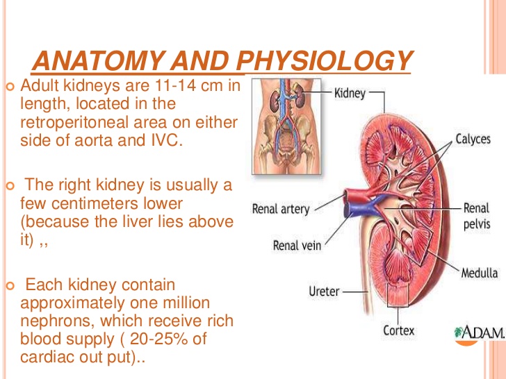
Penetrating Injury: A penetrating injury occurs when an object pierces the skull and enters brain tissue. As the first line of defense, the skull is particularly vulnerable to injury. Skull fractures occur when the bone of the skull cracks or breaks. A depressed skull fracture occurs when pieces of the broken skull press into the tissue of the brain. A penetrating skull fracture occurs when something pierces the skull, such as a bullet, leaving a distinct and localized injury to brain tissue. Skull fractures can cause cerebral contusion.

Brain trauma occurs when a person has an injury to the brain, and can be mild or severe. When a person sustains trauma to the brain, he or she may lose motor functions along with cognitive and physical abilities. Physicians use the Glasgow Coma Scale to determine the extent of brain trauma. This is a neurological scale that measures the level of a person’s consciousness. The Rancho Los Amigos Scale is used to monitor the recovery of the brain.

There are several different types of brain injuries. A mild injury may cause temporary symptoms while a severe injury could require years of rehabilitation. The two most common types of brain trauma are:

1. Traumatic Brain Injuries 

2. Acquired Brain Injuries.

1-Traumatic brain injury occurs from external force on the head or neck. These injuries can occur from blows to the head or aggressive twisting of the neck. Some ways this could happen include falls, motor vehicle accidents, sports, and vigorous shaking. In infants, Shaken Baby Syndrome is a type of traumatic brain injury.

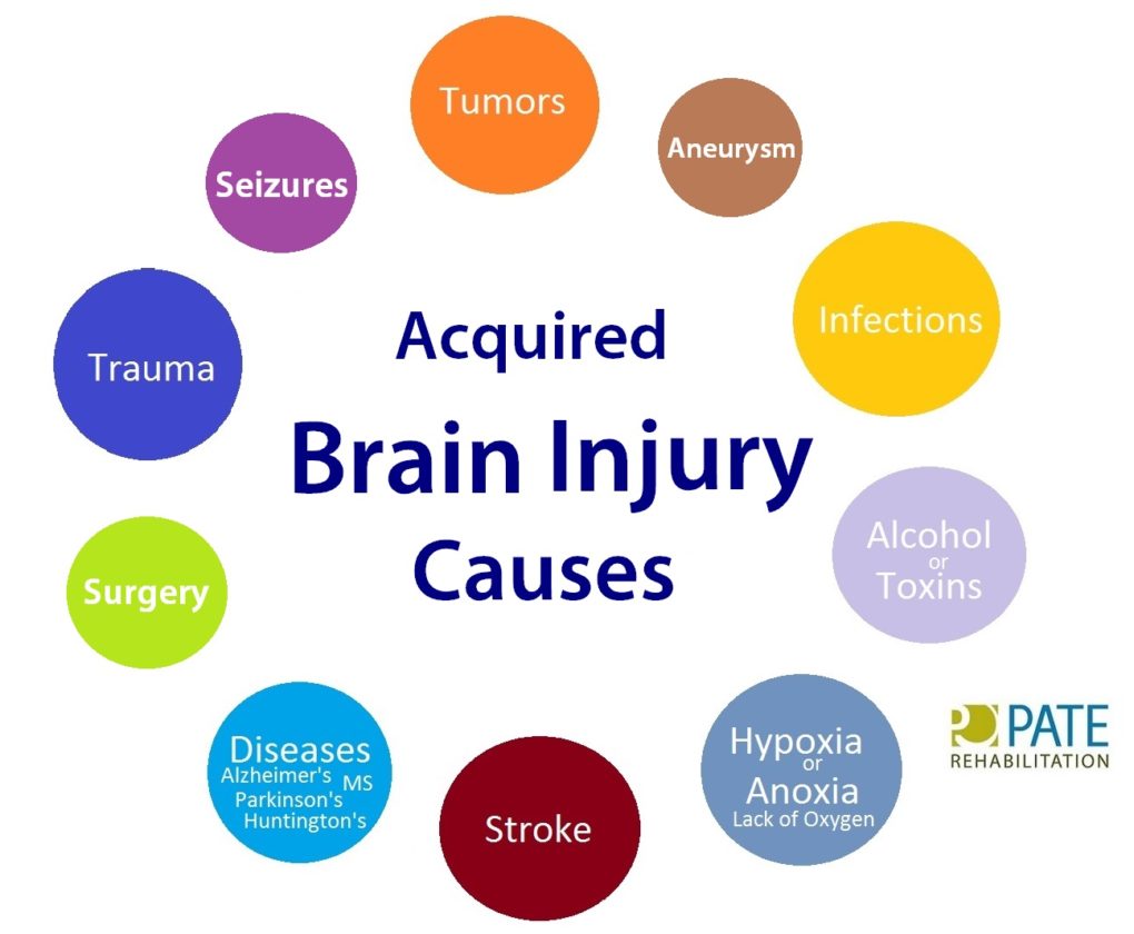
2-An acquired brain injury means simply you got this injury after you were born and it was caused by a condition or illness after birth. This type of injury can result from several different causes like strokes, toxic poisoning or brain tumors. Degenerative diseases and lack of oxygen may also cause this type of brain trauma. Here are some examples of acquired brain injuries:

-Bleeding in the brain which can lead to brain injury.   Blood Vessels in the brain can rupture resulting in an intra-cerebral hemorrhagic (one of the causes of a stroke, its what we called Hemorrhagic Stroke). Symptoms may include headaches, loss of vision, weakness to one side of the body and eye pain to even garbled speech.

Other Causes of Brian Injury:

-Anoxia is another insult to the brain that can cause injury. Anoxia is a condition in which there is an absence of oxygen supply to an organ’s tissues, even if there is adequate blood flow to the tissue.  Common causes of anoxia are near drowning, choking, suffocation, strangulation, heart attacks, lung damage, or very low blood pressure.  They all decrease oxygen intake to the red blood cells that feed the organs nutrients (being oxygen) to our body.  Anoxia is starvation to our body tissues.  

-Hypoxia:

Hypoxia refers to a decrease in oxygen supply rather than a complete absence of oxygen, and ischemia is inadequate blood supply, as is seen in cases in which the brain swells that causes compressing on the brain vessels near where the swelling is. In any of these cases, without adequate oxygen, a biochemical cascade called the ischemic cascade is unleashed, and the cells of the brain can die within several minutes. This type of injury is often seen in near-drowning victims, in heart attack patients, or in people who suffer significant blood loss from other injuries that decrease blood flow to the brain.

-Toxemia:

This occurs due to poisoning from chemical or biological factors that can damage the brain. Toxemia can be caused by drugs, chemicals, gases or even toxic foods.

-Viruses and bacteria. An infection of the brain can be very damaging like:

*Meningitis is a inflammation of the lining around the brain or spinal cord, usually due to infection; Neck stiffness, headache, fever, and confusion are common symptoms.

*Encephalitis (en-sef-uh-LIE-tis) is inflammation of the brain. Viral infections are the most common cause of the condition. Encephalitis can cause flu-like symptoms, such as a fever or severe headache. It can also cause confused thinking, seizures, or problems with senses or movement.

*HIV can lead to brain injury. HIV, can affect the brain in different ways. HIV-meningoencephalitis is infection of the brain and the lining of the brain by the HIV virus. It occurs shortly after the person is first infected with HIV and may cause headache, neck stiffness, drowsiness, confusion and/or seizures. HIV-encephalopathy (HIV-associated dementia) is the result of damage to the brain by longstanding HIV infection.  It is a form of dementia and occurs in advanced HIV infection. Mild Neurocognitive Disorder is problems with thinking and memory in HIV, however is not as severe as HIV-encephalopathy. Unlike HIV-encephalopathy it can occur early in HIV infection and is not a feature of Aquired Immune Deficiency Syndrome – AIDS.

*Lastly, Herpes. There are two types of herpes simplex virus (HSV). Either type can cause encephalitis. HSV type 1 (HSV-1) is usually responsible for cold sores or fever blisters around your mouth, and HSV type 2 (HSV-2) commonly causes genital herpes. Encephalitis caused by HSV-1 is rare, but it has the potential to cause significant brain damage or death.

*Other herpes viruses. Other herpes viruses that may cause encephalitis include the Epstein-Barr virus, which commonly causes infectious mononucleosis, and the varicella-zoster virus, which commonly causes chickenpox and shingles.*Viral infections due to blood sucking insects like mosquitoes and ticks to animals with rabies a rapid progression to encephalitis once symptoms begin. Rabies is a rare cause of encephalitis in the U.S.

When a person is diagnosed with a brain trauma, doctors will decide if rehabilitation is needed.

Rehabilitation programs may vary depending on the type of brain injury and estimated recovery time. Treatment usually consists of physical therapy and daily activities. In extreme cases, patients may need to learn how to read and write again.

Therapy for brain trauma typically takes place on an outpatient basis or through an assisted living facility. Therapy may last several weeks, months or even years, and sometimes the patient is not able to make a full recovery.

It may not always be obvious when a person has sustained a brain injury. The patient may have hit his or her head and not have symptoms until a few hours later. Some signs of a possible brain injury are headaches, confusion and loss of memory. If brain trauma is not treated, it could cause permanent damage or death.

Brain injuries can affect the patient and the patient’s family, with emotional and financial hardship. When problems arise with treatment or financial issues, a brain injury lawyer or specialist may need to intervene.

REVISED 2/25/2021 By Elizabeth Lynch RN BSN Cardiac/Stroke Certified (RN 34.5 Years)