Archives

QUOTE FOR THURSDAY:

“There are two broad types of brain injuries penetrating and non-penetrating brain injuries:

  1. Penetrating TBI (also known as open TBI) happens when an object pierces the skull (e.g., a bullet, shrapnel, bone fragment, etc.) and enters the brain tissue. Penetrating TBI typically damages only part of the brain.
  2. Non-penetrating TBI (also known as closed head injury or blunt TBI) is caused by an external force strong enough to move the brain within the skull. Causes include falls, motor vehicle crashes, sports injuries, blast injury, or being struck by an object.”

National Institute of Neurological Disorders and Stroke (Traumatic Brain Injury (TBI) | National Institute of Neurological Disorders and Stroke)

Part II National Brain Injury Awareness Month!

Overview

Traumatic brain injury (TBI) is sudden damage to the brain caused by a blow or jolt to the head. Common causes include car or motorcycle crashes, falls, sports injuries, and assaults. Injuries can range from mild concussions to severe permanent brain damage. While treatment for mild TBI may include rest and medication, severe TBI may require intensive care and life-saving surgery. Those who survive a brain injury can face lasting effects in their physical and mental abilities as well as emotions and personality. Most people who suffer moderate to severe TBI will need rehabilitation to recover and relearn skills.

What is a traumatic brain injury?

TBI is an injury to the brain caused by a blow or jolt to the head from blunt or penetrating trauma. The injury that occurs at the moment of impact is known as the primary injury. Primary injuries can involve a specific lobe of the brain or can involve the entire brain. Sometimes the skull may be fractured, but not always. During the impact of an accident, the brain crashes back and forth inside the skull causing bruising, bleeding, and tearing of nerve fibers (Fig. 1). Immediately after the accident the person may be confused, not remember what happened, have blurry vision and dizziness, or lose consciousness. At first the person may appear fine, but their condition can decline rapidly. After the initial impact occurs, the brain undergoes a delayed trauma – it swells – pushing itself against the skull and reducing the flow of oxygen-rich blood. This is called secondary injury, which is often more damaging than the primary injury.

TBI

Figure 1. During impact to the head, the soft brain crashes back and forth against the inside of the hard skull causing bruising, bleeding, and shearing of the brain.

Traumatic brain injuries are classified according to the severity and mechanism of injury:

  • Mild: person is awake; eyes open. Symptoms can include confusion, disorientation, memory loss, headache, and brief loss of consciousness.
  • Moderate: person is lethargic; eyes open to stimulation. Loss of consciousness lasting 20 minutes to 6 hours. Some brain swelling or bleeding causing sleepiness, but still arousable.
  • Severe: person is unconscious; eyes do not open, even with stimulation. Loss of consciousness lasting more than 6 hours.

Types of traumatic brain injuries

  • Concussion is a mild head injury that can cause a brief loss of consciousness and usually does not cause permanent brain injury.
  • Contusion is a bruise to a specific area of the brain caused by an impact to the head; also called coup or contrecoup injuries. In coup injuries, the brain is injured directly under the area of impact, while in contrecoup injuries it is injured on the side opposite the impact.
  • Diffuse axonal injury (DAI) is a shearing and stretching of the nerve cells at the cellular level. It occurs when the brain quickly moves back and forth inside the skull, tearing and damaging the nerve axons. Axons connect one nerve cell to another throughout the brain, like telephone wires. Widespread axonal injury disrupts the brain’s normal transmission of information and can result in substantial changes in a person’s wakefulness.
  • Traumatic Subarachnoid Hemorrhage (tSAH) is bleeding into the space that surrounds the brain. This space is normally filled with cerebrospinal fluid (CSF), which acts as a floating cushion to protect the brain. Traumatic SAH occurs when small arteries tear during the initial injury. The blood spreads over the surface of the brain causing widespread effects.
  • Hematoma is a blood clot that forms when a blood vessel ruptures. Blood that escapes the normal bloodstream starts to thicken and clot. Clotting is the body’s natural way to stop the bleeding. A hematoma may be small or it may grow large and compress the brain. Symptoms vary depending on the location of the clot. A clot that forms between the skull and the dura lining of the brain is called an epidural hematoma. A clot that forms between the brain and the dura is called a subdural hematoma. A clot that forms deep within the brain tissue itself is called an intracerebral hematoma. Over time the body reabsorbs the clot. Sometimes surgery is performed to remove large clots.

Although described as individual injuries, a person who has suffered a TBI is more likely to have a combination of injuries, each of which may have a different level of severity. This makes answering questions like “what part of the brain is hurt?” difficult, as more than one area is usually involved.

Secondary brain injury occurs as a result of the body’s inflammatory response to the primary injury. Extra fluid and nutrients accumulate in an attempt to heal the injury. In other areas of the body, this is a good and expected result that helps the body heal. However, brain inflammation can be dangerous because the rigid skull limits the space available for the extra fluid and nutrients. Brain swelling increases pressure within the head, which causes injury to parts of the brain that were not initially injured. The swelling happens gradually and can occur up to 5 days after the injury.

What are the symptoms?

Depending on the type and location of the injury, the person’s symptoms may include:

  • Loss of consciousness
  • Confusion and disorientation
  • Memory loss / amnesia
  • Fatigue
  • Headaches
  • Visual problems
  • Poor attention / concentration
  • Sleep disturbances
  • Dizziness / loss of balance
  • Irritability / emotional disturbances
  • Feelings of depression
  • Seizures
  • Vomiting

Diffuse injuries (such as a concussion or diffuse axonal injury) will typically cause an overall decreased level of consciousness. Whereas, focal injuries (such as an ICH or a contusion) will have symptoms based on the brain area affected (Fig. 2).

Lobes of the brain that can get affected by TBI:

brain anatomy

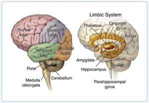
Figure 2. The brain is composed of three parts: the brainstem, cerebellum, and cerebrum, which is divided into lobes. The table lists the lobes of the brain and their normal functions as well as problems that may occur when injured. While an injury may occur in a specific area, it is important to understand that the brain functions as a whole by interrelating its component parts.

Every patient is unique and some injuries can involve more than one area or a partial section, making it difficult to predict which specific symptoms the patient will experience.

QUOTE FOR WEDNESDAY:

“Common Causes of Traumatic Brain Injury:

▪Falls
▪Motor vehicle crashes
▪Sports related injuries
▪Explosive blast/military
combat injuries

Common Causes of Non-Traumatic Brain Injury:

▪Stroke
▪Near-drowning
▪Aneurysm
▪Tumor
▪ Infectious disease that affects
the brain
▪Lack of oxygen supply to the brain”

Brain Injury Association of America (Brain Injury Awareness Month – Brain Injury Association of America)


Part I National Brain Injury Awareness Month

  BRAIN INJURY IMAGE  brain Lobe Regions


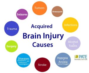
An acquired brain injury (ABI) is an injury to the brain that is not hereditary, congenital, degenerative, or induced by birth trauma. Essentially, this type of brain injury is one that has occurred after birth. The injury results in a change to the brain’s neuronal activity, which affects the physical integrity, metabolic activity, or functional ability of nerve cells in the brain.

There are two types of acquired brain injury: traumatic and non-traumatic.

A traumatic brain injury (TBI) is defined as an alteration in brain function, or other evidence of brain pathology, caused by an external force. Traumatic impact injuries can be defined as closed (or non-penetrating) or open (penetrating).

Often referred to as an acquired brain injury, a non-traumatic brain injury causes damage to the brain by internal factors, such as a lack of oxygen, exposure to toxins, pressure from a tumor, etc.

A Brain Injury is damage to the brain that results in a loss of function such as mobility or feeling.

Traumatic Brain Injuries can result from a closed head injury or a penetrating head injury.

Closed Injury:  A closed injury occurs when the head suddenly and violently hits an object but the object does not break through the skull.

Penetrating Injury: A penetrating injury occurs when an object pierces the skull and enters brain tissue. As the first line of defense, the skull is particularly vulnerable to injury. Skull fractures occur when the bone of the skull cracks or breaks. A depressed skull fracture occurs when pieces of the broken skull press into the tissue of the brain. A penetrating skull fracture occurs when something pierces the skull, such as a bullet, leaving a distinct and localized injury to brain tissue. Skull fractures can cause cerebral contusion.

Brain trauma occurs when a person has an injury to the brain, and can be mild or severe. When a person sustains trauma to the brain, he or she may lose motor functions along with cognitive and physical abilities. Physicians use the Glasgow Coma Scale to determine the extent of brain trauma. This is a neurological scale that measures the level of a person’s consciousness. The Rancho Los Amigos Scale is used to monitor the recovery of the brain.

There are several different types of brain injuries. A mild injury may cause temporary symptoms while a severe injury could require years of rehabilitation. The two most common types of brain trauma are:

1. Traumatic Brain Injuries 

2. Acquired Brain Injuries.

1-Traumatic brain injury occurs from external force on the head or neck. These injuries can occur from blows to the head or aggressive twisting of the neck. Some ways this could happen include falls, motor vehicle accidents, sports, and vigorous shaking. In infants, Shaken Baby Syndrome is a type of traumatic brain injury.

2-An acquired brain injury means simply you got this injury after you were born and it was caused by a condition or illness after birth. This type of injury can result from several different causes like strokes, toxic poisoning or brain tumors. Degenerative diseases and lack of oxygen may also cause this type of brain trauma. Here are some examples of acquired brain injuries:

-Bleeding in the brain which can lead to brain injury.   Blood Vessels in the brain can rupture resulting in an intra-cerebral hemorrhagic (one of the causes of a stroke, its what we called Hemorrhagic Stroke). Symptoms may include headaches, loss of vision, weakness to one side of the body and eye pain to even garbled speech.

Other Causes of Brian Injury:

-Anoxia is another insult to the brain that can cause injury. Anoxia is a condition in which there is an absence of oxygen supply to an organ’s tissues, even if there is adequate blood flow to the tissue.  Common causes of anoxia are near drowning, choking, suffocation, strangulation, heart attacks, lung damage, or very low blood pressure.  They all decrease oxygen intake to the red blood cells that feed the organs nutrients (being oxygen) to our body.  Anoxia is starvation to our body tissues.  

-Hypoxia:

Hypoxia refers to a decrease in oxygen supply rather than a complete absence of oxygen, and ischemia is inadequate blood supply, as is seen in cases in which the brain swells that causes compressing on the brain vessels near where the swelling is. In any of these cases, without adequate oxygen, a biochemical cascade called the ischemic cascade is unleashed, and the cells of the brain can die within several minutes. This type of injury is often seen in near-drowning victims, in heart attack patients, or in people who suffer significant blood loss from other injuries that decrease blood flow to the brain.

-Toxemia:

This occurs due to poisoning from chemical or biological factors that can damage the brain. Toxemia can be caused by drugs, chemicals, gases or even toxic foods.

-Viruses and bacteria. An infection of the brain can be very damaging like:

*Meningitis is a inflammation of the lining around the brain or spinal cord, usually due to infection; Neck stiffness, headache, fever, and confusion are common symptoms.

*Encephalitis (en-sef-uh-LIE-tis) is inflammation of the brain. Viral infections are the most common cause of the condition. Encephalitis can cause flu-like symptoms, such as a fever or severe headache. It can also cause confused thinking, seizures, or problems with senses or movement.

*HIV can lead to brain injury. HIV, can affect the brain in different ways. HIV-meningoencephalitis is infection of the brain and the lining of the brain by the HIV virus. It occurs shortly after the person is first infected with HIV and may cause headache, neck stiffness, drowsiness, confusion and/or seizures. HIV-encephalopathy (HIV-associated dementia) is the result of damage to the brain by longstanding HIV infection.  It is a form of dementia and occurs in advanced HIV infection. Mild Neurocognitive Disorder is problems with thinking and memory in HIV, however is not as severe as HIV-encephalopathy. Unlike HIV-encephalopathy it can occur early in HIV infection and is not a feature of Aquired Immune Deficiency Syndrome – AIDS.

*Lastly, Herpes. There are two types of herpes simplex virus (HSV). Either type can cause encephalitis. HSV type 1 (HSV-1) is usually responsible for cold sores or fever blisters around your mouth, and HSV type 2 (HSV-2) commonly causes genital herpes. Encephalitis caused by HSV-1 is rare, but it has the potential to cause significant brain damage or death.

*Other herpes viruses. Other herpes viruses that may cause encephalitis include the Epstein-Barr virus, which commonly causes infectious mononucleosis, and the varicella-zoster virus, which commonly causes chickenpox and shingles.*Viral infections due to blood sucking insects like mosquitoes and ticks to animals with rabies a rapid progression to encephalitis once symptoms begin. Rabies is a rare cause of encephalitis in the U.S.

When a person is diagnosed with a brain trauma, doctors will decide if rehabilitation is needed.

Rehabilitation programs may vary depending on the type of brain injury and estimated recovery time. Treatment usually consists of physical therapy and daily activities. In extreme cases, patients may need to learn how to read and write again.

Therapy for brain trauma typically takes place on an outpatient basis or through an assisted living facility. Therapy may last several weeks, months or even years, and sometimes the patient is not able to make a full recovery.

It may not always be obvious when a person has sustained a brain injury. The patient may have hit his or her head and not have symptoms until a few hours later. Some signs of a possible brain injury are headaches, confusion and loss of memory. If brain trauma is not treated, it could cause permanent damage or death.

Brain injuries can affect the patient and the patient’s family, with emotional and financial hardship. When problems arise with treatment or financial issues, a brain injury lawyer or specialist may need to intervene.

REVISED 2/25/2021 By Elizabeth Lynch RN BSN Cardiac/Stroke Certified (RN 34.5 Years)


QUOTE FOR TUESDAY:

“Based on the most recent data available, in the United States in 2021, 141,902 new colorectal cancers were reported and in 2022, 52,967 people died from colorectal cancer.

From 2017 to 2021, about 1 in 3 colorectal cancer cases were diagnosed at a localized stage, meaning the cancer had not spread outside the colon or rectum. Almost 4 in 10 colorectal cancers were found at a regional stage (the cancer had spread to nearby lymph nodes, tissues, or organs), and about 2 in 10 were found at a distant stage (the cancer had spread to distant parts of the body).

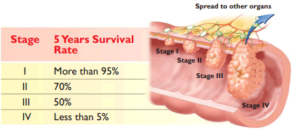
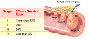
Overall, 64% of colorectal cancer patients had not died from their cancer 5 years later. However, survival varied by stage at diagnosis.

Survival is higher when colorectal cancer is found before it spreads to other parts of the body. Screening tests can prevent colorectal cancer or find it early, when treatment works best.”

Center for Disease Control and Prevention – CDC (U.S. Cancer Statistics Colorectal Cancer Stat Bite | U.S. Cancer Statistics | CDC)


Part III Colonrectal Cancer Awareness Month-Treatments from stages II to IV.


Stage II colorectal cancer is divided into three subcategories: IIA, IIB and IIC.

The difference between the categories lies in the extent to which the cancer has spread.

  • Stage IIA (T3, N0, M0): The cancer has grown into the outermost layers of the colon or rectum, but has not grown through them. It has not reached nearby organs or lymph nodes, and has not spread to distant organs.
  • Stage IIB (T4a, N0, M0): The cancer has grown through all of the layers of the colon or rectum, but has not grown into other organs or tissues.
  • Stage IIC (T4b, N0, M0): The cancer has grown through all of the layers of the colon or rectum, and has grown into nearby organs or tissues. The cancer has not spread to the lymph nodes Surgery
  • Initial treatment for stage II colon cancer is surgery to remove the section of colon that contains the tumor and surrounding tissue with its blood vessels and lymph nodes.
  • The most commonly recommended protocols for patients diagnosed with stage II colon cancer:

Colectomy

A colectomy (or colon resection) is abdominal surgery that removes the section of colon where the tumor is located, tissue containing blood and lymph vessels surrounding the colon (mesentery), healthy tissue margins on either side and, if possible, at least 12 lymph nodes.  Then the remaining ends of colon are reconnected with sutures or staples.  This connection is called an anastomosis.

There are two types of surgery:

  • Open colectomy:  An incision is made in the abdomen, surgery performed through the opening, and the incision closed with sutures and/or staples.
  • Laparoscopic colectomy:  Three small keyhole incisions are made in the abdomen to insert a lighted instrument and specially designed surgical instruments that can be manipulated within the abdomen.  Sometimes an incision is made just long enough for the surgeon’s hand to assist during laparoscopy. This is also known as minimally invasive surgery.If your lymph nodes are cancer-free (also known as node-negative), your diagnosis is stage II colon cancer
  • The tumor, tissue on either side of it (the margins), and fat and lymph nodes attached to the colon are removed for further study by a pathologist.  The pathologist evaluates the cancer cells in the tumor itself, looks for cancer in the margins and other tissue, and studies as many lymph nodes as possible in order to provide an accurate diagnosis.

Chemotherapy

Treatment of node-negative stage II colon cancer is controversial. While surgery to remove the tumor in the colon is universally accepted as initial treatment, the value of chemotherapy after that surgery to keep cancer from recurring (coming back) is hard for patients and doctors to judge.

It’s estimated that between four and five percent of patients with stage II colon cancer will benefit from chemotherapy. However, there are also side effects, some severe, associated with chemotherapy. Very few patients will die as a result of chemotherapy.

Because of the risks of treatment, researchers are looking for ways to identify patients who are at higher risk for recurrence, who are most likely to benefit from chemotherapy.

Some factors have been identified that lead to higher risk for stage II patients including:

  • T4 tumors that extend beyond the outer wall of the colon into adjacent tissues and organs
  • Too few lymph nodes removed and examined (less than 12)
  • Cancer cells in blood and lymph vessels surrounding the tumor (not the same as lymph nodes)
  • Undifferentiated or poorly differentiated tumors
  • Perforation (a hole) of the colon by the tumor
  • A tumor that obstructs (closes off) the colonFor high-risk stage II patients, the number needed to prevent one recurrence or death is smaller, probably 15 to 30 patients.
  • It may help your decision to think about the problem in terms of numbers:  In order to prevent one recurrence or death from all cases of stage II colon cancer, 25 to 50 patients need to receive chemotherapy. One in six of those patients will have a severe side effect; one in 100 to 200 will die as a result of treatment.

Chemotherapy regimens for high-risk stage II colon cancer:

  • FOLFOX:  combination treatment with infusional 5-FU (fluorouracil), leucovorin, and oxaliplatin
  • FLOX: combination with bolus 5-FU, leucovorin, and oxaliplatin (severe diarrhea is more common with FLOX than FOLFOX but outcomes are similar)
  • Xeloda (capecitabine): oral “prodrug” which is converted to 5-FU in the tumor
  • 5-FU and leucovorinThere are some indicators of a patient’s risk of recurrence of their cancer, but no clear information that higher risk means they may benefit from therapy  — thus there are research efforts underway to better define “risk” and develop treatments that will benefit the higher risk patient in a predictable way.
  • Stage III colorectal cancer treatments:In this article
  • Your doctor can discuss the advantages and disadvantages of the different chemotherapy regimens if you decide to proceed with chemotherapy after your surgery.  Chemotherapy usually lasts about six months.

Stage III Colorectal Cancer Treatments

Stage III colorectal cancers have spread outside the colon to one or more lymph nodes (small structures that are found throughout the body that produce and store cells that fight infection). Tumors within the colon wall, which also involve the lymph nodes are classified as stage IIIA, while tumors that have grown through the colon wall and have spread to one to four lymph nodes are classified as stage IIIB cancers. Those tumors, which have spread to more than four lymph nodes are classified as stage IIIC colon cancers.

Treatment involves:

  • Surgery to remove the tumor and all involved lymph nodes if possible.
  • After surgery, the patient will receive chemotherapy with 5-FU, leucovorin and oxaliplatin, capecitabine with oxaliplatin or capecitabine alone.
  • Radiation may be needed if the tumor is large and invading the tissue surrounding the colon.

The five-year survival rate for stage III colon cancer is about 64%. Patients with one to four positive lymph nodes have a higher survival rate than people with more than five positive lymph nodes.

 Stage IV Colorectal cancer treatments:

Stage IV colorectal cancers have spread outside the colon to other parts of the body, such as the liver or the lungs. Cancer that has spread is also called “metastatic.” The tumor can be any size and may or may not include affected lymph nodes (small structures that are found throughout the body that produce and store cells that fight infection).

Treatment may include:

  • Removal of the cancer surgically or another surgical procedure to bypass the colon cancer and hook up healthy colon (an anastomosis).
  • Surgery to remove parts of other organs such as the liver, lungs, and ovaries, where the cancer may have spread.
  • Chemotherapy to relieve symptoms and improve survival.
  • Erbitux, Avastin, or Vectibix in combination with standard chemotherapy, depending upon tumor characteristics.
  • Zaltrap is a drug also approved for use with chemotherapy in cases where the cancer has progressed or is resistant to treatment.
  • Stivarga is a targeted therapy approved in patients whose cancer has progressed after previous therapy.
  • Clinical trials of new chemotherapy regimens, or immunological therapy.
  • Radiation to relieve symptoms.

The five-year survival rate for stage IV colon cancer is nearly 8% or less.

QUOTE FOR MONDAY:

“After someone is diagnosed with colorectal cancer, doctors will try to figure out if it has spread, and if so, how far. This process is called staging. The stage of a cancer describes how much cancer is in the body. It helps determine how serious the cancer is and how best to treat it. Doctors also use a cancer’s stage when talking about survival statistics.

The earliest stage of colorectal cancers is called stage 0 (a very early cancer), and then range from stages I (1) through IV (4). As a rule, the lower the number, the less the cancer has spread.

The staging system most often used for colorectal cancer is the American Joint Committee on Cancer (AJCC) TNM system, which is based on 3 key pieces of information:

  • The extent (size) of the tumor (T): How far has the cancer grown into the wall of the colon or rectum? These layers,from the inner to the outer.
  • The spread to nearby lymph nodes (N): Has the cancer spread to nearby lymph nodes?
  • The spread (metastasis) to distant sites (M): Has the cancer spread to distant lymph nodes or distant organs such as the liver or lungs
  • The system described below is the most recent AJCC system effective January 2018. It uses the pathologic stage (also called the surgical stage), which is determined by examining tissue removed during an operation. This is also known as surgical staging. This is likely to be more accurate than clinical staging, which takes into account the results of a physical exam, biopsies, and imaging tests, done before surgery.
  • Numbers or letters after T, N, and M provide more details about each of these factors. Higher numbers mean the cancer is more advanced. Once a person’s T, N, and M categories have been determined, this information is combined in a process called stage grouping to assign an overall stage.”.

American Cancer Society (Colorectal Cancer Stages | Rectal Cancer Staging | Colon Cancer Staging | American Cancer Society)

Part II Colonrectal Cancer Awareness Month – Treatment on stages O and I of colonrectal cancer.

colorectal-cANCER PERCENTAGE IN RISKSCOLORECTAL CANCER 2

Part II Treatment & Care

Many colon cancer treatment options are available for colorectal cancer, including surgery, chemotherapy, and radiation. Here’s what to expect from each type of treatment and tips for recovery.

Polyp Removal and Other Precancerous Conditions

Precancerous conditions of the colon or rectum are changes to cells that make them more likely to develop into cancer. These conditions are not yet cancer, but there is a higher chance these abnormal changes will become colorectal cancer.

The most common precancerous conditions of the colon or rectum area:

adenomas

hereditary colorectal syndromes

If you have a precancerous condition, you will likely have regular follow-up and screening tests to find cancer as early as possible if it develops. Some precancerous conditions can be treated with surgery to help reduce the risk that they will become cancer.

Colorectal Cancer Treatment

Stages of the cancer with TNM system for colorectal cancer helps determine the RX.

The most commonly used colorectal cancer staging system is known as the TNM system, which has been established by the American Joint Committee on Cancer. The TNM staging system looks at three key factors to determine the stage of cancer:

  • Tumor (T) looks at how far the primary tumor has grown into the wall of the colon or rectum, and if it has expanded into nearby areas.
  • Lymph node (N) examines the extent of the cancer spread to nearby lymph nodes.
  • Metastasis (M) refers to whether cancer has spread to other parts of the body, such as the liver, lungs or brain.

A number (0-4 stages) or the letter X is assigned to each factor. Using this colorectal cancer staging system, a higher number indicates increasing severity. For instance, a T1 score indicates a smaller tumor than a T2 score. The letter X means the information could not be assessed.

Stages of colorectal cancer diagnosis occurs in conjunction with the following TNM categories:

  • T1-T2: If the cancer has grown through the muscularis mucosa and into the submucosa, it is considered T1. Or, if the cancer has grown into the muscularis propria, then it is classified as T2.
  • N0: The cancer has not spread to the lymph nodes.
  • M0: There has been no spreading to organs or other nearby areas
  • Stage I colorectal cancer treatments

Once the T, N and M scores have been assigned, an overall stage is determined, and thus treatment options can be explored.

Here’s a quick rundown of the options available for colorectal cancer treatment from surgery to cutting-edge biologic therapy.

Colorectal Cancer: Treatment by Stages

STAGE 0 (IN SITU) Colorectal Cancer Treatment

Surgery for colon cancer

  • Polypectomy: snaring and removing polyps containing cancer during a colonoscopy.
  • Local excision: removal of flat colon growths “piecemeal” during colonoscopy.
  • Open abdominal surgery to remove cancer, part of colon, and nearby lymph nodes in high risk situations where:
    • There is a spread to polyp stalk
    • There is spread to lymphatic vessels (not lymph nodes)
    • Cells look very abnormal under the microscope (high grade)
    • Surgical margins (edge of tissue) contain cancer cells or can’t be evaluated or contain cancer cells.
    • Local excision would be too time-consuming or difficult to perform.

Chemotherapy

Chemotherapy is not recommended for stage 0 colon cancer.

Stage I Colorectal Cancer Treatment

Surgery

Colectomy (resection): Abdominal surgery to remove the section of colon where the tumor is located, tissue containing blood and lymph vessels surrounding the colon (mesentery), healthy tissue margins on either side, and at least 12 lymph nodes, if possible. Then the remaining ends of colon are reconnected with sutures or staples. This connection is called an anastomosis.

Open colectomy: An incision is made in the abdomen, surgery performed through the opening, and the incision closed with sutures and/or staples.

Laparoscopic colectomy: Three small keyhole incisions are made to insert a lighted instrument and specially designed surgical instruments that can be manipulated within the abdomen. Sometimes an incision is made just long enough for the surgeon’s hand to assist during laparoscopy.

Chemotherapy

Chemotherapy is not recommended for stage I colon cancer.

Part III on continuation of treatments of other stages of this condition (Stage II & III)

QUOTE FOR THE WEEKEND:

“Colon cancer is a growth of cells that begins in a part of the large intestine called the colon. The colon is the first and longest part of the large intestine. The large intestine is the last part of the digestive system. The digestive system breaks down food for the body to use.

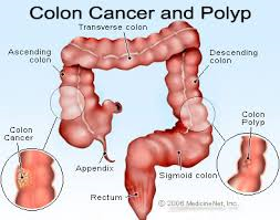
Colon cancer typically affects older adults, though it can happen at any age. It usually begins as small clumps of cells called polyps that form inside the colon. Polyps generally aren’t cancerous, but some can turn into colon cancers over time.

Polyps often don’t cause symptoms. For this reason, doctors recommend regular screening tests to look for polyps in the colon. Finding and removing polyps helps prevent colon cancer.”

MAYO  CLINIC (Colon cancer – Symptoms and causes – Mayo Clinic)

Part I Colonrectal Cancer Awareness Month

colorectal cancer2 colorectal cancer1

Colorectal cancer is cancer that develops in the tissues of the colon and/or rectum. The colon and the rectum are both found in the lower part of the gastrointestinal (digestive) system. They form a long, muscular tube called the large intestine (or large bowel). The colon absorbs food and water and stores waste. The rectum is responsible for passing waste from the body.

If the cancer began in the colon, which is the first four to five feet of the large intestine, it may be referred to as colon cancer. If the cancer began in the rectum, which is the last several inches of the large intestine leading to the anus, it is called rectal cancer.

Colorectal cancer starts in the inner lining of the colon and/or rectum, slowly growing through some or all of its layers. It typically starts as a growth of tissue called a polyp. A particular type of polyp, called an adenoma, can then develop into cancer.

Adenocarcinoma is the most common type of colorectal cancer. Other colorectal cancers include gastrointestinal carcinoid tumors, gastrointestinal stromal tumors, primary colorectal lymphoma, leiomyosarcoma, melanoma and squamous cell carcinoma.

Cancer is a disease in which cells in the body grow out of control. When cancer starts in the colon or rectum, it is called colorectal cancer. Sometimes it is called colon cancer, for short.

Colorectal cancer affects men and women of all racial and ethnic groups, and is most often found in people aged 50 years or older. In the United States, it is the third most common cancer for men and women.

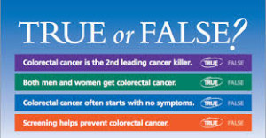
Of cancers that affect both men and women, colorectal cancer is the second leading cancer killer in the United States, but it doesn’t have to be.

Colorectal cancer screening saves lives.

Screening can find precancerous polyps—abnormal growths in the colon or rectum—so that they can be removed before turning into cancer. Screening also helps find colorectal cancer at an early stage, when treatment often leads to a cure. About nine out of every 10 people whose colorectal cancers are found early and treated appropriately are still alive five years later.

If you are aged 50 or older, get screened now. If you think you may be at higher than average risk for colorectal cancer, speak with your doctor about getting screened early.

While screening rates have increased in the U.S., not enough people are getting screened for colorectal cancer. In 2012, 65% of U.S. adults were up-to-date with colorectal cancer screening; 7% had been screened, but were not up-to-date; and 28% had never been screened.

Your risk of getting colorectal cancer increases as you get older. More than 90% of cases occur in people who are 50 years old or older.

Other risk factors include having:

*Inflammatory bowel disease, Crohn’s disease, or ulcerative colitis.

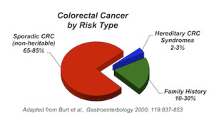
*A personal or family history of colorectal cancer or colorectal polyps.

*A genetic syndrome such as familial adenomatous polyposis (FAP)hereditary non-polyposis colorectal cancer (Lynch syndrome).

Lifestyle factors that may contribute to an increased risk of colorectal cancer include—

  • Lack of regular physical activity.
  • Low fruit and vegetable intake.
  • A low-fiber and high-fat diet.
  • Overweight and obesity.
  • Alcohol consumption.
  • Tobacco use. *Rectal bleeding or blood in your stool.
  • *Diagnosing colorectal cancer:Treatment & Care
  • Tests. Finding colon cancer early is key to beating it. That’s why doctors recommend a yearly fecal occult blood test, which tests for invisible blood in the stool, an early sign of colon cancer. One of the best tools for detecting colorectal cancer is a colonoscopy.
  • *Persistent abdominal discomfort, such as cramps, gas or pain.
  • *A change in your bowel habits, including diarrhea or constipation or a change in the consistency of your stool, that lasts longer than four weeks.
  • *Signs and symptoms of colon-rectal cancer include:
  • Many colon cancer treatment options are available for colorectal cancer, including surgery, chemotherapy, and radiation. Here’s what to expect from each type of treatment and tips for recovery.

*Treatment= Get details on treatment on Part II tomorrow’s article Thurs.

Colon Polyp Removal and Other Precancerous Conditions

Learn how colon polyps are removed and why it’s so important to stay on top of these and other precancerous conditions.

Colorectal Cancer Treatment

Here’s a quick rundown of the options available for colorectal cancer treatment from surgery to cutting-edge biologic therapy.

Colon Cancer: Treatment by Stage

Here you’ll find detailed information on how the various stages of colon cancer are treated — from stage 0 to stage IV and also recurrent colon cancer.

Rectal Cancer Treatment by Stage

Here you’ll find detailed information on how the various stages of rectal cancer are treated — from stage 0 to stage IV and also recurrent rectal cancer.

Colon Cancer Chemotherapy

Learn about the different ways chemotherapy is used to treat colon cancer and rectal cancer and the side effects of commonly used chemotherapy drugs.