Archives

QUOTE FOR THE WEEKEND:

“Early symptoms of Alzheimer’s disease include forgetting recent events or conversations. Over time, Alzheimer’s disease leads to serious memory loss and affects a person’s ability to do everyday tasks.

There is no cure for Alzheimer’s disease. In advanced stages, loss of brain function can cause dehydration, poor nutrition or infection. These complications can result in death.

But medicines may improve symptoms or slow the decline in thinking. Programs and services can help support people with the disease and their caregivers.”

MAYO CLINIC (Alzheimer’s disease – Symptoms and causes – Mayo Clinic)

Part II Alzheimer’s Disease Awareness – diagnosing and treatment.

The National Institute of aging states the following procedures also may be used to diagnose dementia:

  • “Cognitive and neuropsychological tests. These tests are used to assess memory, problem solving, language skills, math skills, and other abilities related to mental functioning.
  • Laboratory tests. Testing a person’s blood and other fluids , as well as checking levels of various chemicals, hormones, and vitamins, can help find or rule out possible causes of symptoms.
  • Brain scans. These tests can identify strokes, tumors, and other problems that can cause dementia. Scans also identify changes in the brain’s structure and function. The most common scans are:
    • Computed tomography (CT), which uses x rays to produce images of the brain and other organs
    • Magnetic resonance imaging (MRI), which uses magnetic fields and radio waves to produce detailed images of body structures, including tissues, organs, bones, and nerves
    • Positron emission tomography (PET), which uses radiation to provide pictures of brain activity
  • Psychiatric evaluation. This evaluation will help determine if depression or another mental health condition is causing or contributing to a person’s symptoms.
  • Genetic tests. Some dementias are caused by a known gene defect. In these cases, a genetic test can help people know if they are at risk for dementia. It is important to talk with a genetic counselor before and after getting tested, along with family members and the doctor.

QUOTE FOR FRIDAY:

“Dementia is a general term for loss of memory, language, problem-solving and other thinking abilities that are severe enough to interfere with daily life. Alzheimer’s is the most common cause of dementia.

Some risk factors for dementia, such as age and genetics, cannot be changed. But researchers continue to explore the impact of other risk factors on brain health and prevention of dementia.

Protecting your brain health — how well you think, learn and remember — is important at every age. Building brain-healthy habits can help you live better now and protect your memory and thinking for the future. Brain health is important for everyone, including people with a family history of dementia. It is never too early or too late to take action for your brain health.”

Alzheimer’s Association (What is Dementia? Symptoms, Causes & Treatment | alz.org)


Part I What is actually Alzheimer’s Disease & the symptoms?

What structurally happens to the brain and what happens to the individual in brain thinking diagnosed with this disease:

Alzheimer’s disease is an irreversible, progressive brain disorder that slowly destroys memory and thinking skills and, eventually, the ability to carry out the simplest tasks. In most people with the disease—those with the late-onset type—symptoms first appear in their mid-60s. Early-onset Alzheimer’s occurs between a person’s 30s and mid-60s and is very rare. Alzheimer’s disease is the most common cause of dementia among older adults.

The disease is named after Dr. Alois Alzheimer. In 1906, Dr. Alzheimer noticed changes in the brain tissue of a woman who had died of an unusual mental illness. Her symptoms included memory loss, language problems, and unpredictable behavior. After she died, he examined her brain and found many abnormal clumps (now called amyloid plaques) and tangled bundles of fibers (now called neurofibrillary, or tau, tangles).

These plaques and tangles in the brain are still considered some of the main features of Alzheimer’s disease. Another feature is the loss of connections between nerve cells (neurons) in the brain. Neurons transmit messages between different parts of the brain, and from the brain to muscles and organs in the body. Many other complex brain changes are thought to play a role in Alzheimer’s, too.

This damage initially appears to take place in the hippocampus, the part of the brain essential in forming memories. As neurons die, additional parts of the brain are affected. By the final stage of Alzheimer’s, damage is widespread, and brain tissue has shrunk significantly.

How many affected in the United States by Alzheimer’s Disease:

Estimates vary, but experts suggest that as many as 5.5 million Americans age 65 and older may have Alzheimer’s. Many more under age 65 also have the disease. Unless Alzheimer’s can be effectively treated or prevented, the number of people with it will increase significantly if current population trends continue. This is because increasing age is the most important known risk factor for Alzheimer’s disease.

Symptoms:

Memory problems are typically one of the first signs of Alzheimer’s, though initial symptoms may vary from person to person. A decline in other aspects of thinking, such as finding the right words, vision/spatial issues, and impaired reasoning or judgment, may also signal the very early stages of Alzheimer’s disease. Mild cognitive impairment (MCI) is a condition that can be an early sign of Alzheimer’s, but not everyone with MCI will develop the disease.

People with Alzheimer’s have trouble doing everyday things like driving a car, cooking a meal, or paying bills. They may ask the same questions over and over, get lost easily, lose things or put them in odd places, and find even simple things confusing. As the disease progresses, some people become worried, angry, or violent.

Alzheimer’s disease is not a normal part of aging.

Memory problems are typically one of the first warning signs of cognitive loss.

According to the National Institute on Aging, in addition to memory problems, someone with Alzheimer’s disease may experience one or more of the following signs:

  • Memory loss that disrupts daily life, such as getting lost in a familiar place or repeating questions.
  • Trouble handling money and paying bills.
  • Difficulty completing familiar tasks at home, at work or at leisure.
  • Decreased or poor judgment.
  • Misplaces things and being unable to retrace steps to find them.
  • Changes in mood, personality, or behavioral.


QUOTE FOR THURSDAY:

“How common is pancreatic cancer?

The American Cancer Society’s estimates for pancreatic cancer in the United States for 2025 are:

  • About 67,440 people ( 34,950 men and 32,490 women) will be diagnosed with pancreatic cancer.
  • About 51,980 people ( 27,050 men and 24,930 women) will die of pancreatic cancer.

Lifetime risk of pancreatic cancer

The average lifetime risk of pancreatic cancer is about 1 in 56 in men and about 1 in 60 in women. But each person’s chances of getting this cancer can be affected by certain risk factors.”

American Cancer Society (Facts About Pancreatic Cancer | American Cancer Society)

Part IV Pancreatic Cancer – Staging and Treatment


In 2025


Pancreatic Cancer, its incidence cuts across all racial and socio-economic barriers and is nearly always fatal. 90% die within the 1st yr of diagnosis.

STAGING OF PANCREATIC CANCER

Stage is a term used in cancer treatment to describe the extent of the cancer’s spread. The stages of pancreatic cancer are from 0 to IV.

The best treatment for pancreatic cancer depends on how far it has spread, or its stage. The stages of pancreatic cancer are easy to understand. What is difficult is attempting to stage pancreatic cancer without resorting to major surgery. In practice, doctors choose pancreatic cancer treatments based upon imaging studies, surgical findings, and an individual’s general state of well being.

Stages of Pancreatic Cancer

Stage is a term used in cancer treatment to describe the extent of the cancer’s spread. The stages of pancreatic cancer are used to guide treatment and to classify patients for clinical trials. The stages of pancreatic cancer are:

  • Stage 0: No spread. Pancreatic cancer is limited to top layers of cells in the ducts of the pancreas. The pancreatic cancer is not visible on imaging tests or even to the naked eye.
  • Stage I: Local growth. Pancreatic cancer is limited to the pancreas, but has grown to less than 2 centimeters across (stage IA) or greater than 2 but no more than 4 centimeters (stage IB).
  • Stage II: Local spread. Pancreatic cancer is over 4 centimeters and is either limited to the pancreas or there is local spread where the cancer has grown outside of the pancreas, or has spread to nearby lymph nodes. It has not spread to distant sites.
  • Stage III: Wider spread. The tumor may have expanded into nearby major blood vessels or nerves, but has not metastasized to distant sites.
  • Stage IV: Confirmed spread. Pancreatic cancer has spread to distant organs.

Determining pancreatic cancer’s stage is often tricky. Imaging tests like CT scans and ultrasound provide some information, but knowing exactly how far pancreatic cancer has spread usually requires surgery.

Since surgery has risks, doctors first determine whether pancreatic cancer appears to be removable by surgery (resectable). Pancreatic cancer is then described as follows:

  • Resectable: On imaging tests, pancreatic cancer hasn’t spread (or at least not far), and a surgeon feels it might all be removable. About 10% of pancreatic cancers are considered resectable when first diagnosed.
  • Locally advanced (unresectable): Pancreatic cancer has grown into major blood vessels on imaging tests, so the tumor can’t safely be removed by surgery.
  • Metastatic: Pancreatic cancer has clearly spread to other organs, so surgery cannot remove the cancer.

If pancreatic cancer is resectable, surgery followed by chemotherapy or radiation or both may extend survival.

Treating Resectable Pancreatic Cancer

People whose pancreatic cancer is considered resectable may undergo one of three surgeries:

Whipple procedure (pancreaticoduodenectomy): A surgeon removes the head of the pancreas and sometimes the body of the pancreas, parts of the stomach and small intestine, some lymph nodes, the gallbladder, and the common bile duct. The remaining organs are reconnected in a new way to allow digestion. The Whipple procedure is a difficult and complicated surgery. Surgeons and hospitals that do the most operations have the best results.

About half the time, once a surgeon sees inside the abdomen, pancreatic cancer that was thought to be resectable turns out to have spread, and thus be unresectable. The Whipple procedure is not completed in these cases.

Distal pancreatectomy: The tail and/or portion of the body of the pancreas are removed, but not the head. This surgery is uncommon for pancreatic cancer, because most tumors arising outside the head of the pancreas within the body or tail are unresectable.

Total pancreatectomy: The entire pancreas and the spleen is surgically removed. Although once considered useful, this operation is uncommon today.

Chemotherapy or radiation therapy or both can also be used in conjunction with surgery for resectable and unresectable pancreatic cancer in order to:

  • Shrink pancreatic cancer before surgery, improving the chances of resection (neoadjuvant therapy)
  • Prevent or delay pancreatic cancer from returning after surgery (adjuvant therapy)

Chemotherapy includes cancer drugs that travel through the whole body. Chemotherapy (“chemo”) kills pancreatic cancer cells in the main tumor as well as those that have spread widely. These chemotherapy drugs can be used for pancreatic cancer:

  • 5-fluorouracil (5-FU) or capecitabine
  • Gemcitabine

Both 5-FU and gemcitabine are given into the veins during regular visits to an oncologist (cancer doctor). An oral drug, capecitabine, may be substituted for 5-FU, especially with radiation.

In radiation therapy, a machine beams high-energy X-rays to the pancreas to kill pancreatic cancer cells. Radiation therapy is done during a series of daily treatments, usually over a period of weeks.

Both radiation therapy and chemotherapy damage some normal cells, along with cancer cells. Side effects can include nausea, vomiting, appetite loss, weight loss, and fatigue as well as toxicity to the blood cells. Symptoms usually cease within a few weeks after radiation therapy is complete.

The best treatment for pancreatic cancer depends on how far it has spread, or its stage. The stages of pancreatic cancer are easy to understand. What is difficult is attempting to stage pancreatic cancer without resorting to major surgery. In practice, doctors choose pancreatic cancer treatments based upon imaging studies, surgical findings, and an individual’s general state of well being.

Treating Metastatic Pancreatic Cancer

In metastatic pancreatic cancer, surgery is used only for symptom control, such as for pain, jaundice, or gastric outlet obstruction. Radiation may be used for symptom relief, as well.

Chemotherapy can also help improve pancreatic cancer symptoms and survival. Gemcitabine has been the most wildly used chemotherapy drug for treating metastatic pancreas cancer. Other drug combinations include gemcitabine with erlotinib, gemcitabine with capecitabine, gemcitabine with cisplatin, and gemcitabine with nab-paclitaxel. If you’re in fairly good health you may receive FOLFIRINOX (5-FU/leucovorin/oxaliplatin/irinotecan). Other combinations include gemcitabine alone or with another agent like (nab)-paclitaxel or capecitabine. Next line drug combinations to treat pancreatic cancer include oxaliplatin/fluoropyrimidine, or irinotecan liposome (Onivyde) in combination with fluorouracil plus leucovorin.

Palliative Treatment for Pancreatic Cancer

As pancreatic cancer progresses, the No. 1 priority of treatment will shift from extending life to alleviating symptoms, especially pain. Numerous treatments can help protect against the discomfort from advanced pancreatic cancer:

  • Procedures like bile duct stents can relieve jaundice, thus reducing itching and loss of appetite associated with bile obstruction.
  • Opioid analgesics and a nerve block called a celiac plexus block can help relieve pain.
  • Antidepressants and counseling can help treat depression common in advanced pancreatic cancer.

Clinical Trials for Pancreatic Cancer

New pancreatic cancer treatments are constantly being tested in clinical trials. You can find out about clinical trials for the latest treatments for pancreatic cancer on the websites of the American Cancer Society and the National Cancer Institute


QUOTE FOR WEDNESDAY:

“The pancreas makes enzymes from a type of cell called an exocrine cell. Most of your pancreas is made up of exocrine cells.

A very small part of the pancreas is made of endocrine cells. These cells make hormones, such as insulin.

Pancreatic tumors start in either exocrine or endocrine cells. More than 9 out of 10 pancreatic cancers are exocrine tumors, not neuroendocrine tumors.

There are about 20 different types of tumors can grow in the pancreas.

These tumors often start in the pancreas ducts, which are small channels that carry digestive enzymes to the intestines.

Adenocarcinoma (A-deh-noh-KAR-sih-NOH-muh): This is the most common kind of exocrine pancreatic cancer. Also called ductal carcinoma, it starts in the lining of pancreas ducts.

Acinar cell carcinoma (ACC): This type of adenocarcinoma starts in exocrine cells and is a very rare cancer.

Adenosquamous carcinoma (A-deh-noh-SKWAY-mus KAR-sih-NOH-muh): This rare exocrine pancreatic cancer is hard to diagnose. It’s made of squamous cell carcinoma and ductal adenocarcinoma. This tumor can be more aggressive than adenocarcinoma.

Pancreatoblastoma: This tumor is very rare. It affects young children and grows slowly.

Squamous cell carcinoma: This tumor is mostly made of squamous cells. It’s very rare and hard to treat, because the cancer is found after it has spread.

Solid pseudopapillary tumor (SPT) or Solid pseudopapillary neoplasm (SPN): Very rare, they make up only about 1 out of every 100 pancreatic tumors. SPT and SPN mostly affect young women and are easier to treat than other pancreatic cancers.

Pancreatic neuroendocrine (islet cell) tumors

Pancreatic neuroendocrine tumors are also known as islet cell tumors or islet cell carcinoma. They’re a type of neuroendocrine tumor found in the pancreas.

Pancreatic tumors that are benign (not cancer)

Pancreatic cysts are growths filled with fluid that start in the pancreas. Most pancreatic cysts are benign (not cancer). They’re not likely to harm you or cause symptoms. But some are precancerous and can turn into pancreatic cancer.”

Memorial Sloan Kettering Cancer Center (Types of Pancreatic Tumors | Memorial Sloan Kettering Cancer Center)


Part III Continuation on Pancreatic Cancer Diagnostic Testing!

6.) Angiography

This is an x-ray test that looks at blood vessels. A small amount of contrast dye is injected into an artery to outline the blood vessels, and then x-rays are taken.

An angiogram can show if blood flow in a particular area is blocked by a tumor. It can also show abnormal blood vessels (feeding the cancer) in the area. This test can be useful in finding out if a pancreatic cancer has grown through the walls of certain blood vessels.  Usually the catheter is put into an artery in your inner thigh and threaded up to the pancreas.

Blood Tests

Several types of blood tests can be used to help diagnose pancreatic cancer or to help determine treatment options if it is found.

Liver function tests: Jaundice (yellowing of the skin and eyes) is often one of the first signs of pancreatic cancer. Doctors often get blood tests to assess liver function in people with jaundice to help determine its cause. Certain blood tests can look at levels of different kinds of bilirubin (a chemical made by the liver) and can help tell whether a patient’s jaundice is caused by disease in the liver itself or by a blockage of bile flow (from a gallstone, a tumor, or other disease).

Tumor markers: Tumor markers are substances that can sometimes be found in the blood when a person has cancer. Tumor markers that may be helpful in pancreatic cancer are:

  • CA 19-9
  • Carcinoembryonic antigen (CEA), which is not used as often as CA 19-9

Neither of these tumor marker tests is accurate enough to tell for sure if someone has pancreatic cancer. Levels of these tumor markers are not high in all people with pancreatic cancer, and some people who don’t have pancreatic cancer might have high levels of these markers for other reasons. Still, these tests can sometimes be helpful, along with other tests, in figuring out if someone has cancer.

In people already known to have pancreatic cancer and who have high CA19-9 or CEA levels, these levels can be measured over time to help tell how well treatment is working. If all of the cancer has been removed, these tests can also be done to look for signs the cancer may be coming back.

Other blood tests: Other tests, like a CBC or chemistry panel, can help evaluate a person’s general health (such as kidney and bone marrow function). These tests can help determine if they’ll be able to withstand the stress of a major operation.

Biopsy

A person’s medical history, physical exam, and imaging test results may strongly suggest pancreatic cancer, but usually the only way to be sure is to remove a small sample of tumor and look at it under the microscope. This procedure is called a biopsy. Biopsies can be done in different ways.

Percutaneous (through the skin) biopsy: For this test, a doctor inserts a thin, hollow needle through the skin over the abdomen and into the pancreas to remove a small piece of a tumor. This is known as a fine needle aspiration (FNA). The doctor guides the needle into place using images from ultrasound or CT scans.

Endoscopic biopsy: Doctors can also biopsy a tumor during an endoscopy. The doctor passes an endoscope (a thin, flexible, tube with a small video camera on the end) down the throat and into the small intestine near the pancreas. At this point, the doctor can either use endoscopic ultrasound (EUS) to pass a needle into the tumor or endoscopic retrograde cholangiopancreatography (ERCP) to place a brush to remove cells from the bile or pancreatic ducts.

Surgical biopsy: Surgical biopsies are now done less often than in the past. They can be useful if the surgeon is concerned the cancer has spread beyond the pancreas and wants to look at (and possibly biopsy) other organs in the abdomen. The most common way to do a surgical biopsy is to use laparoscopy (sometimes called keyhole surgery). The surgeon can look at the pancreas and other organs for tumors and take biopsy samples of abnormal areas.

Some people might not need a biopsy

Rarely, the doctor might not do a biopsy on someone who has a tumor in the pancreas if imaging tests show the tumor is very likely to be cancer and if it looks like surgery can remove all of it. Instead, the doctor will proceed with surgery, at which time the tumor cells can be looked at in the lab to confirm the diagnosis. During surgery, if the doctor finds that the cancer has spread too far to be removed completely, only a sample of the cancer may be removed to confirm the diagnosis, and the rest of the planned operation will be stopped.

If treatment (such as chemotherapy or radiation) is planned before surgery, a biopsy is needed first to be sure of the diagnosis.


QUOTE FOR TUESDAY:

Despite advances in pancreatic cancer the disease is on track to become the second-leading cause of cancer-related deaths before 2030.

LOS ANGELES, Calif. – (January 17, 2024) – The five-year survival rate for pancreatic cancer has increased to 13%, up for the third year in a row according to the American Cancer Society’s Cancer Facts & Figures 2024 released today. A decade ago, the five-year survival rate was only 6% so the increase signals important progress with more patients living longer. The trend in survival rates is primarily driven by patients diagnosed with localized disease and better management of patients diagnosed at earlier stages.

“This is good news. We are seeing more patients being diagnosed at earlier stages and they are living longer,” said Julie Fleshman, JD, MBA, president and CEO of the Pancreatic Cancer Action Network (PanCAN). “However, not enough progress is being made for patients diagnosed with metastatic disease and we need to continue to find better treatment options for those patients.”

Pancreatic Cancer Action Network (Five-Year Survival Rate for Pancreatic Cancer Increased to 13% Signaling More Progress and More Hope for Patients – Pancreatic Cancer Action Network)

Part II on Pancreatic Cancer – Diagnostic Testing from the start.


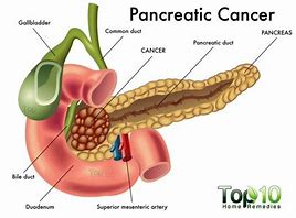
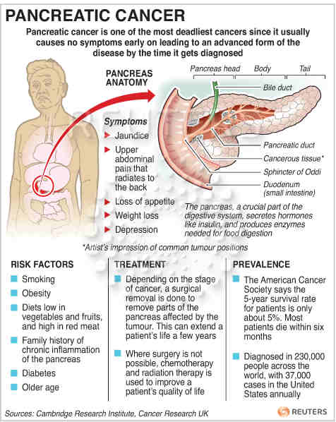
The top of pancreas attaches to the gall bile duct (allowing it into the head of the pancreas) and than there is the mesenteric artery.  Blood supply to the liver, pancreas and gallbladder is via the celiac artery (or celiac axis or celiac trunk). The celiac artery also supplies the duodenum, stomach and esophagus (the foregut and its derviatives). The pancreas is also supplied to some extent by the superior mesenteric artery that goes through the head of the pancreas.  This is how metastasis occurs (spreading) of pancreatic cancer can occur.  These arteries allow cancerous cells thorough the head into the bile duct into the blood stream and metastasis can now happen This can’t occur in the tail of the pancreas, its not attached to anything; which is the best place for it to occur & be diagnosed versus the head of the pancreas due to location.

Pancreatic cancer is hard to find early. The pancreas is deep inside the body, so early tumors can’t be seen or felt by health care providers during routine physical exams. People usually have no symptoms until the cancer has become very large or has already spread to other organs.

For certain types of cancer, screening tests or exams are used to look for cancer in people who have no symptoms (and who have not had that cancer before). But for pancreatic cancer, no major professional groups currently recommend routine screening in people who are at average risk. This is because no screening test has been shown to lower the risk of dying from this cancer, unfortunately.

Genetic History is one of the most common risk factors in getting most cancers, including Pancreatic.  Some people might be at increased risk of pancreatic cancer because of a family history of the disease (or a family history of certain other cancers). Sometimes this increased risk is due to a specific genetic syndrome.

Genetic testing looks for the gene changes that cause these inherited conditions and increase pancreatic cancer risk. The tests look for these inherited conditions, not pancreatic cancer itself. Your risk may be increased if you have one of these conditions, but it doesn’t mean that you have (or definitely will get) pancreatic cancer. 

Knowing if you are at increased risk can help you and your doctor decide if you should have tests to look for pancreatic cancer early, when it might be easier to treat. But determining whether you might be at increased risk is not simple. The American Cancer Society strongly recommends that anyone thinking about genetic testing talk with a genetic counselor, nurse, or doctor (qualified to interpret and explain the test results) before getting tested. It’s important to understand what the tests can − and can’t − tell you, and what any results might mean, before deciding to be tested.

For people in families at high risk of pancreatic cancer, newer tests for detecting pancreatic cancer early may help. The two most common tests used are an endoscopic ultrasound or MRI. These tests are not used to screen the general public, but might be used for someone with a strong family history of pancreatic cancer or with a known genetic syndrome that increases their risk. Doctors have been able to find early, treatable pancreatic cancers in some members of high-risk families with these tests.

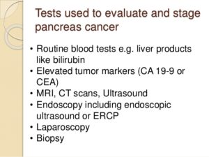
Tests for Pancreatic Cancer and even other Cancers:

A.)  Doctor’s Visit

The M.D. is usually the first thing done and the M.D. will ask about your medical history to learn more about your symptoms. The doctor might also ask about possible risk factors, including smoking and your family history.  Your doctor will also do a physical examine you to look for signs of pancreatic cancer or other health problems.

Doctors are also studying other new tests to try to find pancreatic cancer early.  Interested families at high risk may wish to take part in studies of these new screening tests.

B.)  Imaging Testing=It can be used:

  • To look for suspicious areas that might be cancer
  • To learn how far cancer may have spread
  • To help determine if treatment is working
  • To look for signs of cancer coming back after treatment

1.) CT Scan-detailed cross-sectional images of the body/pancreas.  Their are special types of CT known as a multiphase CT scan or a pancreatic protocol CT scan. During this test, different sets of CT scans are taken over several minutes after you get an injection of an intravenous (IV) contrast.  CT-guided needle biopsy: CT scans can also be used to guide a biopsy needle into a suspected pancreatic tumor.

2.) MRI(magnetic resonance imagery)-uses radio waves and strong magnets instead of x-rays to make detailed images of parts of your body. Most doctors prefer to look at the pancreas with CT scans, but an MRI might also be done.  Special types of MRI scans that can also be used are:

-MR cholangiopancreatography (MRCP), which can be used to look at the pancreatic and bile ducts, is described below in the section on cholangiopancreatography.

-MR angiography (MRA), which looks at blood vessels, is mentioned below in the section on angiography.

3. ) Ultrasound (US) tests

These tests use sound waves to create images of organs such as the pancreas. The two most commonly used types for pancreatic cancer:

A-Abdominal ultrasound – If it’s not clear what might be causing a person’s abdominal symptoms, this might be the first test done because it is easy to do and it doesn’t expose a person to radiation. But if signs and symptoms are more likely to be caused by pancreatic cancer, a CT scan is often more useful.

B-Endoscopic ultrasound (EUS): This test is more accurate than abdominal US and can be very helpful in diagnosing pancreatic cancer. This test is done with a small US probe on the tip of an endoscope, which is a thin, flexible tube that doctors use to look inside the digestive tract and to get biopsy samples of a tumor (more invasive but more detailed in results of the pancreas).

4.) Cholangiopancreatography

Abdominal This is an imaging test that looks at the pancreatic ducts and bile ducts to see if they are blocked, narrowed, or dilated. These tests can help show if someone might have a pancreatic tumor that is blocking a duct. They can also be used to help plan surgery.  If signs and symptoms are more likely to be caused by pancreatic cancer, a CT scan is often more useful.

A – Endoscopic ultrasound (EUS): This test is more accurate than abdominal US and can be very helpful in diagnosing pancreatic cancer. This test is done with a small US probe on the tip of an endoscope, which is a thin, flexible tube that doctors use to look inside the digestive tract and to get biopsy samples of a tumor.

SPYGLASS. This novel technology provides a direct view of the bile duct system, enabling our doctors to visualize lesions and narrowed areas (strictures) in the ducts and to biopsy them to see if they are cancerous. This approach ensures highly accurate sampling of the area in question. It is an excellent tool to use with confocal endomicroscopy.

B – Magnetic resonance cholangiopancreatography (MRCP):This is a non-invasive way to look at the pancreatic and bile ducts using the same type of machine used for standard MRI scans. Unlike ERCP, it does not require an infusion of a contrast dye. Because this test is non-invasive, doctors often use MRCP if the purpose is just to look at the pancreatic and bile ducts. But this test can’t be used to get biopsy samples of tumors or to place stents in ducts; like ERCP. can do also.

5.) Percutaneous transhepatic cholangiography (PTC): In this procedure, the doctor puts a thin, hollow needle through the skin of the belly and into a bile duct within the liver. A contrast dye is then injected through the needle, and x-rays are taken as it passes through the bile and pancreatic ducts. As with ERCP, this approach can also be used to take fluid or tissue samples or to place a stent into a duct to help keep it open. Because it is more invasive (and might cause more pain), PTC is not usually used unless ERCP has already been tried or can’t be done for some reason.

5.) Positron emission tomography (PET) scan

For a PET scan, you are injected with a slightly radioactive form of sugar, which collects mainly in cancer cells. A special camera is then used to create a picture of areas of radioactivity in the body.

This test is sometimes used to look for spread from exocrine pancreatic cancers.

PET/CT scan: Special machines can do both a PET and CT scan at the same time.

Pancreatoscopy. Here what is used is a small camera to visualize the pancreatic duct.

This test can help determine the stage (extent) of the cancer.  It might be able to detect metastasis (spreading beyond the pancreas).  See the top anatomy picture provided to understand this better by knowing the location of the organ to other parts of the body, if needed.