Part I Stroke (CVA) Awareness – what it is the types of strokes and the (including what a TIA is) risk factors for putting you at risk for a stroke!

stroke 1b

stoke 1a


Statistics to know about strokes:

Stroke was the third leading cause of death in the United States. Presently the CDC says under its statistics that Stroke is the fifth leading cause of death for Americans, but the risk of having a stroke varies with many factors.  Remember strokes have declined since 2003 based on CDC risk factors now compared to risk factors CDC listed in 2014.

More than 140,000 people die each year from stroke in the United States, that’s 1 out of every 20 deaths now.

Stroke costs the United States an estimated $34 billion each year.  This total includes the cost of health care services, medicines to treat stroke, and missed days of work.

Stroke is the leading cause of serious, long-term disability in the United States. Each year, approximately 795,000 people suffer a stroke.

About 600,000 of these are first attacks, and 185,000 are recurrent attacks. Nearly three-quarters of all strokes occur in people over the age of 65.  Stroke reduces mobility in more than half of stroke survivors age 65 and over.

The risk of having a stroke more than doubles each decade after the age of 55.

Strokes can & do occur at ANY age. Nearly one fourth of strokes occur in people under the age of 65.  Stroke risk increases with age, but strokes can—and do—occur at any age.

In 2009, 34% of people hospitalized for stroke were less than 65 years old.

Stroke death rates are higher for African-Americans than for whites, even at younger ages.

On average, someone in the United States has a stroke every 40 seconds.

Stroke accounted for about one of every 17 deaths in the United States in 2006. Stroke mortality for 2005 was 137,000.

From 1995–2005, the stroke death rate fell ~30 percent and the actual number of stroke deaths declined ~14 percent.  It still has declined from 2005 by CDC statistics which is good.

While stroke death rates have declined for decades among all race/ethnicities, Hispanics have seen an increase in death rates since 2013.

What is a Stroke?

A Stroke is a disease that affects the arteries leading to and within the brain. It is the No. 5 cause of death and a leading cause of disability in the United States.  A stroke occurs when a blood vessel which carry oxygen and nutrients to the brain is either blocked by a clot or bursts (or ruptures). When that happens, part of the brain cannot get the blood (and oxygen) it needs, so this in the end causes brain cells to die.

There is a stroke noted as a mini stroke which is a transient (temporary) ischemic attack= TIA, which we went over last Friday.  If you want to review it go right ahead, its listed under 5/08/2020 article.  A TIA is different than  strokes.  First some call it a mini stroke but remember the symptoms of a TIA are similar to stroke symptoms listed below for actual strokes; the difference for the patient is that they are completely reversible.  Take angina for example, in this case the pt has the heart affected but the symptoms are completely reversible, just a different organ.  The organs (the heart for angina and the brain for TIA) are simply having the symptoms of an infarction of the organ that is involved but both are due to lack of 0xygen, called ischemia.

Getting back to actual strokes lets review types of strokes.

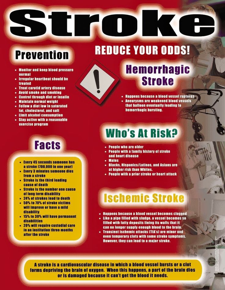
There are 2 types of strokes:

Ischemic Stroke which are strokes that occur through an obstruction of blood flow by a clot called a thrombus.

Hemorrhagic stroke by a blood vessel rupturing and preventing blood flow to the brain.

In the case of a stroke its a infarction to the brain due to lack of oxygen to the organ we call the brain, only the symptoms are not reversible but they can decrease in the intensity of the damage the caused in time with treatment (PT and OT) in time.  For some the symptoms are almost completely gone, again it depends on the intensity of the stroke to the brain, how bad was it with the symptoms it caused.

What are the risk factors for a stroke?

Non modiafiable risks meaning you can’t change them are:

Age:  Stroke occurs in all age groups.  Studies show the risk of stroke doubles for each decade between the ages of 55 and 85.  But strokes also can occur in childhood or adolescence.  Although stroke is often considered a disease of aging, the risk of stroke in childhood is actually highest during the perinatal period, which encompasses the last few months of fetal life and the first few weeks after birth.

-Gender:  Men have a higher risk for stroke, but more women die from stroke.  Men generally do not live as long as women, so men are usually younger when they have their strokes and therefore have a higher rate of survival.

-Race:  People from certain ethnic groups have a higher risk of stroke.  For African Americans, stroke is more common and more deadly—even in young and middle-aged adults—than for any ethnic or other racial group in the United States.  Studies show that the age-adjusted incidence of stroke is about twice as high in African Americans and Hispanic Americans as in Caucasians.  An important risk factor for African-Americans is sickle cell disease, which can cause a narrowing of arteries and disrupt blood flow. The incidence of the various stroke subtypes also varies considerably in different ethnic groups.

Family history of stroke:  Stroke seems to run in some families.  Several factors may contribute to familial stroke.  Members of a family might have a genetic tendency for stroke risk factors, such as an inherited predisposition for high blood pressure (hypertension) or diabetes.  The influence of a common lifestyle among family members also could contribute to familial stroke.

Modiafiable Risk Factors meaning you CAN change them:

High Blood Pressure (hypertension)

RX: DIET & EXERCISE & MEDS that a MD would decide.

High Cholesterol

RX: DIET and if necessary MEDS that a MD would decide.

Diabetes Mellitus

RX: DIET & EXERCISE & MEDS that a MD would decide.

Cigarette Smoking

RX: QUIT

Carotid Artery Disease

RX: DIET & EXERCISE & MEDS  even possible SURGERY that a MD would decide.

Atrial Fibrillation

RX: DIET & EXERCISE & MEDS even possible SURGERY that a MD would decide.

Unhealthy Diet   RX: DIET

Physical Inactivity and Obesity

RX: DIET & EXERCISE & possibly even MEDS that a MD would decide.

Go to your DOCTOR before doing any program and let your MD tell you what type of a program would be best for you especially if you are diagnosed with disease (EX. Diabetes, Cardiac, etc…).

QUOTE FOR WEDNESDAY:

“Urothelial carcinoma accounts for the vast majority of bladder cancer cases in the United States, about 90%. It is sometimes called transitional cell carcinoma (TCC) because the urothelial cells from which it develops are also known as transitional cells. This is because they may expand when the bladder is full and contract when it is empty.

These urothelial (transitional) cells line the bladder and, as more cancer cells grow, they may form a cancerous tumor.

The exact cause of bladder cancer is not known, but researchers have identified some factors that may increase a person’s lifetime risk for developing this cancer.

These risk factors may be linked to DNA changes in the body, causing mutations in the parts of cells that control growth. These mutations change how bladder cells behave, allowing them to grow abnormally, which may cause cancer cells to form.

Gene changes that occur during a person’s life sometimes happen randomly, without a known cause. Other times, exposure to chemicals that cause cancer, such as tobacco smoke, may be what prompts cells in the bladder to mutate. About 50% of people diagnosed with bladder cancer have a history of smoking.”

City of Hope (What is Bladder Cancer & Its Causes | City of Hope)


Part III What are the bladder cancer types? Learn the type of tumors, what urothelial carcinoma (UTUC) is?

The differences between UTUC, Small cell carcinoma, squamous cell carcinoma, adenocarcinoma, and NMIBC and Urothelial Carcinoma of the Bladder are as follows:

  • UTUC (Urothelial Carcinoma of the Ureter): Develops in mesoderm-derived epithelium and has lymphatic drainage patterns that vary by anatomical location.
  • Small cell carcinoma: Has oat-grain shaped cells and is found in cancers like lung, prostate, and pancreatic neuroendocrine tumors.
  • Squamous cell carcinoma: Cells look flat and are often arranged like tiles on a floor.
  • Adenocarcinoma: Arises from glandular cells and is found in organs like the lungs, breast, and colon.
  • NMIBC (Non-Malignant Invasive Bladder Carcinoma): A precursor lesion that is not yet malignant but may progress to bladder cancer.

NMIBC and Urothelial Carcinoma of the Bladder

About 9 out of 10 bladder cancers are urothelial carcinoma (also called transitional cell carcinoma). They start in the cells on the surface of the bladder’s inner linings. Most urothelial carcinomas are a form of non-muscle invasive bladder cancer (NMIBC). That means the tumor stays within the bladder’s inner lining.

Urothelial carcinoma also has rarer subtypes, called variants. Each one has a different treatment. We identify the variant based on how the cells look under a microscope. The variants are called:

  • Plasmacytoid
  • Nested
  • Micropapillary
  • Lipoid cell
  • Sarcomatoid
  • Microcystic
  • Lymphoepithelioma-like
  • Inverted papilloma-like
  • Clear cell

Cancer occurs when cells in the bladder start to grow out of control. Most tumours develop on the inner layer of the bladder. Some can grow into the deeper bladder layers. As cancer grows through these layers into the wall, it becomes harder to treat. The lining, where tumours initiate, is also found in the inner layers of the kidneys, ureters, and urethra. So, similar cancers can occur in these areas, though much less frequently.

Three types of bladder cancer may form, and each type of tumor can be present in one or more areas of the bladder, and more than one type can be present at the same time:

  • Papillary tumors stick out from the bladder lining on a stalk. They tend to grow into the bladder cavity, away from the bladder wall, instead of deeper into the layers of the bladder wall.
  • Sessile tumors lie flat against the bladder lining. Sessile tumors are much more likely than papillary tumors to grow deeper into the layers of the bladder wall.
  • Carcinoma in situ (CIS) is a cancerous patch of bladder lining, often referred to as a “flat tumor.” The patch may look almost normal or may look red and inflamed. CIS is a type of nonmuscle-invasive bladder cancer that is of higher grade and increases the risk of recurrence and progression. At diagnosis, approximately 10% of patients with bladder cancer present with CIS.

While the majority of bladder cancers (approximately 90-95%) arise in the bladder, the urothelial cells that line the bladder are found in other locations in the urinary system. Sometimes these urothelial cancers can occur in the lining of the kidney or in the ureter that connects the kidney to the bladder. This is known as upper tract urothelial cancer (UTUC) correspond to a subset of urothelial cancers that arise in the urothelial cells in the lining of the kidney (called the renal pelvis) or the ureter (the long, thin tube that connects that kidney to the bladder).

Upper Tract Urothelial Carcinoma, or UTUC, is urothelial carcinoma that occurs in the renal pelvis or ureter(s). Approximately 5-7% of urothelial cancer can occur in the inner lining of the kidney, called the calyx and renal pelvis. It could also occur in one or both of the ureter(s), tubes that lead from each of your kidneys to the bladder.

Types of urothelial carcinoma:

  • Non-invasive: More than half the people have this type, where the cancer remains in the urothelial cells that line the renal pelvis or ureters.
  • Invasive: the rest have this type, where the cancer has grown beyond those urothelial cells. Or it may have spread to other parts of the body.

Just as with bladder cancer, UTUC tumors can be low grade or high grade. The grade of the UTUC is important to know as you and your doctor choose the best treatment for your cancer. The grade of the tumor is determined by a pathologist who examines the cells under a microscope. Doctors may also use imaging studies to help them stage UTUC.

  • Low grade UTUC: In low grade UTUC, the tumors are typically noninvasive and are less aggressive.
  • High grade UTUC: High grade UTUC can be more aggressive. It may spread to other parts of your urinary tract, or to other parts of your body.

Ask your doctor to explain the details about your diagnosis and pathology report.

Understanding your UTUC combined with your overall health, will help your doctor recommend the best treatment options for your cancer.

Urothelial carcinoma (yoo-REE-thrul KAR-sih-NOH-muh) is by far the most common type of bladder cancer in the United States. Others are rarer. This section has information on some of these rarer types. Some rare bladder cancers are more common outside of the United States.

Squamous Cell Carcinoma of the Bladder

This cancer begins in the thin, flat squamous cells that can form in the bladder after chronic inflammation (swelling) and infection. It’s most often found in areas, such as the Middle East, where a parasitic infection called schistosomiasis is common. In North America and Europe, squamous cell carcinoma is the second most common bladder cancer. It accounts for about 5 out of every 100 cases.

Adenocarcinoma of the Bladder

This rare form of bladder cancer accounts for about 1 out of every 100 cases of the disease. denocarcinoma (A-deh-noh- KAR-sih-NOH-muh) can be caused by certain bladder problems you’re born with. It’s also caused by chronic infection and inflammation.

Small Cell Carcinoma of the Bladder

This form of the disease can spread very quickly. It’s often, but not always, found at an advanced stage, after it has metastasized (spread). Small cell bladder cancers usually need a combination of treatments, including chemotherapy, surgery, and radiation therapy.

Small cell carcinoma starts in small, nerve-like cells in the bladder called neuroendocrine (NOOR-oh-EN-doh-krin) cells. It makes up about 1 out of every 100 cases of bladder cancers.

QUOTE FOR TUESDAY:

“Finding cancer early, when it’s small and hasn’t spread, often allows for more treatment options. Some early cancers may have signs and symptoms that can be noticed, but that’s not always the case.

After a cancer diagnosis, staging provides important information about the extent (amount) of cancer in the body and the likely response to treatment.

Bladder cancer signs and symptoms may include:

  • Blood in urine (hematuria), which may cause urine to appear bright red or cola colored, though sometimes the urine appears normal and blood is detected on a lab test
  • Frequent urination
  • Painful urination
  • Back pain
  • Feeling tired or weak (advanced)
  • Swelling in the feet (advanced)
  • Bone pain (advanced)”

American Cancer Society  (Bladder Cancer Signs and Symptoms | American Cancer Society)


Part II Bladder Cancer – the prevention, how its diagnosed, the staging. treatments and after treatments!


Prevention of bladder cancer:

Although there’s no guaranteed way to prevent bladder cancer, you can take steps to help reduce your risk. For instance:

  • Don’t smoke. If you don’t smoke, don’t start. If you smoke, talk to your doctor about a plan to help you stop. Support groups, medications and other methods may help you quit.
  • Take caution around chemicals. If you work with chemicals, follow all safety instructions to avoid exposure.
  • Choose a variety of fruits and vegetables. Choose a diet rich in a variety of colorful fruits and vegetables. The antioxidants in fruits and vegetables may help reduce your risk of cancer.

How bladder cancer is diagnosed could include the following:

  • Using a scope to examine the inside of your bladder (cystoscopy). To perform cystoscopy, your doctor inserts a small, narrow tube (cystoscope) through your urethra. The cystoscope has a lens that allows your doctor to see the inside of your urethra and bladder, to examine these structures for signs of disease. Cystoscopy can be done in a doctor’s office or in the hospital.
  • Removing a sample of tissue for testing (biopsy). During cystoscopy, your doctor may pass a special tool through the scope and into your bladder to collect a cell sample (biopsy) for testing. This procedure is sometimes called transurethral resection of bladder tumor (TURBT). TURBT can also be used to treat bladder cancer.
  • Examining a urine sample (urine cytology). A sample of your urine is analyzed under a microscope to check for cancer cells in a procedure called urine cytology.
  • Imaging tests. Imaging tests, such as computerized tomography (CT) urogram or retrograde pyelogram, allow your doctor to examine the structures of your urinary tract.During a CT urogram, a contrast dye injected into a vein in your hand eventually flows into your kidneys, ureters and bladder. X-ray images taken during the test provide a detailed view of your urinary tract and help your doctor identify any areas that might be cancer.Retrograde pyelogram is an X-ray exam used to get a detailed look at the upper urinary tract. During this test, your doctor threads a thin tube (catheter) through your urethra and into your bladder to inject contrast dye into your ureters. The dye then flows into your kidneys while X-ray images are captured.

Determining the extent of the cancer

After confirming that you have bladder cancer, your doctor may recommend additional tests to determine whether your cancer has spread to your lymph nodes or to other areas of your body.

Tests may include:

  • CT scan
  • Magnetic resonance imaging (MRI)
  • Positron emission tomography (PET)
  • Bone scan
  • Chest X-ray

Staging of Bladder Cancer:

Your doctor uses these diagnostic tests listed above for information from these procedures to assign your cancer a stage.

The stages of bladder cancer are indicated by Roman numerals ranging from 0 to IV. The lowest stages indicate a cancer that’s confined to the inner layers of the bladder and that hasn’t grown to affect the muscular bladder wall. The highest stage — stage IV — indicates cancer that has spread to lymph nodes or organs in distant areas of the body, like a lot of other cancers are staged I through IV.

Treatments of bladder cancer:

If cancer invades the muscles of the bladder, doctors will usually treat it with chemotherapy to shrink the tumor, followed by surgery to remove the bladder. However, a recent clinical trial found that adding immunotherapy to chemotherapy may allow certain patients to avoid surgery.

Bladder cancer treatment may include: Surgery, to remove the cancer cells. Chemotherapy in the bladder (intravesical chemotherapy), to treat cancers that are confined to the lining of the bladder but have a high risk of recurrence or progression to a higher stage.

Approaches to bladder cancer surgery might be used could include:

  • Transurethral resection of bladder tumor (TURBT). TURBT is a procedure to diagnose bladder cancer and to remove cancers confined to the inner layers of the bladder — those that aren’t yet muscle-invasive cancers. During the procedure, a surgeon passes an electric wire loop through a cystoscope and into the bladder. The electric current in the wire is used to cut away or burn away the cancer. Alternatively, a high-energy laser may be used.Because doctors perform the procedure through the urethra, you won’t have any cuts (incisions) in your abdomen.As part of the TURBT procedure, your doctor may recommend a one-time injection of cancer-killing medication (chemotherapy) into your bladder to destroy any remaining cancer cells and to prevent cancer from coming back. The medication remains in your bladder for a period of time and then is drained.
  • Cystectomy. Cystectomy is surgery to remove all or part of the bladder. During a partial cystectomy, your surgeon removes only the portion of the bladder that contains a single cancerous tumor.A radical cystectomy is an operation to remove the entire bladder and the surrounding lymph nodes. In men, radical cystectomy typically includes removal of the prostate and seminal vesicles. In women, radical cystectomy may involve removal of the uterus, ovaries and part of the vagina.Radical cystectomy can be performed through an incision on the lower portion of the belly or with multiple small incisions using robotic surgery. During robotic surgery, the surgeon sits at a nearby console and uses hand controls to precisely move robotic surgical instruments.
  • Neobladder reconstruction. After a radical cystectomy, your surgeon must create a new way for urine to leave your body (urinary diversion). One option for urinary diversion is neobladder reconstruction. Your surgeon creates a sphere-shaped reservoir out of a piece of your intestine. This reservoir, often called a neobladder, sits inside your body and is attached to your urethra. The neobladder allows most people to urinate normally. A small number of people difficulty emptying the neobladder and may need to use a catheter periodically to drain all the urine from the neobladder.
  • Ileal conduit. For this type of urinary diversion, your surgeon creates a tube (ileal conduit) using a piece of your intestine. The tube runs from your ureters, which drain your kidneys, to the outside of your body, where urine empties into a pouch (urostomy bag) you wear on your abdomen.
  • Continent urinary reservoir. During this type of urinary diversion procedure, your surgeon uses a section of intestine to create a small pouch (reservoir) to hold urine, located inside your body. You drain urine from the reservoir through an opening in your abdomen using a catheter a few times each day.

Chemotherapy drugs can be given:

  • 1-Through a vein (intravenously). Intravenous chemotherapy is frequently used before bladder removal surgery to increase the chances of curing the cancer. Chemotherapy is going in your system generally through the blood stream and chemo may also be used to kill cancer cells that might remain after surgery. In certain situations, chemotherapy may be combined with radiation therapy.
  • 2-Directly into the bladder (intravesical therapy). During intravesical chemotherapy, a tube is passed through your urethra directly to your bladder. The chemotherapy is placed in the bladder for a set period of time before being drained. It can be used as the primary treatment for superficial bladder cancer, where the cancer cells affect only the lining of the bladder and not the deeper muscle tissue.

Radiation therapy:

Radiation therapy uses beams of powerful energy, such as X-rays and protons, to destroy the cancer cells. Radiation therapy for bladder cancer usually is delivered from a machine that moves around your body, directing the energy beams to precise points.

Radiation therapy is sometimes combined with chemotherapy to treat bladder cancer in certain situations, such as when surgery isn’t an option or isn’t desired at that time or ever depending on your stage of cancer.

Immunotherapy:

Immunotherapy is a drug treatment that helps your immune system to fight cancer.

Immunotherapy can be given:

  • Directly into the bladder (intravesical therapy). Intravesical immunotherapy might be recommended after TURBT for small bladder cancers that haven’t grown into the deeper muscle layers of the bladder. This treatment uses bacillus Calmette-Guerin (BCG), which was developed as a vaccine used to protect against tuberculosis. BCG causes an immune system reaction that directs germ-fighting cells to the bladder.
  • Through a vein (intravenously). Immunotherapy can be given intravenously for bladder cancer that’s advanced or that comes back after initial treatment. Several immunotherapy drugs are available. These drugs help your immune system identify and fight the cancer cells.

Targeted therapy:

Targeted therapy drugs focus on specific weaknesses present within cancer cells. By targeting these weaknesses, targeted drug treatments can cause cancer cells to die. Your cancer cells may be tested to see if targeted therapy is likely to be effective.

Targeted therapy may be an option for treating advanced bladder cancer when other treatments haven’t helped.

Bladder preservation:

In certain situations, people with muscle-invasive bladder cancer who don’t want to undergo surgery to remove the bladder may consider trying a combination of treatments instead. Known as trimodality therapy, this approach combines TURBT, chemotherapy and radiation therapy.

First, your surgeon performs a TURBT procedure to remove as much of the cancer as possible from your bladder while preserving bladder function. After TURBT, you undergo a regimen of chemotherapy along with radiation therapy.

If, after trying trimodality therapy, not all of the cancer is gone or you have a recurrence of muscle-invasive cancer, your doctor may recommend a radical cystectomy.

After bladder cancer treatment:

Bladder cancer may recur, even after successful treatment. Because of this, people with bladder cancer need follow-up testing for years after successful treatment. What tests you’ll have and how often depends on your type of bladder cancer and how it was treated, among other factors.

In general, doctors recommend a test to examine the inside of your urethra and bladder (cystoscopy) every three to six months for the first few years after bladder cancer treatment. After a few years of surveillance without detecting cancer recurrence, you may need a cystoscopy exam only once a year. Your doctor may recommend other tests at regular intervals as well.

People with aggressive cancers may undergo more-frequent testing. Those with less aggressive cancers may undergo testing less often.


QUOTE FOR MONDAY:

“Bladder cancer is a common type of cancer that begins in the cells of the bladder. The bladder is a hollow muscular organ in your lower abdomen that stores urine.

Bladder cancer most often begins in the cells (urothelial cells) that line the inside of your bladder. Urothelial cells are also found in your kidneys and the tubes (ureters) that connect the kidneys to the bladder. Urothelial cancer can happen in the kidneys and ureters, too, but it’s much more common in the bladder.

Most bladder cancers are diagnosed at an early stage, when the cancer is highly treatable. But even early-stage bladder cancers can come back after successful treatment. For this reason, people with bladder cancer typically need follow-up tests for years after treatment to look for bladder cancer that recurs.”

MAYO Clinic (Bladder cancer – Symptoms and causes – Mayo Clinic)

Part I Bladder Cancer-including signs/symptoms, how common is this cancer, types of bladder cancer, the survival rate, and risk factors!

Bladder cancer is a common type of cancer that begins in the cells of the bladder. The bladder is a hollow muscular organ in your lower abdomen that stores urine.

Bladder cancer most often begins in the cells (urothelial cells) that line the inside of your bladder. Urothelial cells are also found in your kidneys and the tubes (ureters) that connect the kidneys to the bladder. Urothelial cancer can happen in the kidneys and ureters, too, but it’s much more common in the bladder.

Most bladder cancers are diagnosed at an early stage, when the cancer is highly treatable. But even early-stage bladder cancers can come back after successful treatment like many other cancers. For this reason, people with bladder cancer typically need follow-up tests for years after treatment to look for bladder cancer that recurs.

Signs and symptoms of Bladder Cancer may include the following:

  • Blood in urine (hematuria), which may cause urine to appear bright red or cola colored, though sometimes the urine appears normal and blood is detected on a lab test
  • Frequent urination
  • Painful urination
  • Back pain

Bladder cancer begins when cells in the bladder develop changes (mutations) in their DNA. A cell’s DNA contains instructions that tell the cell what to do. The changes tell the cell to multiply rapidly and to go on living when healthy cells would die. The abnormal cells form a tumor that can invade and destroy normal body tissue. In time, the abnormal cells can break away and spread (metastasize) through the body.

How common is Bladder cancer:

Urinary Bladder Cancer is the sixth most common type of cancer in the United States. In 2024, approximately 83,190 people will receive a diagnosis of bladder cancer, and some 16,840 people are expected to die from it, according to estimates by the Surveillance, Epidemiology, and End Results Program (SEER) of the National Cancer Institute (NCI).

Bladder cancer is most likely to affect white men. In fact, the incidence rate of bladder cancer is four times higher among men than among women. It is also twice as high in white males as among Black, Hispanic or Asian/Pacific Islander men.

The NCI explains that there are three major types of bladder cancer. The name of each type of cancer indicates the kind of cells that become malignant.

  • Transitional cell carcinoma begins in cells in the innermost tissue layer of the bladder.
  • Squamous cell carcinoma begins in the squamous cells. This type may form after long-term infection or irritation.
  • Adenocarcinoma begins in glandular (secretory) cells in the lining of the bladder.

Cancer in the lining of the bladder is called superficial bladder cancer. Cancer that has spread through the lining of the bladder and has invaded the muscle wall of the organ, or has spread to nearby organs and lymph nodes, is called invasive bladder cancer.

Survival Rate:

Approximately 78 percent of people diagnosed with bladder cancer survived five years or more after diagnosis between 2012 and 2019, according to federal estimates.

Types of bladder cancer:

Different types of cells in your bladder can become cancerous. The type of bladder cell where cancer begins determines the type of bladder cancer. Doctors use this information to determine which treatments may work best for you.

Types of bladder cancer include:

  • Urothelial carcinoma. Urothelial carcinoma, previously called transitional cell carcinoma, occurs in the cells that line the inside of the bladder. Urothelial cells expand when your bladder is full and contract when your bladder is empty. These same cells line the inside of the ureters and the urethra, and cancers can form in those places as well. Urothelial carcinoma is the most common type of bladder cancer in the United States.
  • Squamous cell carcinoma. Squamous cell carcinoma is associated with chronic irritation of the bladder — for instance, from an infection or from long-term use of a urinary catheter. Squamous cell bladder cancer is rare in the United States. It’s more common in parts of the world where a certain parasitic infection (schistosomiasis) is a common cause of bladder infections.
  • Adenocarcinoma. Adenocarcinoma begins in cells that make up mucus-secreting glands in the bladder. Adenocarcinoma of the bladder is very rare.

Some bladder cancers include more than one type of cell.

Risk factors:

Factors that may increase bladder cancer risk include:

  • Smoking. Smoking cigarettes, cigars or pipes may increase the risk of bladder cancer by causing harmful chemicals to accumulate in the urine. When you smoke, your body processes the chemicals in the smoke and excretes some of them in your urine. These harmful chemicals may damage the lining of your bladder, which can increase your risk of cancer.
  • Increasing age. Bladder cancer risk increases as you age. Though it can occur at any age, most people diagnosed with bladder cancer are older than 55.
  • Being male. Men are more likely to develop bladder cancer than women are.
  • Exposure to certain chemicals. Your kidneys play a key role in filtering harmful chemicals from your bloodstream and moving them into your bladder. Because of this, it’s thought that being around certain chemicals may increase the risk of bladder cancer. Chemicals linked to bladder cancer risk include arsenic and chemicals used in the manufacture of dyes, rubber, leather, textiles and paint products.
  • Previous cancer treatment. Treatment with the anti-cancer drug cyclophosphamide increases the risk of bladder cancer. People who received radiation treatments aimed at the pelvis for a previous cancer have a higher risk of developing bladder cancer.
  • Chronic bladder inflammation. Chronic or repeated urinary infections or inflammations (cystitis), such as might happen with long-term use of a urinary catheter, may increase the risk of a squamous cell bladder cancer. In some areas of the world, squamous cell carcinoma is linked to chronic bladder inflammation caused by the parasitic infection known as schistosomiasis.
  • Personal or family history of cancer. If you’ve had bladder cancer, you’re more likely to get it again. If one of your blood relatives — a parent, sibling or child — has a history of bladder cancer, you may have an increased risk of the disease, although it’s rare for bladder cancer to run in families. A family history of Lynch syndrome, also known as hereditary nonpolyposis colorectal cancer (HNPCC), can increase the risk of cancer in the urinary system, as well as in the colon, uterus, ovaries and other organs.

QUOTE FOR THE WEEKEND:

“According to a 2023 report, the prevalence of ALS in the United States is about 9.1 cases per 100,000 people.

ALS (amyotrophic lateral sclerosis), commonly known as Lou Gehrig’s disease, is a rare neurological disorder affecting approximately 30,000 people in the U.S. It can run in families, but most cases have no family connection.      According to the National Organization for Rare Disorders, ALS develops in 1.5 to 3 people per 100,000 in the United States and Europe each year.

ALS affects the brain cells (neurons) that control your muscles. As ALS progresses, people lose voluntary control of their muscles and various bodily functions, including standing, chewing food, and speaking clearly.
The ALS Association suggests that men are about 20% more likely than women to develop ALS, though as people get older, the sex differences tend to even out.
The ALS Association reports that ALS is more common among non-Hispanic individuals, with white adults about twice as likely as Black adults to develop ALS.”

Part III ALS awareness month-What is the Rx?


Over the past decade, understanding of the multiple destructive pathways that lead to neuronal death in amyotrophic lateral sclerosis (ALS) has greatly improved.  There is still no cure for ALS unfortunately with all the technology medicine has but there are therapies.

Home Care at some point in middle to beginning of late stage.

This would include a Home Health Assistant followed under a Visiting Nurse/RN with an Attending Doctor for follow up on the pt care=A whole team of medical care including as soon as needed:

PT and OT:

Physical therapy (PT) is the use of exercises and treatments to improve physical movement and overall mobility. Occupational therapy (OT) is similar but is focused on developing or maintaining the physical skills needed to perform everyday tasks. For example, a physical therapist might prescribe stretching exercises to limit discomfort and preserve range of motion or use pool therapy to help you walk and improve joint function. An occupational therapist may help you find new ways to brush your teeth or recommend equipment that can make your activities of daily living easier to perform.

Speech Therapy:

For people living with ALS with bulbar symptoms, ALS leads to speech problems when it attacks bulbar neurons. These are the nerve cells responsible for bringing messages from the lower parts of the brain (bulbar region) to the muscles that move the lips, tongue, soft palate (back of roof of mouth), jaw, and vocal folds (voice box). As nerves are lost to the disease, the muscles they control become weak and tight. This causes dysarthria-difficulty speaking).  A speech-language pathologist can help with both speaking and swallowing difficulties. This may include finding devices to help you communicate as your speech becomes harder for others to understand.

Respiratory Therapy:

A respiratory therapist can teach you new techniques for breathing and coughing, helping you keep your airway and lungs clear and healthy. When mechanical ventilators are needed, they can help you evaluate the options and choose the best ventilator for your needs.  An ALS diagnosis is shocking and frightening, but as with any disease, knowledge is power. Being aware of symptoms and how you can prepare for – and manage – them is key to quality of life and often, for peace of mind. The impact of ALS on breathing is one of the most daunting aspects of the disease and one for which you and your family can and should prepare for early in the ALS journey.

Psychotherapy:

Feeling sad or scared after being diagnosed with ALS is completely natural.

In time the patient with ALS has there life completed turned upside down when they need assistance with there activitities of daily living from independent in their ADLs.  It’s not unusual to feel depressed or anxious after getting diagnosed with ALS. If you have difficulty coping with the mental and emotional side of ALS, a counselor or psychiatrist can help.

Potential symptoms of depression include:  Prolonged feelings of sadness, hopelessness, worthlessness, anxiety, or guilt.   Irritability or angry outbursts over small matters. Changes in sleeping patterns, including insomnia or sleeping too much.  Changes in appetite.

Medications:

There are a number of medications that can help treat the various symptoms of ALS and new drugs are being developed all the time. Talk with your doctor or therapist to find out what is currently available and whether any such medications might be right for you.

There are currently four drugs approved by the U.S. Food and Drug Administration to treat ALS (Radicava, Rilutek, Tiglutik, and Nuedexta). Studies all over the world, many funded by The ALS Association, are ongoing to develop more treatments and a cure for ALS.

Radicava™ (edaravone)

The FDA approved Radicava™ in 2017, less than a year after Mitsubishi Tanabe Pharma America submitted a new drug application, making it the first new treatment specifically for ALS in 22 years.

Rilutek (riluzole, now generic)

This was the first FDA-approved drug available to treat ALS — in 1995. It inhibits glutamate release and prolongs life approximately three months. Riluzole is the generic name of Rilutek.

Tiglutik (thickened riluzole)

The first and only thickened liquid form of riluzole, Tiglutik was approved by the FDA in September 2018. This formulation contrasts with the oral pill form of riluzole that has been on the market for ALS for more than 20 years. It is designed to avoid potential problems of crushing tablets.

Nuedexta®

Indicated for the treatment of pseudobulbar affect (PBA), which is characterized by frequent, involuntary, and often sudden episodes of crying and/or laughing that are exaggerated and/or don’t match how you feel. PBA occurs secondary to a variety of otherwise unrelated neurologic conditions. Nuedexta® (dextromethorphan HBr and quinidine sulfate) was FDA-approved in 2011.


QUOTE FOR FRIDAY:

“Amyotrophic lateral sclerosis (ALS) is a progressive neurological disorder that affects motor neurons in the brain and spinal cord, leading to the degeneration and death of these crucial nerve cells [1]. This fatal condition, often referred to as Lou Gehrig’s disease, causes a gradual loss of voluntary muscle control, impacting essential functions such as walking, talking, chewing, and breathing [2]. ALS typically manifests between the ages of 40 and 70,

Notably, ALS does not typically affect cognitive function, sensory perception, or bladder control [2]. While the exact cause remains unknown for most cases, approximately 10% are attributed to genetic factors, classified as familial ALS. The remaining 90% are considered sporadic, occurring randomly without a clear hereditary link [3]. Despite ongoing research, there is currently no cure for ALS, and the average life expectancy after diagnosis ranges from 3 to 5 years, although some individuals may survive for a decade or more [1].”

ALS United Ohio (ttps://alsohio.org/als-vs-ms-amyotrophic-lateral-sclerosis-vs-multiple-sclerosis/)