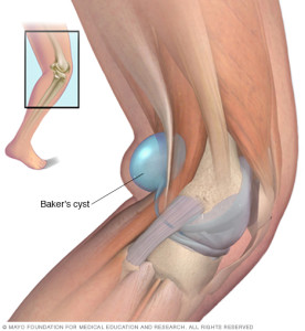
A Baker cyst is swelling caused by fluid from the knee joint protruding to the back of the knee. The back of the knee is also referred to as the popliteal area of the knee. A Baker cyst is sometimes referred to as a popliteal cyst. When an excess of knee joint fluid is compressed by the body weight between the bones of the knee joint, it can become trapped and separate from the joint to form the fluid-filled sac of a Baker cyst. The name of the cyst is in memory of the physician who originally described the condition, the British surgeon William Morrant Baker (1839-1896).
- A Baker cyst is swelling caused by fluid from the knee joint protruding to the back of the knee.
- Baker cysts are common and can be caused by virtually any cause of joint swelling (arthritis).
- A Baker cyst may not cause symptoms or be associated with knee pain and/or tightness behind the knee, especially when the knee is extended or fully flexed.
- Baker cysts can rupture and become complicated by spread of fluid down the leg between the muscles of the calf (dissection).
- Baker cysts can be treated with medications, joint aspiration and cortisone injection, and surgical operation, usually arthroscopic surgery.SIGNS & SYMPTOMS OF BAKER CYSTS:Baker cysts can become complicated by spread of fluid down the leg between the muscles of the calf (dissection). The cyst can rupture, leaking fluid down the inner leg to sometimes cause the appearance of a painless bruise under the inner ankle. Baker cyst dissection and rupture are frequently associated with swelling of the leg and can mimic phlebitis of the leg. A ruptured Baker cyst typically causes rapid-onset swelling of the leg.How is a Baker cyst treated? Baker cysts often resolve with aspiration (removal) of excess knee fluid in conjunction with cortisone injection. Medications are sometimes given to relieve pain and inflammation.
- When cartilage tears or other internal knee problems are associated, physical therapy or surgery can be the best treatment option. During a surgical operation, the surgeon can remove the swollen tissue (synovium) that leads to the cyst formation. This is most commonly done with arthroscopic surgery.
- DIAGNOSING BAKER CYSTS: Baker cysts can be diagnosed by the doctor’s examination and confirmed by imaging tests (either ultrasound, injection of contrast dye into the knee followed by imaging, called an arthrogram, or MRI scan) if necessary.
- A Baker cyst may cause no symptoms or be associated with knee pain and/or tightness behind the knee, especially when the knee is extended or fully flexed. Baker cysts are usually visible as a bulge behind the knee that is particularly noticeable on standing and when compared to the opposite uninvolved knee. They are generally soft and minimally tender.
- Baker cysts are not uncommon and can be caused by virtually any cause of joint swelling (arthritis). The excess joint fluid (synovial fluid) bulges to the back of the knee to form the Baker cyst. The most common type of arthritis associated with Baker cysts is osteoarthritis, also called degenerative arthritis. Baker cysts can occur in children with juvenile arthritis of the knee. Baker cysts also can result from cartilage tears (such as a torn meniscus), rheumatoid arthritis, and other knee problems.

Is it dangerous to have that cyst iam 45yrs it comes and goes what do i do
Get it evaluated and A Baker’s cyst is caused by swelling in the knee. The swelling is due to an increase in the fluid that lubricates the knee joint (synovial fluid). When pressure builds up, fluid bulges into the back of the knee.
Baker’s cyst commonly occurs with:
•A tear in the meniscal cartilage of the knee
•Knee arthritis (in older adults).
Also rheumatoid arthritis and other problems.
So when it swells up get it checked if not even before.
Shining a light through the cyst (translumination) can show that the growth is fluid filled.
Often no treatment is needed. The health care provider can watch the cyst over time. If painful or limiting your mobility of the lower extremity get evaluated by an MD.